From the Department of Gastroenterology, Division of Gastrosurgery and Coloproctology, Hospital das Clínicas, Faculty of Medicine, University of São Paulo – São Paulo/SP, Brazil.
E-mail: [email protected] Received for publication on
September 16, 2003.
CASE REPORT
UMBILICAL MASS AS THE SOLE PRESENTING
SYMPTOM OF PANCREATIC CANCER: A CASE
REPORT
Fábio Crescentini, Fernanda Deutsch, Carlos Walter Sobrado and Sérgio de Araújo
CRESCENTINI F et al. Umbilical mass as the sole presenting sign of pancreatic cancer: a case report. Rev. Hosp. Clín. Fac. Med. S. Paulo 59(4):198-202, 2004.
Umbilical nodes are rare. The metastatic involvement of the region was first described in 1846. Sister Mary Joseph was the first observer to establish the correlation between carcinomas and umbilical nodes. The umbilical node may be the sole presenting sign of cancer and is usually associated with advanced disease and poor prognosis.
A 64-year-old woman, previously healthy, presented vague abdominal discomfort and a hard umbilical nodule for 1 week, which was first diagnosed as an incarcerated umbilical hernia. She underwent a new clinical assessment and biopsy. After immunohistochemical analysis and computerized tomography, she was diagnosed with pancreatic cancer.
The clinical staging showed advanced disease with distant metastasis. She received palliative chemotherapy. After 8 months, she was alive in poor clinical condition.
Clinical suspicion should lead to a careful additional evaluation whenever an umbilical nodule presents with malignant signs.
KEY WORDS: Sister Mary Joseph’s nodule. Umbilical mass. Unknown primary site. Cancer.
Tumors of the umbilicus are rare. The metastatic involvement of the re-gion due to visceral carcinomas is even less frequent.1,2 The first medical report
of such a condition was by Walshe et al. in 1846, through the Tanchov’s re-view of data. The author found only 2 cases of umbilical involvement among 9118 deaths from cancer in the period of 1830 to1840.¹ In 1864 Storer et al. reported the first complete case study of a metastatic umbilical nodule due to gastric adenocarcinoma.³ However, the association of umbilical masses and visceral carcinomas was firmly es-tablished after the clinical observa-tions of a nurse from the Mayo Clinic (Rochester, USA). Her name was attrib-uted to this clinical feature, which was
designated as “Sister Mary Joseph’s” sign.
Even though it is a rare clinical finding, an umbilical mass may be the sole presentation of malignant tumors.4,5 Shetty¹ reviewed all cases
re-lated to “Sister Mary Joseph’s nodule” published until 1989. This review found a total of 265 cases and 85 nod-ules from unknown primary tumors. Concerning pancreatic tumors, only 12 cases had been reported.3-10 We found
only 1 description¹¹ since 1989, and in 1998 the metastatic involvement of the umbilicus from a pancreatic cancer was exhibited as the image of the month in Gastroenterology¹². We un-dertook this bibliographic research us-ing the PubMed database and the fol-lowing keywords: “Sister Mary Joseph’s nodule”, unknown primary tumor, pancreatic cancer, umbilical metastasis.
DESCRIPTION OF THE CASE
A 64-year-old woman presented with a 3-month history of vague ab-dominal discomfort in the epigastric region, with no irradiation sites, slowly progressive, and not related to food in-take. She had noticed an umbilical nodule the previous week. She did not present with weight loss, fever, or other systemic symptoms. She denied hav-ing cardiorespiratory discomfort or gastrointestinal complaints. Her refer-ral doctor (generefer-ral practice) first diag-nosed the mass as an incarcerated um-bilical mass. A surgeon re-examined and re-evaluated the patient. She pre-sented in good general condition, well nourished, no palpable masses at the cervical, abdominal, or thoracic re-gions. On physical examination, a peri-umbilical nodule of 2 by 2 cm was noted, which was hyper-pigmented, indurated, discretely painful, and pro-duced a fetid discharge.
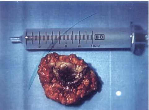
Routine laboratory investigation showed normal results. The sonographic scan of the abdomen showed no abnormalities. At the first physical and laboratory investigation, the patient presented evidence of tumoral disease, with unknown pri-mary tumor. Therefore, the umbilical nodule was resected for diagnostic purposes and an anatomic-pathologic analysis was performed (Figure 1), which showed a mucinous, poorly dif-ferentiated metastatic adenocarci-noma, suggestive of a GI tract (stom-ach or pancreas) or ovarian cancer. Im-munohistochemical analysis was posi-tive for cytokeratin 7 and negaposi-tive for cytokeratin 20. This finding suggested a primary site at stomach or pancreas.
Due to the anatomic-pathologic findings, an abdominal CT scan was performed. The exam showed a cystic and solid tumor of 6 cm in the pancre-atic body (Figure 2). For staging pur-poses, a thoracic CT scan was also per-formed, and several pulmonary
nod-ules, diagnosed as metastases, were en-countered (Figure 3). The brain CT scan showed no abnormalities.
The patient was referred to the medical oncology service for pallia-tive chemotherapy because of ad-vanced pancreatic cancer with
perito-neal dissemination and lung metastases. Even though the patient was alive 8 months after the diagno-sis, she progressively deteriorated and presented in poor clinical condition. She had undergone thoracocentesis 3 times for relief of severe dyspnea.
Figure 2 – Abdominal CT scan with IV contrast (cystic lesion with solid components in the pancreatic body).
DISCUSSION
The finding of an umbilical nod-ule as the only clinical manifestation of a disease leads to several possible diagnoses including umbilical hernia, granuloma, attachments of the urachus, pilonidal sinus, omphalomesenteric duct abnormalities, endometriosis, and benign and malignant tumors.7-13
Be-nign lesions are diagnosed in the vast majority of cases, and the finding of the metastatic involvement of the um-bilical region is rare.8-14 Shetty et al.
reviewed all cases of malignant disease at the umbilicus from 1830 to 1989 and found only 265 cases previously published; 85 cases of umbilical me-tastasis from unknown primary origin were found.¹
Several modes of spreading to the umbilicus have been discussed. Metas-tasis to the umbilicus may occur due to proximity to the tumor, hematogenic and lymphatic dissemi-nation, or via umbilical ligaments. The most prevalent form of umbilical in-volvement is related to direct invasion of peritoneal metastasis. The retro-grade flux from superficial and deep
lymphatic systems originated at axil-lary, inguinal, and para-aortic nodes may lead to umbilical involvement. Another possible form involves venous communication between the lateral thoracic veins and internal mammary vein with the portal circulation.14
Therefore, theoretically, all types of cancer may disseminate to the umbili-cal region. However, the most preva-lent primary sites are from intra-ab-dominal origin. Shetty (1990) re-viewed cases of umbilical metastasis previously published and found that 42% originated at the abdomen or pel-vis, mainly from the GI tract, classified as follows: gastric cancer 17%, large bowel 6%, pancreas 6%, gallbladder 3%, and small bowel 1%. He also found a large prevalence among tumors from the female genital tract, account-ing for 9% of all cases. Other primary sites, such as lung, cervix, fallopian tube, and melanoma were rare and rep-resented 1% of patients. However, this data should be analyzed cautiously since it was partially obtained before modern radiological techniques. Nearly 30% of cases had an unknown primary site.¹ In the present case report,
the lymphatic spread from the pancre-atic body to the umbilicus originated mostly through the splenic and para-aortic nodes, as described by Donatini et al.15 Although there were no signs
of carcinomatosis in the reported case, the umbilical involvement due to proximity to the primary site should always be investigated.
The metastatic umbilical nodule, known as Sister Mary Joseph’s nodule, is morphologically firm, an indurated plaque or nodule with a vascular ap-pearance, and may be fissured and ul-cerated with some fetid discharge. However, it may also present as an un-characteristic diffuse hardening of the umbilical region or as a profound node.16 Steck et al. observed that in
45% of their patients, the umbilical node was the only clinical sign of can-cer. This figure demonstrates the im-portance of an evaluation of all um-bilical lesions, especially in patients after the fifth decade of life.¹³
should be properly indicated and may not replace the full anatomic-pathologic analysis. Moreover, radio-logical exams, such as ultrasound imaging, CT scan, or MRI have a poor cost-benefit relationship due to their low diagnostic power. Those methods should be preserved for staging pur-poses or for special cases when the pa-thologist evaluation is not possible.
The metastatic spread to the
um-bilical region representnaan advanced stage of the primary disease and wors-ened prognosis. Consequently, pallia-tive treatment is usually the only re-maining therapeutic option. However, as previously discussna, an umbilical nodule may be the only presenting sign of cancer, enhancing the diagno-sis before the appearance of more exu-berant features, such as ascites, pulmo-nary masses, and bone metastasis,
which are responsible for decreased quality of life and overall survival even after the administration of pallia-tive treatment.
Even though it is a rare finnang, an umbilical mass may be the first mani-festation of neoplastic disease, as was observed in the present case. Therefore, the clinical suspicion and diagnostic evaluation are extremely important for therapeutic and prognostic purposes.
RESUMO
CRESCENTINI F e col. Nódulo um-bilical como única apresentação clínica de tumor pancreático: relato de caso. Rev. Hosp. Clín. Fac. Med. S. Paulo 59(4):198-202,
2004.
Nódulos umbilicais são raros. Des-de 1846, o comprometimento metas-tático da região vem sendo descrito. A Irmã Mary Joseph foi a primeira a re-lacionar o aparecimento de nódulos umbilicais com carcinomas. Esses nó-dulos podem ser a única manifestação
de câncer, normalmente associada a estadios avançados e pior prognóstico. Uma senhora de 64 anos, previa-mente hígida, apresentava desconfor-to abdominal inespecífico e apareci-mento de nódulo umbilical endureci-do há uma semana. O diagnóstico ini-cial foi hérnia umbilical encarcerada. Após reavaliação, o nódulo foi biop-siado, cujo exame anátomo-patológi-co demonstrou carcinoma anátomo-patológi-com sítio primário desconhecido. Á análise imuno-histoquímica e tomografia, o diagnóstico foi carcinoma de
pâncre-as. O estadiamento demonstrou doen-ça avandoen-çada, com metastáses à distân-cia. A paciente foi submetida a qui-mioterapia paliativa. Após 8 meses, encontrava-se em mau estado geral.
A suspeita clínica deve originar avaliação clínica cuidadosa, auxiliada por exames subsidiários, sempre que um nódulo umbilical apresentar sinais de malignidade.
UNITERMOS: Nódulo Sister Mary Jpseph. Massa umbilical. Tu-mor primário desconhecido. Cancer.
REFERENCES
1. Shetty MR. Metastatic tumors of the umbilicus: a review 1830-1989. J Surg Oncol 1990;45:212-5.
2. Edoute Y, Malberger E, Kuten A. Umbilical metastasis diagnosed by fine needle aspiration. J Surg Oncol 1990; 45:56-58. 3. Barrow MV. Metastatic tumors of the umbilicus. J Chron Dis
1966; 19:1113-17.
4. Bukovsky I, Lifshits Y, Langer R. Umbilical mass as a presenting symptom of endometrial adenocarcinoma. Gynecol Obstet Invest 1979;17:229-30.
5. Shvili D, Halevy S, Sandbank M. Umbilical metastasis as the presenting sign of pancreatic adenocarcinoma. Cutis 1983;5:555-6.
6. Charoenkul V, DelCampo A, Derby A, Hodgson WJ, McElhinney AJ. Tumors of the umbilicus. Mt Sinai J Med 1977;44:257-62.
7. Scarpa FJ, Dineen JP, Boltax RS. Visceral neoplasia presenting at the umbilicus. J Surg Oncol 1979;11:351-9.
8. Chatterjee SN, Bauer HM. Umbilical metastasis from carcinoma of the pancreas. Arch Dermatol 1980;166:954-5.
9. Sharaki M, Abdel-Kadner M. Umbilical deposits from internal malignancy. Clin Oncol 1981;7:351-5.
10. Heatley MK, Toner PG. Sister Mary Joseph’s nodule: a study of the incidence of biopsied umbilical secondary tumors in a defined population. Br J Surg 1989;76:728-9.
11. Foster MA, Gering SA, Bradley YC. Sister Mary Joseph’s sign from metastatic disease of the pancreas. J Am Coll Surg 2001;192:130.
13. Steck WD, Helwig EB. Tumors of umbilicus. Cancer 1965;18:907-15.
14. Powell FC, Cooper AJ, Massa MC, Goellner JR, Su WP. Sister Mary Joseph’s nodule: A clinical and histologic study. J Am Acad Dermatol 1984;10:610-5.
15. Donatini B, Hidden G. Routes of lymphatic drainage from the pancreas: a suggested segmentation. Surg Radiol Anat 1992; 14:35-42.
16. Khan AJ, Cook B. Metastatic carcinoma of umbilicus: “Sister Mary Joseph’s nodule”. Cutis 1997;60:297-8.