129
J Vasc Bras. 2013 Jun; 12(2):129-132
O R I G I N A L
A R T I C L E
Postoperative determination of the
degree of carotid artery stenosis
Determinação pós-operatória do grau de estenose na artéria carótida
J. Janzen1, P. Chenais1, W. Janzen1, F. Schibler2, J. Schmidli3
Abstract
his article describes the VascMorph 1a prototype software and reports irst results obtained with postoperative determination of the degree of stenosis in the carotid artery.
Keywords: degree of stenosis; atherosclerosis; carotid artery.
Resumo
Este artigo descreve o programa protótipo VascMorph 1a e apresenta os primeiros resultados obtidos com a determinação pós-operatória do grau de estenose na artéria carótida.
Palavras-chave: grau de estenose; aterosclerose; artéria carótida.
1 VascPath, Bern, Switzerland. 2 University of Bern – UNIBE, Switzerland.
3 Department of Vascular Surgery, University Hospital Bern – UNIBE, Switzerland
Postoperative determination of the degree of carotid artery stenosis
130 J Vasc Bras. 2013 Jun; 12(2):129-132
a correction factor is used to adjust ECST/NASCET values5.
POSTOPERATIVE DETERMINATION OF THE DEGREE OF CAROTID ARTERY STENOSIS (VASCMORPH 1A)
Differently from sonographic methods, in this paper the VascMorph 1a prototype software is proposed. This method represents a planimetric method that can be used postoperatively.
SAMPLE PREPARATION
Endarterectomy specimens were laminated after conventional formalin fixation and calcification from proximal to distal at a distance of 3-5 mm.
Subsequently, sections were embedded in parafin
and stained using the Elastica van Gieson method.
ASSESSMENT OF VASCULAR PATHOLOGY Arterial cross sections were assessed following routine microscopic diagnosis (Figure 2), as follows:
• Characterization of plaque coniguration and compo -sition (foam cells, cholesterol crystals, hemorrhage, inlammatory iniltrates, connective tissue cap, etc.); • Deinition of the degree of atherosclerosis according
to Stary’s classiication6 ; and
• Determination of the arterial type (elastic muscular, transition zone)7.
DETERMINATION OF THE DEGREE OF CAROTID ARTERY STENOSIS
Carotid artery stenosis was identiied and digitally
photographed (Figure 2) and uploaded to the VascMorph 1a software (Figure 3). After marking the ideal lumen (Figure 4) and the residual lumen (Figure 5), the degree of stenosis was calculated using the formula below:
Ideal volume – Residual volume × 100% Degree of stenosis (%) =
Ideal volume
COMPARISON OF PRE- AND POSTOPERATIVE MEASUREMENTS
A total of 56 endarterectomy samples were investigated. Preoperatively, the degree of stenosis was determined using magnetic resonance angiography. In these investigations, the mean degree of stenosis was 65.32% (standard deviation: 11.939; standard error of the mean: 1.595).
Postoperative determination of the degree of stenosis using VascMorph 1a revealed statistically higher results (p=0.005; t test), with a mean of
73.95% (standard deviation: 13.2073; standard error of the mean: 1.7649).
INTRODUCTION
Vascular surgeons and angiologists have often posed the question about the degree of stenosis of operated vascular specimens. With this goal in mind, the VascMorph 1a prototype software has been developed. The software performs a planimetric procedure which allows to determine the percentage
of stenosis. The VascMorph 1a was irst tested in
atherosclerotic carotid artery samples1.
The present paper describes the assessment of a total of 56 endarterectomy samples provided by the Department of Vascular Surgery of the University Hospital of Bern (Head of Department: Prof. Dr. J. Schmidli). Materials obtained from 20 women (35.7%) and 36 men (64.3%) were examined; mean age was 78.6 years among women and 74.9 years among men.
PREOPERATIVE DETERMINATION OF THE DEGREE OF CAROTID ARTERY STENOSIS
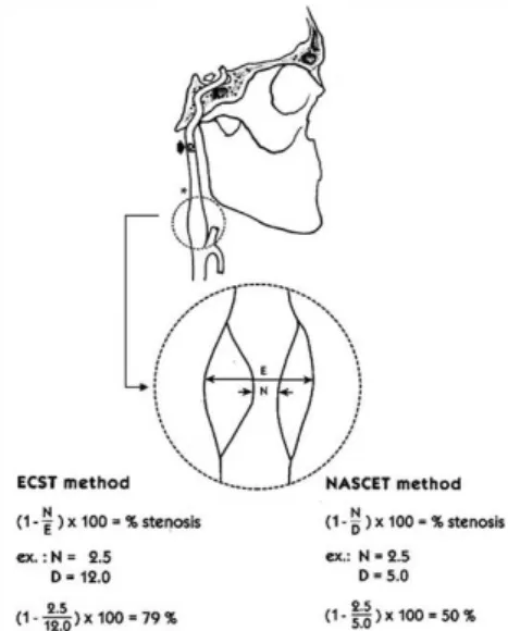
In order to examine preoperative stenosis, different sonographic and angiographic methods were used (e.g. color-coded duplex sonography, intra-arterial subtraction angiography, magnetic resonance angiography) according to the European Carotid Surgery Trial (ECST) or the North American Symptomatic Carotid Endarterectomy Trial (NASCET). In such diameter-related measurement methods, among other things, an idealized circular shape is assumed for both the vessel lumen and the stenosis. The degree of stenosis is not determined using planimetry, but rather based on differences in
the respective low rates (Figure 1)2-4. Occasionally,
J. Janzen, P. Chenais et al.
131
J Vasc Bras. 2013 Jun; 12(2):129-132
DISCUSSION
The VascMorph 1a prototype software is a promising tool for microscopic vascular diagnosis. The software not only yields reports on the degree of stenosis of paraffin embedded materials but also allows comparison with values determined preoperatively. The results of our pilot study (n=56)
demonstrated a signiicantly higher degree of stenosis calculated in parafin-embedded materials when
compared to pre-operative values obtained with magnetic resonance angiography, suggesting that calculations made using the planimetric method are more accurate than those made using the diameter-based method, because idealized circular-atherosclerotic stenoses are more complex in nature. In the diameter-based method, assumption of the stenosis circular shape causes slit-shaped, elliptical, and scalloped lumina to be ignored. Conversely, in the VascMorph 1a prototype software, different plaque morphologies were easily imaged.
It is worthy to note that about 5-10% shrinkage may occur as a result of histopathological treatment; this phenomenon equally affects the lumen and thin vascular walls. Conversely, in relatively rigid endarterectomy specimens which hold a high
proportion of minerals, no signiicant shrinkage is
expected. In our view, the distortion in measured values is quite low, and does not fall under the current literature classification related to postoperative perfusion of blood vessels8-11.
ACKNOWLEDGMENTS
We are thankful to Dipl.-Math. Brigitta Gahl for helping us with statistics and to Mr. Rafay Siddiqui, MSc, for his assistance in designing the manuscript.
Figure 2. Postoperative aspect and histological cross-sections of specimens.
Figure 3. Image of carotid artery stenosis uploaded to Vasc-Morph 1a.
Figure 4. Marking of ideal lumen parallel to internal elastic membrane.
Postoperative determination of the degree of carotid artery stenosis
132 J Vasc Bras. 2013 Jun; 12(2):129-132
8. Bahr GF, Bloom G, Friberg U. Volume changes of tissues in physiological fluids during fixation in osmium tetroxide or formaldehyde and during subsequent treatment. Exp Cell Res. 1957;12:342-3. http://dx.doi.org/10.1016/0014-4827(57)90148-9 9. Beneš V, Netuka D, Mandys V, et al. Comparison between
degree of carotid stenosis observed at angiography and in histological examination. Acta Neurochir (Wien). 2004;146:671-7. PMid:15197610. http://dx.doi.org/10.1007/s00701-004-0279-3 10. Bond MG, Insull W, Glagov S, et al. Clinical diagnosis of
atherosclerosis. Quantitative methods of evaluation. New York, Heidelberg, Berlin: Springer; 1983. p. 283-303. http://dx.doi. org/10.1007/978-1-4684-6277-7
11. Schwartz JN, Kong Y, Hackel DB, Bartel AG. Comparison of angiographic and postmortem indings in patients with coronary artery disease. Am J Cardiol. 1975;36:174-8. http://dx.doi. org/10.1016/0002-9149(75)90522-6
Correspondence
J. Janzen VascPath Switzerland PO Box 350 CH-3000 Bern 22 – Switzerland E-mail: info@janlab.ch
*All authors have read and approved the inal version submitted to J Vasc Bras.
REFERENCES
1. J a n z e n J , C h é n a i s P, S c h m i d l i J . P o s t o p e r a t i v e Stenosegradbestimmung der Arteria carotis (VascMorph 1a), Posterpräsentationen, 99. In: Jahreskongress der Schweizerischen Gesellschaft für Chirurgie; 2012; Davos. Bern: Unionstagung der Schweizerischen Gesellschaften für Gefässkrankkheiten; 2012. 2. Alexandrov AV, Bladin CF, Maggisano R, Norris JW. Measuring
carotid stenosis. Time for a reappraisal. Stroke. 1993;24:1292-6. PMid:8362420. http://dx.doi.org/10.1161/01.STR.24.9.1292 3. Veith FJ, editor. Current critical problems in vascular surgery. St.
Louis: Quality Medical Publishing; 1994. p. 214. PMid:12320197. 4. European Carotid Surgery Trialists’ Collaborative Group.
Randomised trial of endarterectomy for recently symptomatic carotid stenosis: inal results of the MRC European Carotid Surgery Trial (ECST). Lancet. 1998;351:1379-87. http://dx.doi.org/10.1016/ S0140-6736(97)09292-1
5. Arning C, Widder B, Von Reutern GM, et al. Ultraschallkriterien zur Graduierung von Stenosen der A. carotis interna – Revision der DEGUM-Kriterien und Transfer in NASCET-Stenosierungsgrade. Ultraschall Med. 2010;31:251-7. PMid:20414854. http://dx.doi. org/10.1055/s-0029-1245336
6. Stary HC. Atlas of atherosclerosis: progression and regression. New York: Parthenon; 1999. p. 10-125.

