A R T I C L E
Midterm results of endovascular treatment
of iliac artery lesions: analysis of 59 cases
Resultados a médio prazo do tratamento endovascular
das lesões de artérias ilíacas: análise de série de 59 casos*
Leonardo Ghizoni Bez1,2
Abstract
Background:Endovascular treatment of peripheral arterial occlusive disease has become increasingly frequent in the past few years. Because it is a less invasive procedure, lower morbidity and mortality rates are associated with this form of treatment. Objectives: To describe the endovascular procedures performed in iliac arteries for the treatment of peripheral arterial occlusive disease. Methods: his retrospective study assesses 59 cases of iliac artery angioplasty performed according to a speciic protocol from January 2004 to February 2010. Results:Mean age of patients was 62 years (minimum: 42, maximum: 89). hirty seven were male (62.72%) and 22 female (37.28%). he main indications for treatment were moderate to severe intermittent claudication in 30 cases (50.84%) and rest pain or trophic lesions (critical ischemia) in 29 cases (49.15%). Postoperative follow-up included ankle-brachial index measurements and a duplex ultrasound at 30 days, 3 months, 6 months, 12 months, and every 6 months thereafter. Minimum follow-up time was 3 months, and maximum, 72 months (6 years), with primary and secondary patency rates of 91.37 and 94.82%, respectively. Conclusions:he results of this case series, combined with literature review results, allow to conclude that the endovascular approach is an efective and safe option to treat peripheral arterial occlusive disease in iliac arteries.
Keywords: peripheral arterial disease; angioplasty; iliac artery; atherosclerosis.
Resumo
Contexto: O tratamento da doença arterial oclusiva periférica por via endovascular vem apresentando aumento progressivo nos últimos anos. Por se tratar de procedimento pouco invasivo, possui a vantagem de propiciar menos morbimortalidade. Objetivos:O presente trabalho tem como objetivo veriicar as intervenções endovasculares no território das artérias ilíacas para doença arterial oclusiva periférica. Métodos: Trata-se de estudo retrospectivo, avaliando, através de protocolo especíico de coleta de dados, 59 casos de angioplastias de artérias ilíacas realizadas no período de janeiro de 2004 a fevereiro de 2010. Resultados: A idade média dos pacientes foi de 62 anos (mínima: 42, máxima: 89), sendo 37 do sexo masculino (62,72%) e 22 do sexo feminino (37,28%). As principais indicações para tratamento foram a claudicação intermitente limitante ou incapacitante em 30 casos (50,84%) e a manifestação de dor em repouso ou lesão tróica (isquemia crítica) em 29 casos (49,15%). Acompanharam-se os pacientes no pós-operatório, com medidas do índice tornozelo-braço e duplex-scan, aos 30 dias, três meses, seis meses, 12 meses e, posteriormente, de seis em seis meses. O seguimento mínimo foi de três meses e o máximo de 72 meses (seis anos), com perviedade primária de 91,37% e secundária de 94,82%. Conclusões: Os resultados desta série de casos e a revisão da literatura permitiram concluir que a abordagem endovascular é uma opção eicaz e segura para o tratamento da doença arterial oclusiva periférica no território das artérias ilíacas.
Palavras-chave: doença arterial periférica; angioplastia; artéria ilíaca; aterosclerose.
1Hospital Felício Rocho, Belo Horizonte, Belo Horizonte, MG, Brazil.
2Service of Vascular Surgery, Hospital do Instituto de Previdência dos Servidores do Estado de Minas Gerais – IPSEMG, Belo Horizonte, MG, Brazil.
Financial support: None.
Conlicts of interest: No conlicts of interest declared concerning the publication of this article. Submitted on: 03.11.11. Accepted on: 03.05.13.
he study was carried out at Hospital Felício Rocho, Belo Horizonte, Minas Gerais, Brazil.
speciic protocol presented to patients at the time of hospitalization. The protocol included data about patient age and sex, clinical severity of arterial disease (claudication, rest pain or tissue lesion), comorbidities and risk factors of atherosclerosis (smoking, diabetes, hypertension, dyslipidemia), laboratory tests and diagnostic procedures and vascular assessment. All patients signed an informed consent term at the time of hospitalization and granted permission for the performance of procedures. The consent term was the one used in the hospital where the patients underwent operation, approved by the Ethics Committee of the institutions where the study was conducted.
The patients underwent operations in two tertiary hospitals: a private hospital with 400 beds and a cardiovascular service; and a public hospital with 500 beds and a Residence Program in Vascular Surgery and a C-arm in the operating room for the performance of vascular interventions.
The procedures were performed under local anesthesia and sedation; the patients were hospitalized after angioplasty and discharged in the next morning. Antiplatelet therapy with 200 mg/ day of acetylsalicylic acid (AAS) and 75 mg/day of clopidogrel was initiated three to ive days before the intervention. The use of 75 mg/day of clopidogrel was continued for 30 days after the operation, combined with 200 mg/day of AAS. After 30 days, clopidogrel was discontinued, but AAS at 200 mg/ day was maintained.
All 59 patients underwent postoperative follow-up, which included clinical examinations, ankle-brachial index measurements and a duplex ultrasound or CT angiogram at 30 days, 3 months, 6 months, 12 months, and every 6 months thereafter. Follow-up was performed by the author in the outpatient service of the public hospital and in his private ofice in the case of his private patients.
The protocol for data collection is shown in the supplementary annex (Annex 1).
Description of angioplasty technique
Vascular access
Access to the iliac artery was via retrograde ipsilateral puncture of the common femoral artery. Two techniques can be used for this puncture: puncture of the anterior wall of the artery only, using an arterial puncture needle; or use of a piercing technique and a Jelco 16G catheter.
INTRODUCTION
Peripheral arterial occlusive disease is responsible for thousands of amputations in Brazil every year. In the United States, about 100,000 amputations a year are due to lower extremity atherosclerosis1.
Chronic ischemia of lower limbs secondary to peripheral atherosclerotic disease may lead to intermittent claudication or critical ischemia, whose symptoms include rest pain and trophic lesion. Incidence increases with population age, as well as with risk factors, such as smoking, diabetes and hypercholesterolemia.
Patients with chronic lower limb ischemia have not only a high risk of limb amputation, but also higher morbidity and mortality rates due to ischemic heart and cerebrovascular diseases. Critical ischemia, which includes rest pain and tissue loss, is associated with an annual mortality rate of about 20%2,3. Seldinger, in 1953, Dotter, in 1964, Grutzing,
in 1974, and Palmaz, in 1985, were the pioneers of minimally invasive procedures in vascular surgery. Since their time, new devices, such as guide wires, catheters, balloons and stents, have been developed
for endovascular interventions every year4-7.
The development of endovascular techniques to treat peripheral arterial diseases has enabled the use of minimally invasive methods and reduced morbidity and mortality in comparison with the rates obtained with conventional surgery.
In the region of the iliac artery, endovascular treatment has been associated with low morbidity and mortality when compared with open surgery. Procedures are performed under local anesthesia and percutaneously, which results in less surgical trauma to patients with multiple comorbidities. Patient recovery is rapid, and patency rates in the long term are comparable to those of open surgery.
OBJECTIVE
This study described and evaluated the results of a series of patients that underwent iliac artery angioplasty, as well as mid and long term results according to the following factors:
• primary patency rates; • secondary patency rates; • mortality rates;
• complications associated with procedure; • amputation rates.
METHOD
In patients without a femoral pulse, techniques for puncturing without a pulse were adopted, always using a puncture needle8:
• radioscopy to identify the site at the intersection of the mid third and the medial third of the femur head; • duplex scan guided puncture;
• injection for road-mapping from contralateral ac-cess;
• directions using previous angiograms; • visualization of calciications in the artery; • lateral orientation to guide wire placed in the femo
-ral vein.
In cases of ostial lesions of the common iliac artery, the bilateral puncture of femoral arteries was necessary to avoid the occlusion of the contralateral iliac artery by displacement of plaques from the bifurcation when the kissing balloon technique was used.
Interventions in the aortic bifurcation
This technique was used in cases of bilateral ostial lesions of the common iliac arteries. The technique had the following steps9:
• retrograde right femoral puncture: • placement of 6F or 7F introducer; • retrograde left femoral punctures; • placement of 6F or 7F introducer; • arteriogram;
• road mapping;
• placement of 260-cm standard or stiff hydrophilic guide wires bilaterally through the lesions; and • use the kissing balloon or kissing stent technique
simultaneously.
Unilateral iliac lesion
In cases of unilateral iliac lesions, several techniques were used, as described below.
When the lesion is less than 2 cm from the aortic bifurcation, a contralateral puncture is also necessary.
Contralateral puncture may also be useful for angiographic control during the procedure.
Brachial access may also be used in case of occlusions, because the lesion may often be recanalized more easily; and, also, in cases of lesions of the external iliac arteries close to the inguinal ligament when there is not enough space to position the introducer. A pigtail catheter positioned via brachial access may also be used for angiographic control during the procedures. In any case, this access is used only in selected cases of iliac artery angioplasty.
Use of stents
Stents were used selectively according to indications:
• inadequate result of balloon angioplasty with exten
-sive dissection or limiting low, or with a pressure gradient greater than 11 mmHg;
• occlusions; • ostial lesions; and
• calciied plaques or large ulcerations.
In ostial or calcified lesions, the balloon-expandable stent was used because of its greater radial force and more accurate release. When the lesion reached the common and external iliac arteries, or when the lesions were very long and in tortuous segments, self-expanding nitinol stents were used because of their marked lexibility.
Figures of cases in the series
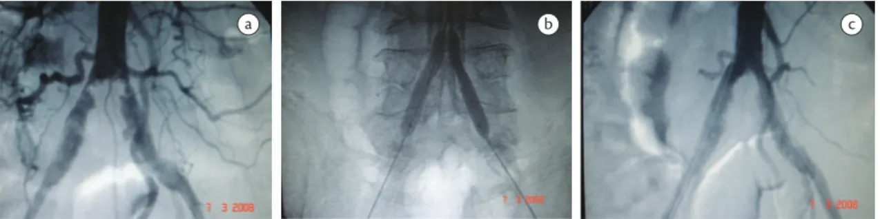
Case 1
Patient with trophic lesion in ifth toe, stenosis of left common iliac artery and subocclusion of left external iliac artery treated with angioplasty and balloon-expandable stent in left common iliac artery and self-expanding stent in the distal third of the common iliac artery and external iliac artery (Figures 1a-c).
of at least one side was achieved (partial technical failure). In one of the patients, with a TASC D lesion, there was no canalization in either side (treatment failure). He died on the 10th postoperative day due to complications of underlying diseases: chronic obstructive pulmonary disease (COPD), diabetes and sepsis.
The patients underwent postoperative follow-up, which included c ankle-brachial index measurements and a duplex ultrasound scan at 30 days, 3 months, 6 months, 12 months, and every 6 months thereafter. Minimum follow-up was three months, and maximum, 72 months (six years), and mean up was 22 months. There was a small loss to follow-up, and it was not possible to calculate late survival.
During postoperative follow-up, there were: • two occlusions (3.38%): one patient with occlusion
at 12 months after the procedure was asymptomatic and was treated only clinically, without another inter
-vention. Another patient had occlusion 30 days after the procedure and underwent aortobifemoral bypass; • three restenosis occurred in the irst 12 months
after angioplasty (5.08%): two underwent another angioplasty, which was successful, and the other, aortobifemoral bypass because there were severe diffuse lesions of the common and external iliac arteries bilaterally; and
• a major amputation (iliac angioplasty followed by posterior distal femoral tibial bypass. Iliac angio
-plasty was patent, but the bypass occluded in the irst week after operation, and the patient underwent amputation of the thigh).
These data show primary patency in 91.37% and secondary patency in 94.82% (Table 1).
A patient had more serious complications: perforation of leg artery in a combined infrapopliteal procedure that required postoperative fasciotomy due to compartment syndrome. The patient had chronic renal failure with hemodialysis and had a coagulation disorder, which contributed to the development of
Case 2
Patient with disabling intermittent claudication and bilateral ostial stenosis of the common iliac artery. Treatment with a balloon-expanding kissing stent (Figures 2a-c).
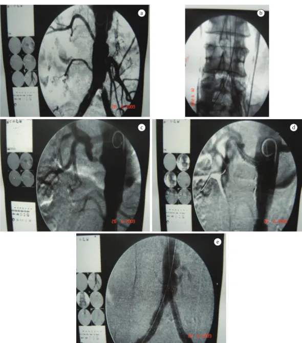
Case 3
Patient had undergone stent angioplasty of left common iliac artery four years before and had in-stent restenosis and stenosis of renal artery. A neobifurcation was prepared with kissing stents and stent angioplasty of renal artery (Figures 3a-e).
Case 4
Patient with trophic lesion of left foot. Aortogram revealed total left common iliac artery occlusion and proximal ulcerated plaque in right common iliac artery. Treatment with balloon-expanding kissing stent (Figures 4a-d).
RESULTS
Mean patient age was 62 years (minimum: 42; maximum: 89); 37 were male patients (62.72%) (Figure 5).
The predominant risk factors were smoking, in 72% of the cases, and diabetes, in 30%.
The main treatment indications were limiting or disabling intermittent claudication in 30 cases (50.84%) and rest pain or trophic lesion (critical ischemia) in 29 cases (49.15%) (Figure 6).
Lesions were classiied as TASC A in 39 cases (66.10%), TASC B in 13 (22.03 %), TASC C in two (3.38%) and TASC D in ive (8.47%) (Figure 7).
Patients had stenosis in the iliac arteries in 42 cases (71.18% of the cases in the series) and occlusions in 17 cases (28.81% of the cases in the series).
Initial technical success was achieved in 56 cases (94.91%). Three were classiied as technical failures: two had bilateral lesions, but recanalization
Figure 3. Case 3: (a) intrastent restenosis originating in left common iliac artery; (b) new bifurcation with balloon-expanding kiss-ing stents; (c) severe stenosis in right renal artery; (d) balloon-expandkiss-ing stent in right renal artery; (e) inal aortogram.
the compartment syndrome and the need to undergo fasciotomy. She recovered satisfactorily.
There were no pseudoaneurysms, retroperitoneal hematoma, arterial occlusions or need of blood transfusion.
There were two deaths in the irst 30 days after operation (3.3% mortality) not associated with the angioplasty. A kidney transplant recipient, who was hospitalized with acute pancreatitis and developed acute ischemia of the right lower limb, required ibrinolysis and angioplasty of the common iliac artery and limb perfusion was restored. Ten days after the procedure, she died due to complications of acute pancreatitis (respiratory failure and sepsis). Another patient, with a TASC D lesion in which recanalization was not achieved, already described above, died due to COPD complications, diabetes and sepsis.
DISCUSSION
Although open surgery, particularly aortobifemoral bypass, apparently results in better patency in the long term than endovascular surgery in the aortoiliac region, the risks of open surgery are much higher than those of endovascular treatment, not only in terms of mortality, but also in regard to morbidity and patient return to usual daily activities. A meta-analysis of 25 studies about aortobifemoral bypass found 4.4% deaths and 12.2% complications, in addition to 91% patency at ive years for patients with claudication and 87% for those with critical ischemia10.
However, studies about the use of an endovascular approach showed that, in a series of 151 cases of angioplasty of the iliac arteries, there were no deaths and only one minor complication (inguinal hematoma), whereas assisted primary and secondary patency was 98% at seven years. Most lesions treated in this series were stenosis and TASC A or B lesions11. Therefore, the approach for each patient
should take into consideration general health and type of lesion.
In a retrospective study with 138 patients with iliac occlusions treated with angioplasty and stents, patency rates were 90%, 85%, 80% and 68% at three, ive, seven and 10 years. According to the authors, an initial endovascular approach seems to be justiiable for most patients, particularly older patients and those with severe comorbidities, because it is less invasive than surgery12. Another retrospective study
with 937 patients found 73% primary patency, 88% assisted primary patency and 90% secondary patency at 10 years, with mortality rates lower than Figure 5. Patient distribution according to sex.
Figure 6. Patient distribution according to degree of ischemia.
Figure 7. Patient distribution according to TASC classiication.
Table 1. Primary and secondary patency rates.
Patency Patency rates (%)
Primary 91.37
• disabling claudication;
• no improvement with clinical treatment; • rest pain; and
• trophic lesion.
In the series of patients in this study, these criteria were used to indicate interventions. The decision about invasive treatment for patients with claudication should also be made based on the individual context of each patient. Age, social life, itness and professional status should be taken into consideration, and the simple application of only a rigid classiication system should be avoided. In some speciic cases, patients with moderate to severe claudication may become candidates for invasive treatment. Quality of life questionnaires may be used to decide whether or not an invasive procedure should be used and to select patients that will have the greatest beneit from that type of intervention19.
Angioplasty may improve the quality of life of patients with claudication when compared with clinical treatment and physical exercises alone20.
The decision about a selective or routine use of stenting to treat iliac arteries remains controversial21.
The Dutch Iliac Stent Trial (DIST) conducted a
prospective and randomized comparative study of selective and routine stenting of 279 patients with claudication, and found similar results in both groups, without statistically signiicant differences22.
However, a meta-analysis comparing six studies with 1,300 patients that underwent angioplasty of the iliac artery without stenting and eight studies with 816 patients that underwent stenting suggested that the use of stents seems to bring immediate success and better long term results23. Another study with a series
of patients investigated the use of routine stenting in 90 lesions of the external iliac artery in patients with claudication and found that primary and secondary patency rates were 84% and 93% at three years of follow-up24.
In our sample, we chose to use stenting selectively in the cases of unsatisfactory results of angioplasty, ostial or calciied lesions or presence of ulcerated plaque. Stents were also used routinely in cases of occlusion.
CONCLUSIONS
According to studies in the literature, angioplasty in iliac arteries is currently accepted as the method of choice for the treatment of lesions in this anatomic site, particularly because of its low morbidity and mortality rates and its good long term results. 1%13. Pedron et al.14 studied a series of 24 cases of
iliac occlusion treated with angioplasty and found technical success rates of 91.7% and patency of 71% at 1 year14. Moreira et al.15 evaluated neointimal
hyperplasia using intravascular ultrasound 8 months after the procedure in 30 patients that underwent angioplasty of the iliac arteries and stenting, and found that it was self-limited and did not lead to signiicant restenosis in any of the cases15.
In our study, most patients had TASC A or B lesions (88.13% of the cases) and stenosis (42 patients, or 71.18% of the cases), and primary (91%) and secondary (94%) patency rates were also high, whereas morbidity and mortality rates were low. These indings suggest that the endovascular approach should be the irst treatment alternative in case of stenosis and TASC A and B lesions. If the endovascular treatment fails for a patient, there remains the option of undergoing another angioplasty procedure or attempting open surgery. Of the three patients with restenosis in our series, two underwent another angioplasty and one, open surgery, as shown in the results of postoperative follow-up. Of the two that had occlusion during follow-up, one underwent surgery, and the other was treated only clinically.
Initial technical and clinical success of angioplasty in the treatment of stenosis in iliac arteries is greater than 90% in practically all publications in the literature. This rate may reach almost 100% in the case of focal stenosis. In contrast, the success rate in the case of occlusions is about 85% and depends on ibrinolysis. The development of advanced materials and the availability of more advanced techniques have resulted in greater technical success when recanalizing occlusions16.
Several recent studies have also conirmed the eficacy and safety of this method. A study with a series of 118 patients with occlusion of the iliac artery treated with primary stenting found a secondary patency of 93% at ive years of follow-up17.
However, the indications of interventions, be them open surgery or endovascular approach, have not changed. In the case of patients with claudication, they should be treated irst clinically, with the indication of smoking cessation, walking, control of risk factors, use of antiplatelet drug, vasodilating drugs and statins. Invasive treatments are indicated in the following conditions, according to recommendations of the Brazilian Society of Angiology and Vascular Surgery18.
11. Kudo T, Chandra FA, Ahn S. Long-term uotcomes and predictors of iliac angioplasty with stenting. J Vasc Surg. 2005 Sep;42(3):466-75. http://dx.doi.org/10.1016/j.jvs.2005.05.002
12. Gandini R, Fabiano S, Chiocchi M, Chiappa R, Simonetti G. Percutaneous treatment in iliac artery occlusion: long-term results. Cardiovasc Intervent Radiol. 2008 Nov-Dec;31(6):1069-76. http://dx.doi.org/10.1007/s00270-008-9386-5
13. Davies MG, Bismuth J, Saad WE, Naoum JJ, Peden EK, Lumsden AB. Outcomes of reintervention for recurrent disease after percutaneous iliac angioplasty and stenting. J Endovasc her. 2011 Apr;18(2):169-80. http://dx.doi.org/10.1583/10-3257.1
14. Pedron C, Ristow AV, Cury-Filho JM, Martin HS, Peixoto CC, Fonseca LMB. Tratamento endovascular da oclusão das artérias ilíacas. Radiol Bras. 2001;34(5):261-265. http://dx.doi.org/10.1590/ S0100-39842001000500004
15. Moreira SM, Kambara A M, Ajzen S, Costa Junior JR . Quantificação volumétrica da hiperplasia neointimal em artérias ilíacas após implante de suporte intravascular metálico. Radiol Bras. 2009;42(4):231-234. http://dx.doi.org/10.1590/ S0100-39842009000400008
16. Saket RR, Rzavi MK, Padidar A, Kee ST, Sze DY, Dake MD. Novel intravascular ultrasound-guided method to create transluminal arterial communications: initial experience in peripheral occlusive disease and aortic dissection. J Endovasc her. 2004;11(3):274-280. http://dx.doi.org/10.1583/03-1133.1
17. Ozkan U, Oguzkurt L, Tercan F. Technique, complication and long-term outcome for endovascular treatment of iliac artery oclusion. Cardiovasc Intervent Radiol. 2010 Feb;33(1):18-24. http://dx.doi. org/10.1007/s00270-009-9691-7
18. Sociedade Brasileira de Angiologia e Cirurgia Vascular - SBACV. Diretrizes SBACV. J Vasc Br. 2005;4.
19. Aquarius AE, Denolet J, Hamming JF, Breek JC, De Vries J. Impaired heath status and invasive treatment in peripheral arterial disease: a prospective 1-year follow-up study. J Vasc Surg. 2005;41:436-42. http://dx.doi.org/10.1016/j.jvs.2004.12.041
20. Greenhalgh RM, Belch JJ, Brown LC. he adjuvant beneit of angioplasty in patients with mild to moderate intermitent claudication managed by supervised exercise, smoking cessation advice and best medical therapy: results from two randomised trials for stenotic femoropopliteal and aortoiliac disease. Eur J Vasc Endovasc Surg. 2008 Dec;36(6):680-8. http://dx.doi.org/10.1016/j. ejvs.2008.10.007
21. Casserly IP, Sachar R, Yadav JS. Manual of peripheral vascular intervention. Lippincott Williams e Wilkins; 2005.
22. Tetteroo E, Van der Graaf Y, Bosch JL. Randomised comparison of primary stent placement versus primary angioplasty followed by selective stent placement in patients with iliac-artery occlusive disease. Dutch Iliac Stent Trial Study Group. Lancet. 1998;351:1153-1159. http://dx.doi.org/10.1016/ S0140-6736(97)09508-1
23. Bosch JL, Hunink MG. Meta-analysis of the results of percutaneous transluminal angioplasty and stent placement for aorto-iliac occlusive disease. Radiology. 1997;204:87-96.
24. Maurel B, Lancelevee J, Jacobi D, Bleuet F, Martinez R, Lermusiaux P. Endovascular treatment of external iliac artery stenoses for claudication with sistematic stenting. Ann Vasc Surg. 2009 Nov-Dec;23(6):722-8. http://dx.doi.org/10.1016/j.avsg.2008.05.019 Findings in this study showed that:
• the primary patency rate for angioplasty in the iliac arteries was 91.3%;
• the secondary patency rate for angioplasty in the iliac arteries was 94.82%;
• the rate of mortality in the irst 30 days after opera
-tion was 3.3%;
• the rates of complications associated with the pro
-cedure were low (1 case of arterial perforation); and • the rate of amputations was low (1 case in the series). Endovascular treatment is an eficacious, long-lasting and safe option for the treatment of peripheral occlusive arterial disease in iliac arteries, particularly TASC A and B lesions with low complication rates and high patency rates, as demonstrated in this study, in which most lesions that underwent treatment received that classiication.
Angioplasty in iliac arteries is a risk, low-mortality procedures with patency results that compare with those achieved when conventional surgery is used.
REFERENCES
1. Hallet JW. Comprehensive vascular and endovascular surgery. 2nd ed. Mosby; 2009.
2. Ouriel K. Peripheral arterial disease. Lancet. 2001;358:12571264. http://dx.doi.org/10.1016/S0140-6736(01)06351-6
3. Trans-atlantic Inter-Society Consensus Working Group. M anagement of p epip heral ar terial dis ea s e . J Va s c Surg. 2007;45(suppl 5):55-567.
4. Seldinger S. Catheter placement of the needle in percutaneous arteriography: A New Technique. Acta Radiol. 1953;39:368. http:// dx.doi.org/10.3109/00016925309136722
5. Dotter CT, Judkins M. Transluminal treatment of arteriosclerotic obstruction. Circulation. 1964;30:654-670. http://dx.doi. org/10.1161/01.CIR.30.5.654
6. G r u n t z i g A , H o p f f h H . Pe r k u t a n e r e k a n a l i s a t i o n chronischer arterieller verschlusse mit einem neven dilatationskatheter: modifikation der Dotter-technik. Dtsch Med Wochenschr. 1974;99:2502-2510. http://dx.doi. org/10.1055/s-0028-1108161
7. Palmaz JC, Richter GM, Noldge G, Kauffmann GW, Wenz W. Intraluminal Palmaz stent implantation. he irst clinical case report on balloon-expanded vascular prosthesis. Radiology. 1987;27:560-63.
8. Moore WS, Ahn SS. Endovascular surgery. 3rd ed. Philadelphia: WBSaunders Company; 2001.
9. Schneider PA. Endovascular skills: guidewire and catheter skills for endovascular surgery. 3nd ed. Informa heathcare USA; 2009.
Annex 1
PROTOCOL FOR PERIPHERAL ANGIOPLASTY – DATA COLLECTION
IDENTIFICATION (attach tag) Name
Sex
Registration number Age
Description of clinical signs and symptoms
Intermittent claudication. distance
_____________
Disabling claudication
Rest pain
Tissue lesion
Acute occlusion Degree: _________
thrombolysis
Deep vein thrombosis
Other:
Risk factors
Smoking
Diabetes
hypertension
Renal failure
Hyperlipidemia
Characteristics of trophic lesion RLL:
LLL:
Pulses Doppler ultrasound
RLL: LLL: RLL: LLL:
Femoral Dorsalis pedis artery
Popliteal Posterior tibial artery
Dorsalis pedis
Posterior tibial Ankle-brachial index
Rutherford
I
II
III
IV
V
VI
Arteriogram / Angio-CT / Angio-MRI / Duplex (lesion description) TASC
A
B
C
D
Lesion anatomy
Iliac arteries Femoral popliteal segment
A 1 B 2 B 3 B 4 C 5 C 6 C 7 D 8 D 9 D 10 D 11 D 12
Stenosis < 3 cm CIA or EIA (unilateral or bilateral) Stenosis, single lesion 3-10 cm
Total stenosis, 2 lesions < 5 cm in CIA or EIA Unilateral occlusion CIA
Bilateral stenosis 5-10 cm CIA and/or EIA Unilateral occlusion EIA
Bilateral occlusion CIA
Multiple, difuse stenosis in CIA, EIA and CFA Unilateral occlusion of CIA and EIA Bilateral occlusion of EIA
Difuse disease afecting aorta and both iliac arteries Iliac stenosis in patient with AAA
A 1
B 2
B 3 B 4 B 5
C 6 C 7 D 8
Stenosis up to 3 cm, except when origin in SFA or distal popliteal artery
Stenosis/occlusion, single lesion 3-5 cm not afecting distal popliteal artery
highly calciied stenosis, up to 3 cm
Multiple stenosis/occlusions, each lesion < 3 cm
Single or multiple lesion in the absence of continuous tibial runof to improve inlow for bypass
Stenosis/occlusion, single lesion greater than 5 cm Multiple stenosis/occlusions 3-5 cm
Complete occlusion of CFA or SFA or PA or TFT
A1 B2 B3 B4 C5 C6 C7 D8 D9
Stenosis, single lesion smaller than 1 cm in tibial or ibular arteries
Multiple focal stenosis, lesions of tibial or ibular arteries, each smaller than 1 cm One or two lesions, focal stenosis, each smaller than 1 cm in the tibial trifurcation Tibial or ibular stenosis associated with femoral popliteal angioplasty
Stenosis, lesion 1-4 cm long
Occlusions 1-2 cm long in tibial or ibular artery Long stenosis, lesion in tibial trifurcation Occlusions > 2 cm in tibial or ibular artery Difuse disease in tibial or ibular artery
Endovascular approach Treatment description: Date:
Surgical team:
Segment treated
aorta
Iliac arteries
Femoral artery
Popliteal artery
Infrapopliteal artery
Carotid artery
a. subclavian artery
Venous graft
Synthetic graft
Arteriovenous istula
AVM
Renal
mesenteric
vein:_________________
Filter of vena cava Access:
Ipsilateral femoral artery
contralateral femoral artery
Anterograde femoral
R brachial
L brachial
radial
axillary
venous
Pre-dilatation Balloon:
Post-dilatation
Balloon:
Stent: ______________________
No. of stents ____
Sizes: ________________________
Expanding balloon
Self-expanding balloon
surgery time (min) ______________
Complications:
Immediate postoperative AB index (24 h): POSTOPERATIVE FOLLOW-UP
1) 30 DAYS ABI
Imaging indings 2) 3 MONTHS
ABI
Imaging indings 3) 6 MONTHS
ABI
Imaging indings 4) 1 YEAR
ABI
Imaging indings 5) SUBSEQUENT FOLLOW-UP RECORD ABI AND IMAGING FINDINGS