Authors
Cassiana Regina de Góes 1 Ana Claudia Soncini Sanches 1
André Balbi 1 Daniela Ponce 1
1 Universidade Estadual
Paulista.
Submitted on: 4/26/2016. Approved on: 10/14/2016.
Correspondence to:
Cassiana Regina de Góes. Universidade Estadual Paulista, Faculdade de Medicina de Botucatu. Av Paranapanema, nº 165, Bairro Braz II, Avaré, SP, Brazil. CEP: 18701-240
E-mail: cassina.goes@yahoo. com.br
Fundação de Amparo à Pesquisa do Estado de São Paulo (FAPESP).
Daily variability of resting energy expenditure in acute kidney
injury patients on dialysis
Variabilidade diária do gasto energético de repouso em pacientes
com lesão renal aguda em tratamento dialítico
Introdução: É imprescindível a correta es-timativa do gasto energético de repouso (GER), que pode apresentar considerável variação diária no paciente crítico com le-são renal aguda (LRA). Objetivo: Avaliar a variabilidade diária do GER medido por calorimetria indireta (CI) em pacientes com LRA e indicação dialítica e identificar as va-riáveis clínicas associadas ao GER. Métodos:
O GER foi medido no dia da indicação do procedimento dialítico e nos quatro dias subsequentes. Também foram avaliados pa-râmetros que podem influenciar o GER. As diferenças diárias foram analisadas pelo mo-delo linear generalizado para medidas repeti-das, com distribuição gama, além da correla-ção de Spearman e regressão linear múltipla.
Resultados: Foram 301 medidas de CI reali-zadas em 114 pacientes, com idade de 60,65 ± 16,9 anos e 68,4% do sexo masculino. O GER médio foi de 2081 ± 645 Kcal, com aumento no dia 5 (2270 ± 556 Kcal), quan-do comparaquan-do aos dias 2 e 3 (2022 ± 754; 2022 ± 660 kcal, respectivamente, p = 0,04); quando normalizado para peso, não houve diferença significante no GER (kcal/kg/dia) durante o acompanhamento. GER correla-cionou-se positivamente com temperatura corporal, contagem total de leucócitos, pro-teína C reativa, volume minuto (VM), fração inspirada de oxigênio (FiO2), aparecimento de nitrogênio ureico (UNA), peso corporal e estatura e inversamente com idade. Após a regressão linear múltipla, somente VM, FiO2 e peso corporal e idade se correlacionaram independentemente. Conclusão: Pacientes com LRA dialíticos apresentam GER está-vel. O GER foi associado independentemen-te com FiO2, VM, peso e idade. Assim, re-quisitos ventilatórios precisam ser avaliados diariamente para que alterações necessárias na prescrição dietética sejam feitas.
RESUMO
Palavras-chave: consumo de energia; le-são renal aguda; metabolismo energético.
Introduction: It is needed for nutrition
prescription correct estimate of resting energy expenditure (REE), which is a challenge given the possible daily variation in critically ill patients with acute kidney injury (AKI). Objective:
To evaluate the daily variability of REE measured by indirect calorimetry (IC) in patients with AKI and dialysis indication and identify clinical variables associated with REE. Methods: The REE was measured on the time of dialysis indication and the subsequent four days. We also evaluated parameters that can influence the REE. The daily differences were analyzed by generalized linear model for repeated measures. We also used Spearman correlation and multiple linear regression. Results: There were 301 IC measurements in 114 patients, mean age of 60.65 ± 16.9 years and 68.4% were male. The average REE was 2081 ± 645 kcal, rising on day 5 (2270 ± 556 kcal) compared to the days 2 and 3 (2022 ± 754; 2022 ± 660 kcal, respectively, p = 0,04). When normalized to weight, there was no significant difference in REE (kcal/kg/day) during follow-up. REE was positively correlated with total leukocyte count, C-reactive protein, minute volume (MV), fraction of inspired oxygen (FiO2) urea nitrogen appearance (UNA), weight and height and inversely with age. After multiple regression, MV, FiO2, weight and age are correlated independently with REE. Conclusion: Patients with AKI have REE stable. The REE was associated independently with FiO2, MV, body weight and age. Thus, ventilatory parameters should be evaluated each day for the necessary dietary changes may be made.
ABSTRACT
Keywords: acute kidney injury; energy
consumption; energy metabolism.
INTRODUCTION
Acute renal injury (AKI) is defined as an abrupt drop in glomerular filtration rate (GFR), resulting in increased nitrogen slag, acid-base balance disorders, fluid and electrolyte disturbances.1 AKI is a common complication in hospitalized patients, and it can affect 10% to 30% of patients admitted to intensive care units (ICUs). Approximately 5% of these patients required renal replacement therapy (RRT).2
AKI usually develops in the context of multiple organ failure in the ICU. Nutritional therapy for these patients has the purpose of reversing or attenuating the negative effects of catabolism and hypermetabolism associated with these acute diseases. The basis of nutritional support is to preserve lean body mass and the immune function, as well as to prevent complications related to under and overfeeding.
Thus, adequate nutritional support is essential in the therapeutic strategy of both AKI and the failure of multiple organs and systems. In addition, when RRT is required, it should be carefully adjusted, given the characteristic metabolic disturbances and the effects of RRT on nutrient balance.3
In order to prescribe the best nutritional intake, a correct estimation of nutritional needs is essential, and indirect calorimetry (IC) is considered the gold standard for measuring energy expenditure (EE) in critically-ill patients.4
However, many factors can alter the EE in ICU patients, such as pain, medications, body temperature, diet, heart rate, among others.5 In addition to these factors, patients with severe AKI may also have an altered EE due to loss of kidney homeostasis and the adverse effects of the chosen RRT.1,3 Thus, only one EE measure alone may not characterize the patient’s correct energy needs.
Studies with critical patients reported a change in daily EE between 4% and 56%, and that more clinically stable patients presented lower variability.6-8 In patients with AKI, few studies evaluated the EE by IC and only one longitudinal study evaluated resting EE (REE) in critically-ill, mechanically ventilated patients, with AKI in continuous RRT.
The authors showed that the resting energy expenditure (REE) measured on the first day was 2.153 ± 380 Kcal and there was an increase of 56 ± 24 cal/d during the study period of 6 days, with an end value of REE of 2.431 ± 498 Kcal (p < 0.0001).9 However, in this study, IC was performed in patients
during RRT and IC guidelines contraindicate this moment for the measurement of EE, due to possible interferences of the dialysis procedure in gas exchange, which would lead to errors in the measurement of EE using IC.5,10,11
The present study aims to evaluate the daily variability of REE measured by IC in patients with AKI and dialytic indication and to identify the clinical variables associated with REE.
METHODS
A prospective cohort study that evaluated patients older than 18 years, from March 2013 to December 2015.
PATIENTS
We included patients admitted to the ICU with a diagnosis of AKI according to the KDIGO criteria,12 clinical symptoms suggestive of acute tubular necrosis (ATN), need for RRT (stage 3), and mechanically ventilated.
We excluded those patients with AKI of other etiologies, renal transplanted or those with chronic renal disease stages 4 and 5 (Glomerular Filtration Rate - GFR - < 30 ml/min estimated by the Modification of Diet in Renal Disease - MDRD,13), considering the patient’s baseline creatinine for the calculation, defined as the serum creatinine value most recently obtained prior to admission, not preceding 12 months of hospitalization. If this value is unknown or obtained 12 months before hospitalization, we considered baseline creatinine as the lowest value observed during follow up.14
Other exclusion criteria were fraction of inspired oxygen (FiO2) greater than 0.60; Expiratory Positive Airway Pressure (PEEP) > 10 cm H2O; Maximum airway pressure > 60 cm H2O; stirring presence; use of neuromuscular blockers; air leakage into the ventilator circuit, around the endotracheal tube cuff, or from a bronchopleural fistula: because these factors lead to inaccuracies in the REE measurement by IC.
This study was approved by the Research Ethics Committee of the institution (protocol 4383/2012). The Term of Consent was signed by the legal guardian of the participant before entry into study.
ENERGY EXPENDITURE MEASUREMENT
Resting energy expenditure (REE) is measured because of the impossibility of achieving the conditions to measure baseline EE of critically-ill patients. To ensure REE measurement, the patient needed to be in supine position and rested for at least 30 minutes prior to measurement; In a thermoneutral environment (22-25 °C) for at least 30 minutes before and during measurement; without having used additional analgesics and sedatives within 30 minutes of initiation of IC; without procedures within 60 minutes of IC initiation; without general anesthesia within 8 hours of IC initiation; And continued parenteral and/or enteral nutrition during the data collection period.
IC was performed using the RMR Quark apparatus (Cosmed, Rome, Italy). The RMR Quark is designed to accurately and instantly measure the energy needs of patients breathing spontaneously or mechanically. For our study, the calorimeter was used connected to the patient’s mechanical ventilator, with connectors in the exhalation line of the ventilator and in the respiratory circuit: in this way, it is possible to collect both inspired and expired gases. The device has a paramagnetic oxygen sensor to measure oxygen concentrations and analyzes them based on infrared absorption for carbon dioxide measurements.
The calorimeter was calibrated before each use. The examination had an average duration of 30 minutes. Patients were expected to reach steady state during the test. The steady-state was defined as a variability of < 10% in the measurements of oxygen consumption (VO2) and carbon dioxide production (VCO2), and < 5% in the respiratory quotient from minute to minute.
In addition to the measured REE, baseline EE (BEE) was estimated using the Harris and Bendict formula.15 For the calculation of the patient’s height equation (cm), it was measured upon admission to the ICU when possible, or using the value documented in the medical record. Weight (kg) was measured using hospital beds, calibrated upon admission, for most of the patients. If the patient had edema at the time of the measurement, according to the medical assessment,
we asked relatives about the patient’s habitual weight, and used it as the real weight in the formula.
We also evaluated parameters that may influence REE, such as ventilatory (minute volume, respiratory rate, PEEP, FIO2), vasoactive drugs and body temperature.
STATISTICAL ANALYSIS
The results were described by median and interquartile range, or mean and standard deviation. The daily differences in the studied parameters were analyzed by the generalized linear model for repeated measures, with gamma distribution.
To evaluate the parameters correlated with REE, and those that could influence its daily variability, all measures of REE, clinical parameters, laboratory, ventilatory and catabolism markers performed during follow-up were used. Spearman’s univariate correlation was used first, and the variables with significant p were placed in multiple linear regression to analyze which was independently associated with REE.
Variables with asymmetric distribution were transformed by the log function for inclusion in the multivariate analysis. The collinearity of the predictive variables was tested by the tolerance (Tolerance) and the Variance Inflation Factor (VIF), and if the tolerance was < 0.1 and/or the FIV > 4, then one of them was removed from the multivariate models.
A p < 0.05 was used as statistically significant.
RESULTS
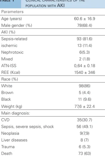
A total of 114 patients were evaluated, with mean age of 60.65 ± 16.9 years and 68.4% were males. Diagnoses of sepsis and cardiovascular disease (CVD) accounted for 79.8% of the hospitalizations. AKI was the etiology associated with sepsis in most patients (81.6%). The specific prognostic index for ATN (ATN-ISS) was 0.64 ± 0.18. The BEE estimated by the Harris-Benedict formula averaged 1,540 ± 346 Kcal; whereas the REE measured by the IC on day 1 was significantly higher (2061 ± 700 Kcal, p
< 0.001). Table 1 shows the clinical characteristics of the general population studied at the time when dialysis was indicated.
The percent increase (or decrease) in REE was calculated to investigate the mean variability in EE from one day to the other. This calculation was done by subtracting the REE from the day of interest by the REE from the previous day, and dividing that difference by the REE from the previous day, times 100 (percentage). This variability had a median of 0.44% (-14, 14).
Table 2 shows the evolution of clinical and laboratory parameters during the 5-day follow-up.
REE correlations with clinical parameters (temperature and dose of VAD), laboratory [serum urea and creatinine, total leukocyte count (WBC) and CRP], ventilatory variables (VM and FiO2) and catabolism (NB and urea nitrogen - UNA). The correlations are presented on Table 3. REE correlated positively and significantly with body temperature, WBC, CRP, MV, FiO2 and BUN, weight and height, and inversely with NB and age.
After multiple linear regression, only MV, FiO2, body weight and age were independently correlated with REE. The result of the regression is shown on Table 4.
DISCUSSION
Information about the energy expenditure assessment of patients with AKI is scarce in the literature. Our study found that the baseline EE estimated by the Harris and Benedict formula15 was significantly lower than that measured by IC. This finding corroborates the indication not to use this formula in critically-ill patients,7,16-18 and in patients with AKI.19
The best way to assess the energy needs of critically-ill patients with AKI is by means of IC.19 In the present study, REE increased as measured by IC on the fifth day of follow-up, compared to days 2 and 3. However, this Significant increase was seen only in REE without normalization for weight. When normalized (kcal/kg/day), there was no significant difference during follow-up.
Increased REE was observed by Scheinkestel et al.9 in patients under RRT, and the mean REE value in the patients in this study was also close to that of the present study (2,153 ± 380 Kcal and 2,081 ± 645 Kcal, respectively). Despite this similarity in EE, our study evaluated the patients during non-dialysis time, as recommended by the guidelines for IC use.5,20,21
This recommendation is because during dialysis, the correction of metabolic acidosis and CO2 Parameters
Age (years) 60.6 ± 16.9
Male gender (%) 78(68.4)
AKI (%)
Sepsis-related 93 (81.6)
ischemic 13 (11.4)
Nephrotoxic 6(5.3)
Mixed 2 (1.8)
ATN-ISS 0,64 ± 0.18
REE (Kcal) 1540 ± 346
Race (%)
White 98(86)
Brown 5 (4.4)
Black 11 (9.6)
Weight (kg) 77,6 ± 22.4
Main diagnosis:
CVD 35(30.7)
Sepsis, severe sepsis, shock 56 (49.1)
Neoplasia 9(7,9)
Liver diseases 8 (7)
Trauma 6 (5.3)
Death 73 (63)
Values shown in frequency, mean and standard deviation or median and interquartile intervals. CVD: cardiovascular disease; AKI: acute kidney injury; ATN-ISS: Individual Severity Score in Acute Tubular Necrosis; BEE: Baseline Energy Expenditure.
TABLE 1 CLINICALCHARACTERISTICSOFTHE POPULATIONWITH AKI
taken off the study because of the suspension of the dialysis procedure, recovery of renal function, or death. Of these, 73 patients left the study due to death (63%).
The mean REE measured by IC was 2,081 ± 645 Kcal (27.7 ± 10 kcal/kg/day). There was an increase in REE on day 5 (2270 ± 556 Kcal) when compared to days 2 and 3 (2,022 ± 754; 2,022 ± 660 kcal, respectively, p = 0.04), only when total ERR (kcal/ day) was found. When normalized by weight, there was no significant difference during follow-up days.
The vasoactive drug dose (VAD), minute ventilation (MV) and body temperature variables remained constant during follow-up. There was a significant decrease in serum urea and creatinine levels from day 3. The fraction of inspired oxygen (FiO2) and C-reactive protein (CRP) also decreased on day 5, when compared to days 1 and 2 (p < 0.05);
Day 1 N = 114
Day 2 N = 76
Day 3 N = 52
Day 4 N = 35
Day 5 N = 24
REE (Kcal) 2061 ± 700 2022 ± 754 a 2022 ± 660 a 2150 ± 539 2270 ± 556
REE (Kcal/Kg/d) 27.9 ± 10.4 26.6 ± 10.6 26.6± 9.6 29.2 ± 8.9 30.4 ± 8.3
REE variability (%)
-5 (-16. 7).
-1.6 (-10. 12).
0 (-11. 17).
8 (-13. 25).
VAD (mcg/Kg/ min)
0.18 (0.06-0.58)
0.1 (0-0.28)
0.04 (0-0.19)
0.06 (0-0.33)
0 (0-0.19)
VM 8.5 ± 2.6 8.3 ± 2.4 9.1 ± 2.6 8.8 ± 2.7 9 ± 3.6
Freq. (resp/min) 16 ± 4 16 ± 5 18 ± 5.5 a,b 18 ± 6 b 19 ± 6 b
PEEP 6 ± 2 6 ± 2 6 ± 2 6 ± 1 6 ± 1
FIO2 40 ± 11.3
a 40 ± 12 a 37.5 ± 10 c 37.6 ± 9.1 34.2 ± 10
Temperature (ºC) 37.7 ± 0.95 37.5 ± 0.8 37.6 ± 0.9 37.6 ± 0.83 37.8 ± 1 Urea (mg/dl) 178 ± 79 160 ± 63 b 142 ± 51 b,c 131.4 ± 46.4 b,c 143 ± 68.7 b
WBC (mm3) 17050
(12300-22400) d
17600 (10600-25500) d
17800 (13700-26100) d
22300 (14800- 29300)
16900 (12500- 22600)
PCR (mg/dl) 27.3 (11- 35.3)
25.2 (7.8-31)
23 (6.7-31) b
26.7 (7.4-36.7)
19.3 (6.6-37.3) b
NB (g/day) -5.51 (-14.12;-0.68)
-7.74 (-18.13; -1.63)
-6.1 (-11.7; 3.1)
-1.27 (-5.8; 1.98) b
-0.81 (-7.98; 3.12) b
BUN (g/day) 13.65 (7.56; 19.12)
18 (9.87; 24.6) b
14.24 (8.6; 22.5)
15.6 (9.5; 23.3)
14.13 (9.93; 20) Values shown in frequency, mean and standard deviation or median and interquartile intervals. REE: Resting Energy Expenditure, VAD: vasoactive drug; FIO2: Fraction of Inspired Oxygen; Mv: Minute volume; RR: respiratory rate; PEEP: expiratory positive airway pressure; Creat: serum creatinine; WBC: white blood-cell count; CRP: C-reactive protein; NB: Nitrogen Balance; BUN: blood urea nitrogen. REE variability was calculated as: (REE day I want to assess - REE previous day/REE previous day. ap > 0.05 when compared to day 5; bp < 0.05 when compared to day 1; cp < 0.05 when compared to day 2; dp < 0.05 when compared to day 4.
TABLE 2 EVOLUTIONOFCLINICALANDLABORATORYPARAMETERSOFPATIENTSINDIALYSISWITHACUTEKIDNEYINJURY DURING 5 DAYS
removal22-24 can result in an underestimation of the EE. In addition, RRT may also lead to a reduction in body temperature and decrease REE.25 Thus, the comparison between these two studies may be impaired.
Koukiasa et al.,26 when evaluating critically-ill patients with non-septic intracranial hemorrhage, also found elevated REE during follow-up on the seventh day of the study. This increase in EE is usually seen in critical ICU patients. The peak metabolic rate is reached during the second week post admission, and some explanation for this are complications or discontinuation of sedation. The increase seen by Koukiasa et al.,26 in EE can be partially explained by increases in body temperature.
In the present study, there was no change in body temperature during follow-up. Only ventilatory parameters such as RR and FiO2 changed on day 5, compared to the initial days.
The daily variability on REE in our study was 0.44% (-14, 14). The variability reported in the
literature is higher. Reid7 obtained a daily variability of 31.7 ± 22.6% and Weissman et al.6 reported a mean variability of 21 ± 16%. Both were studies with critically-ill ICU patients. Evaluating patients with intestinal failure using parenteral nutrition at home, Ławiński et al.27 found a variability closer to that of our study (8% ± 7%), when evaluating two REE measures.
This difference in variability seen in ICU studies may be due to the heterogeneity of the studied population, since EE can be altered by several factors, such as disease severity and course, sedation, mechanical ventilation, infectious complications, neurological deficits, among others.26
In the present study, some parameters correlated with the EE, such as body temperature, WBC, CRP, MV, FiO2, BUN, weight, height and age, and after multiple analysis, MV, FiO2, weight and age remained independently associated with the REE measured by IC.
Spearman correlation coefficient with
REE p
VAD 0.009 0.883
mV 0.246 < 0.001
FiO2 0.183 0.001
Temperature 0.167 0.004
UR 0.105 0.068
Creat 0.100 0.085
WBC 0.159 0.11
CPR 0.105 0.045
NB -0.129 0.05
BUN 0.158 0.026
Weight 0.320 < 0.001
Height 0.158 0.006
Age -0.361 < 0.001
TABLE 3 UNIVARIATEASSOCIATIONBETWEEN
RESTINGENERGYEXPENDITUREANDCLINICAL, LABORATORY, VENTILATORYANDNUTRITIONAL PARAMETERSOFPATIENTSWITHDIALYSIS ACUTEKIDNEYINJURY (N = 301)
REE: rest energy expenditure; VAA: vasoactive agent; UR: serum urea; Creat: serum creatinine; WBC: White blood cell count; CRP: C-reactive protein; NB: nitrogen balance; BUN: Blood Urea Nitrogen; FIO2: fraction of inspired oxygen, mV: minute volume.
Beta Standard error t p Tolerance VIF
mV (l/min) 51.776 19.119 2.708 0.008 0,925 1.081
FiO2 (%) 14.248 5.033 2.831 0.005 0,764 1.309
Temperature (ºC) 96.952 64.394 1.506 0.134 0,859 1.164
Weight (Kg) 5.504 2.608 2.111 0.036 0,761 1.314
Height (m) -6.184 7.046 -0.878 0.382 0,671 1.491
Age (Years) -15.127 3.142 -4.814 0.000 0,915 1.093
CRPLN 39.621 64.314 0.616 0.539 0,797 1.255
UNALN -25.165 71.799 -0.350 0.726 0,781 1.280
WBCLN 73.657 78.287 0.941 0.348 0,828 1.207
TABLE 4 DETERMINANTSOFRESTINGENERGYEXPENDITUREINPATIENTSWITHACUTEKIDNEYINJURY (N = 301)
mV: minute volume; FIO2: fraction of inspired oxygen; WBCNL: Napierian logarithm of total leucocyte count; CRPNL: C-reactive protein Napierian logarithm; UNANL: urea nitrogen appearance Napierian logarithm.
formula28, which considers the inspired volume of O 2 (VO2) and expired CO2 (VCO2). To calculate VO2 and VCO2 by IC, the required measures are inspired and expired concentrations of oxygen (FIO2, FEO2), carbon dioxide (IRCO2, ERCO2) and the volume of inspired and expired air (VI, VE).29
As for MV, its increase may be due to an increase in carbon dioxide volume (VCO2) during the increase of metabolism, according to Kiiski & Takala.30 Kinney et al.31 have shown that the increase in MV is not proportional to the increase in metabolic rate. In addition, it is still possible that fever, by itself,
stimulates the exhaled volume increase, regardless of the increase in oxygen consumption or CO2 production. Despite these variable effects, there is a linear relationship between MV and the metabolic rate in critically-ill patients.32
The linear relationship between weight and REE is known. Body weight and fat-free mass are two variables that directly affect energy expenditure. Changes in body weight (gain and/ or loss) show a direct relationship with increase or reduction in REE.33,34 This association is also seen from REE with age; but in a negative correlation, nonetheless.
Aging is associated with a lower mass of some organs that contribute to energy metabolism and with changes in lean body mass (reduction), and in adipose tissue (increase).35 Studies have reported a progressive decline in REE of about 1-2 % per decade, and that this decline is largely explained by changes in body composition.36-39 In addition, it has also been suggested that the appearance of these REE changes may depend on gender, adiposity, and lean body mass.35,40
The study has some limitations. First, it was performed in a single center, and despite the substantial number of IC measurements, only 24 patients completed the five-day follow-up. In addition, our study presents an evaluation of a specific ICU population: patients with AKI on dialysis, that is, their results cannot be extrapolated to all critical patients or all patients with AKI. Despite these limitations, to our knowledge, this is the first study to evaluate REE variability in patients with AKI, outside the dialysis time.
follow-up. REE in these patients was independently and positively associated with FiO2, MV and weight, and inversely related with age. Thus, ventilatory requirements need to be evaluated daily, so that necessary changes in dietary prescription are made in these patients to avoid the known negative influences of under and overnutrition in critically-ill patients.
REFERENCES
1. Krenitsky J, Rosner MH. Nutritional Support for Patients with Acute Kidney Injury: How Much Protein is Enough or Too Much? Pract Gastroenterol 2011;35:28-42.
2. Uchino S, Kellum JA, Bellomo R, Doig GS, Morimatsu H, Morgera S, et al.; Beginning and Ending Supportive Therapy for the Kidney (BEST Kidney) Investigators. Acute renal failure in critically ill patients: a multinational, multicenter study. JAMA 2005;294:813-8. PMID: 16106006 DOI: http://dx.doi.org/10.1001/jama.294.7.813 3. Fiaccadori E, Regolisti G, Cabassi A. Specific nutritional
problems in acute kidney injury, treated with non-dialysis and dialytic modalities. NDT Plus 2010;3:1-7. DOI: http://dx.doi. org/10.1093/ndtplus/sfp017
4. McClave SA, Taylor BE, Martindale RG, Warren MM, Johnson DR, Braunschweig C, et al.; American Society for Parenteral and Enteral Nutrition. Guidelines for the Provision and Assessment of Nutrition Support Therapy in the Adult Critically Ill Patient: Society of Critical Care Medicine (SCCM) and American Society for Parenteral and Enteral Nutrition (A.S.P.E.N.). JPEN J Parenter Enteral Nutr 2016;40:159-211. 5. Schlein KM, Coulter SP. Best practices for determining resting energy
expenditure in critically ill adults. Nutr Clin Pract 2014;29:44-55. DOI: http://dx.doi.org/10.1177/0884533613515002
6. Weissman C, Kemper M, Hyman AI. Variation in the resting metabolic rate of mechanically ventilated critically ill patients. Anesth Analg 1989;68:457-61. PMID: 2929978 DOI: http:// dx.doi.org/10.1213/00000539-198904000-00006
7. Reid CL. Poor agreement between continuous measurements of energy expenditure and routinely used prediction equations in intensive care unit patients. Clin Nutr 2007;26:649-57. DOI: http://dx.doi.org/10.1016/j.clnu.2007.02.003
8. Vermeij CG, Feenstra BW, van Lanschot JJ, Bruining HA. Day-to-day variability of energy expenditure in critically ill surgical patients. Crit Care Med 1989;17:623-6. PMID: 2736921 DOI: http://dx.doi.org/10.1097/00003246-198907000-00005 9. Scheinkestel CD, Kar L, Marshall K, Bailey M, Davies A,
Nyulasi I, et al. Prospective randomized trial to assess caloric and protein needs of critically Ill, anuric, ventilated patients requiring continuous renal replacement therapy. Nutrition 2003;19:909-16. DOI: http://dx.doi.org/10.1016/S0899-9007(03)00175-8 10. Haugen HA, Chan LN, Li F. Indirect calorimetry: a practical
guide for clinicians. Nutr Clin Pract 2007;22:377-88. DOI: http://dx.doi.org/10.1177/0115426507022004377
11. Singer P, Singer J. Clinical Guide for the Use of Metabolic Carts: Indirect Calorimetry-No Longer the Orphan of Energy Estimation. Nutr Clin Pract 2016;31:30-8. DOI: http://dx.doi. org/10.1177/0884533615622536
12. Kidney Disease: Improving Global Outcomes (KDIGO) Acute Kidney Injury Work Group. KDIGO Clinical Practice Guideline for Acute Kidney Injury. Kidney Inter Suppl 2012;2:1-138. 13. Levey AS, Bosch JP, Lewis JB, Greene T, Rogers N, Roth D. A more
accurate method to estimate glomerular filtration rate from serum creatinine: a new prediction equation. Modification of Diet in Renal Disease Study Group. Ann Intern Med 1999;130:461-70. DOI: http://dx.doi.org/10.7326/0003-4819-130-6-199903160-00002 14. Brito GA, Balbi AL, Abrão JM, Ponce D. Long-term outcome
of patients followed by nephrologists after an acute tubular necrosis episode. Int J Nephrol 2012;2012:361528. DOI: http://dx.doi.org/10.1155/2012/361528
15. Harris JA, Benedict FG. A Biometric Study of Human Basal Metabolism. Proc Natl Acad Sci U S A 1918;4:370-3.
16. Frankenfield D, Hise M, Malone A, Russell M, Gradwell E, Compher C; Evidence Analysis Working Group. Prediction of resting metabolic rate in critically ill adult patients: results of a systematic review of the evidence. J Am Diet Assoc 2007;107:1552-61. PMID: 17761232 DOI: http://dx.doi.org/10.1016/j.jada.2007.06.010 17. Kross EK, Sena M, Schmidt K, Stapleton RD. A comparison of
predictive equations of energy expenditure and measured energy expenditure in critically ill patients. J Crit Care 2012;27:321. e5-12. DOI: http://dx.doi.org/10.1016/j.jcrc.2011.07.084 18. Hickmann CE, Roeseler J, Castanares-Zapatero D, Herrera
EI, Mongodin A, Laterre PF. Energy expenditure in the critically ill performing early physical therapy. Intensive Care Med 2014;40:548-55. PMID: 24477456 DOI: http://dx.doi. org/10.1007/s00134-014-3218-7
19. de Góes CR, Berbel-Bufarah MN, Sanches AC, Xavier PS, Balbi AL, Ponce D. Poor Agreement between Predictive Equations of Energy Expenditure and Measured Energy Expenditure in Critically Ill Acute Kidney Injury Patients. Ann Nutr Metab 2016;68:276-84. DOI: http://dx.doi.org/10.1159/000446708 20. da Rocha EE, Alves VG, da Fonseca RB. Indirect calorimetry:
methodology, instruments and clinical application. Curr Opin Clin Nutr Metab Care 2006;9:247-56. DOI: http://dx.doi. org/10.1097/01.mco.0000222107.15548.f5
21. Lev S, Cohen J, Singer P. Indirect calorimetry measurements in the ventilated critically ill patient: facts and controversies-the heat is on. Crit Care Clin 2010;26(4):e1-9. DOI: http://dx.doi. org/10.1016/j.ccc.2010.08.001
22. Hunt JM, Chappell TR, Henrich WL, Rubin LJ. Gas exchange during dialysis. Contrasting mechanisms contributing to comparable alterations with acetate and bicarbonate buffers. Am J Med 1984;77:255-60. PMID: 6431811 DOI: http:// dx.doi.org/10.1016/0002-9343(84)90700-9
23. Symreng T, Flanigan MJ, Lim VS. Ventilatory and metabolic changes during high efficiency hemodialysis. Kidney Int 1992;41:1064-9. DOI: http://dx.doi.org/10.1038/ki.1992.162 24. Carlon GC, Campfield PB, Goldiner PL, Turnbull AD. Hypoxemia
during hemodialysis. Crit Care Med 1979;7:497-9. PMID: 487846 DOI: http://dx.doi.org/10.1097/00003246-197911000-00004 25. Cadena M, Medel H, Rodriguez F, Flores P, Mariscal A,
Franco M, et al. Isothermic vs thermoneutral hemodiafiltration evaluation by indirect calorimetry. Conf Proc IEEE Eng Med Biol Soc 2008;2008:719-22.
26. Koukiasa P, Bitzani M, Papaioannou V, Pnevmatikos I. Resting Energy Expenditure in Critically Ill Patients With Spontaneous Intracranial Hemorrhage. JPEN J Parenter Enteral Nutr 2015;39:917-21. DOI: http://dx.doi.org/10.1177/0148607114539352
27. Ławiński M, Singer P, Gradowski Ł, Gradowska A, Bzikowska
A, Majewska K. Predicted versus measured resting energy expenditure in patients requiring home parenteral nutrition. Nutrition 2015;31:1328-32. DOI: http://dx.doi.org/10.1016/j. nut.2015.05.002
28. Weir JB. New methods for calculating metabolic rate with special reference to protein metabolism. J Physiol 1949;109:1-9. PMID: 15394301 DOI: http://dx.doi.org/10.1113/jphysiol.1949.sp004363 29. Branson RD, Johannigman JA. The measurement of energy
expenditure. Nutr Clin Pract 2004;19:622-36. DOI: http:// dx.doi.org/10.1177/0115426504019006622
30. Kiiski R, Takala J. Hypermetabolism and efficiency of CO2 removal in acute respiratory failure. Chest 1994;105:1198-203. PMID: 8162749DOI: http://dx.doi.org/10.1378/chest.105.4.1198 31. Kinney JM, Askanazi J, Gump FE, Foster RJ, Hyman AI. Use
of the ventilatory equivalent to separate hypermetabolism from increased dead space ventilation in the injured or septic patient. J Trauma 1980;20:111-9. PMID: 7354492 DOI: http://dx.doi. org/10.1097/00005373-198002000-00001
33. Leibel RL, Rosenbaum M, Hirsch J. Changes in energy expenditure resulting from altered body weight. N Engl J Med 1995;332:621-8. PMID: 7632212 DOI: http://dx.doi. org/10.1056/NEJM199503093321001
34. Weinsier RL, Hunter GR, Zuckerman PA, Redden DT, Darnell BE, Larson DE, et al. Energy expenditure and free-living physical activity in black and white women: comparison before and after weight loss. Am J Clin Nutr 2000;71:1138-46. PMID: 10799376 35. Siervo M, Oggioni C, Lara J, Celis-Morales C, Mathers JC,
Battezzati A, et al. Age-related changes in resting energy expenditure in normal weight, overweight and obese men and women. Maturitas 2015;80:406-13. DOI: http://dx.doi. org/10.1016/j.maturitas.2014.12.023
36. Roberts SB, Rosenberg I. Nutrition and aging: changes in the regulation of energy metabolism with aging. Physiol Rev 2006;86:651-67. DOI: http://dx.doi.org/10.1152/ physrev.00019.2005
37. Krems C, Lührmann PM, Strassburg A, Hartmann B, Neuhäuser-Berthold M. Lower resting metabolic rate in the elderly may not be entirely due to changes in body composition. Eur J Clin Nutr 2005;59:255-62. DOI: http://dx.doi. org/10.1038/sj.ejcn.1602066
38. Alfonzo-González G, Doucet E, Bouchard C, Tremblay A. Greater than predicted decrease in resting energy expenditure with age: cross-sectional and longitudinal evidence. Eur J Clin Nutr 2006;60:18-24. PMID: 16151460 DOI: http://dx.doi. org/10.1038/sj.ejcn.1602262
39. Cooper JA, Manini TM, Paton CM, Yamada Y, Everhart JE, Cummings S, et al. Longitudinal change in energy expenditure and effects on energy requirements of the elderly. Nutr J 2013;12:73. DOI: http://dx.doi.org/10.1186/1475-2891-12-73 40. Manini TM. Energy expenditure and aging. Ageing Res