Braz J Med Biol Res, March 2010, Volume 43(3) 316-323
Hypomagnesemia is a risk factor for nonrecovery of renal
function and mortality in AIDS patients with acute kidney injury
M.S. Biagioni Santos, A.C. Seguro and L. Andrade
ISSN 0100-879X
BIOMEDICAL SCIENCES
AND
CLINICAL INVESTIGATION
www.bjournal.com.br
www.bjournal.com.br
Volume 43 (03) 226-324 March 2010
Institutional Sponsors
Hypomagnesemia is a risk factor for
nonrecovery of renal function and mortality
in AIDS patients with acute kidney injury
M.S. Biagioni Santos
1, A.C. Seguro
1,2and L. Andrade
1,21Instituto de Infectologia Emílio Ribas, São Paulo, SP, Brasil 2Disciplina de Nefrologia, Faculdade de Medicina, Universidade de São Paulo, São Paulo, SP, Brasil
Abstract
The objective of the present study was to determine the prevalence of electrolyte disturbances in AIDS patients developing acute kidney injury in the hospital setting, as well as to determine whether such disturbances constitute a risk factor for nephrotoxic and ischemic injury. A prospective, observational cohort study was carried out. Hospitalized AIDS patients were evaluated for age; gender; coinfection with hepatitis; diabetes mellitus; hypertension; time since HIV seroconversion; CD4 count; HIV viral load; proteinuria; serum levels of creatinine, urea, sodium, potassium and magnesium; antiretroviral use; nephrotoxic drug use; sepsis; intensive care unit (ICU) admission, and the need for dialysis. Each of these characteristics was correlated with the development of acute kidney injury, with recovery of renal function and with survival. Fifty-four patients developed acute kidney injury: 72% were males, 59% had been HIV-infected for >5 years, 72% had CD4 counts <200 cells/mm3, 87% developed elec-trolyte disturbances, 33% recovered renal function, and 56% survived. ICU admission, dialysis, sepsis and hypomagnesemia
were all significantly associated with nonrecovery of renal function and with mortality. Nonrecovery of renal function was sig
-nificantly associated with hypomagnesemia, as was mortality in the multivariate analysis. The risks for nonrecovery of renal
function and for death were 6.94 and 6.92 times greater, respectively, for patients with hypomagnesemia. In hospitalized AIDS patients, hypomagnesemia is a risk factor for nonrecovery of renal function and for in-hospital mortality. To determine whether hypomagnesemia is a determinant or simply a marker of critical illness, further studies involving magnesium supplementation in AIDS patients are warranted.
Key words: Acquired immunodeficiency syndrome; Acute kidney failure; Water-electrolyte imbalance; Magnesium deficiency;
Drug toxicity
Introduction
Correspondence: L. Andrade, Laboratório de Pesquisa Básica, FM, USP, Av. Dr. Arnaldo, 455, Sala 3310, 01246-903 São Paulo, SP, Brasil. Fax: +55-11-3088-2267. E-mail: [email protected]
Received August 18, 2009. Accepted January 7, 2010. Available online January 22, 2010. Published March 12, 2010. It is recognized that renal complications have a
substan-tive impact on the course of AIDS and alter the prognosis of the disease (1,2). In patients with AIDS, kidney disease can occur as a primary manifestation of HIV infection, as a secondary complication of an underlying illness or due to the side effects of antiretroviral therapy (3,4).Kidney disease has emerged as a leading cause of death among AIDS patients in the era of highly active antiretroviral therapy (HAART) (5).
Acute kidney injury (AKI) is one of the most common causes of renal failure in AIDS patients (6), as well as a strong predictor of in-hospital morbidity and mortality in the general population (7). Patients with AIDS frequently require hospitalization and are often treated with drug combinations, factors that contribute to the development of AKI (1),which has been reported to occur in up to 20% of hospitalized
Magnesium, AIDS and acute kidney injury 317
and opportunistic infections of the renal parenchyma (12). Patients with AIDS who develop AKI can also present un-derlying diseases such as HIV-associated nephropathy and other glomerular diseases (13), as well as non-HIV-related kidney diseases such as diabetes and hypertension, as well as hepatitis C- or B-related nephropathy (14).
Electrolyte disturbances are common in AIDS patients (15). Marked changes in electrolyte balance can result from the many and varied drugs used to control AIDS infection or can be secondary to opportunistic infections.
It has been shown that hypokalemia constitutes a risk factor for the development of AKI (16,17). Hypokalemia also causes a marked increase in gentamicin nephrotoxicity (18). In addition, hypokalemia is a common complication of treatment with amphotericin B and potentiates amphotericin B nephrotoxicity (19,20). The antiretroviral drugs zidovu-dine (AZT) and didanosine (ddI) are widely used in AIDS patients, and data suggest that their use in patients with hypokalemia can have adverse effects on renal function (21). It has been shown that both drugs decrease glomerular
filtration rate and renal blood flow in hypokalemic rats, but that only AZT reduces glomerular filtration rate and renal blood flow in rats with hypomagnesemia. Therefore, chronic
AZT and ddI administration can produce AKI in AIDS pa-tients with hypokalemia or hypomagnesemia (21). Seguro et al. (17) showed that hypokalemia also enhances the tubular damage resulting from ischemic injury. In addition, magnesium supplementation has been shown to protect against postischemic AKI (22).
Although hypomagnesemia is also a common distur-bance in AIDS patients, its effects in such patients have not been studied thoroughly.
The aim of the present study was to determine the prevalence of electrolyte disturbances in AIDS patients who develop AKI in the hospital setting, as well as whether such disturbances constitute a risk factor for nephrotoxic and
ischemic injury. We also evaluated the recovery of renal
function and mortality rates in this population of patients.
Material and Methods
Study design and population
This was a prospective cohort study conducted at the Emílio Ribas Institute of Infectology in São Paulo, Brazil. All hospitalized AIDS patients over 18 years of age were eli-gible for inclusion. Those developing AKI between May and August of 2006 were selected for study. Patients presenting serum creatinine levels <1.5 mg/dL were excluded, as were pregnant patients and those with pre-existing chronic renal disease. Clinical, biochemical and demographic data were collected at enrollment and prospectively. The study design was approved by the Ethics in Research Committee of the Emílio Ribas Institute of Infectology and written informed consent was obtained from all patients, who participated in the study.
Data collection
Data related to the following variables were collected: gender, age, coinfection with hepatitis B, coinfection with hepatitis C, diabetes mellitus, hypertension, interval be-tween HIV seroconversion and the occurrence of AKI, use of antiretroviral therapy, nephrotoxic drugs administered in the hospital, sepsis, and dehydration. Laboratory testing included the followingparameters: CD4 count, HIV viral load, serum levels of urea, creatinine, sodium, potassium and magnesium, urinary protein excretion, and urinalysis.
Outcome measures
Each patient was evaluated prospectively throughout the entire hospital stay. The primary outcome measures were survival, duration of AKI, need for renal replacement therapy, and admission to the intensive care unit (ICU).
Comorbidities and laboratory data
Underlying clinical conditions and laboratory test results at the time of AKI development were reviewed in order to determine the potential cause of AKI.
A diagnosis of AKI was made if serum creatinine levels
remained ≥1.5 mg/dL for more than 3 consecutive days dur -ing the hospital stay. Patients present-ing serum creatinine
levels ≥1.5 mg/dL for less than 3 days were considered to
have prerenal azotemia and were excluded from the analy-sis. The criteria for not to consider prerenal azotemia after 3 days were based on the clinical analysis of the nephrolo-gist (all patients were evaluated by the same nephrolonephrolo-gist). The clinical signs of salt depletion were analyzed: postural hypotension, reduction in central venous pressure and skin turgor. Also, we did not consider prerenal azotemia if saline infusion was given for the extracellular volume expansion and the serum levels of creatinine remained the same or even increased despite the treatment. Recovery of renal
function was defined as a return of serum creatinine levels
to <1.5 mg/dL.
Coinfection with hepatitis B was defined as serum
positivity for hepatitis B surface antigen or hepatitis Be
antigen. Coinfection with hepatitis C was defined as serum
positivity for hepatitis C antibodies or detectable hepatitis C virus RNA.
Diabetes mellitus was defined as glucose intolerance
requiring dietary or pharmacologic management. Patients under treatment with antihypertensive medications at the
time of admission were classified as hypertensive. Hypona
-tremia was defined as serum sodium levels <135 mEq/L,
whereas serum sodium levels >145 mEq/L were considered
to be indicative of hypernatremia. Hyperkalemia was defined
as serum potassium levels >5 mEq/L, and hypokalemia
was defined as serum potassium levels <3.5 mEq/L. Hy
-pomagnesemia was defined as serum magnesium levels <1.7 mg/dL, and hypermagnesemia was defined as serum
magnesium levels >2.1 mg/dL.
-scriptase polymerase chain reaction using the standard and ultrasensitive versions of the Amplicor HIV Monitor Assay (Roche Diagnostics, USA).
Statistical analysis
Statistical analysis was based on the frequency of nonrecovery of renal function and on the mortality rate, as well as on the odds ratios estimated using univariate and multivariate logistic regression. In the univariate analysis, factors associated with nonrecovery and mortality were analyzed individually. The initial multivariate model included
all variables that presented an association of P ≤ 0.15 in the univariate analysis. In the final model, only variables that maintained an association of P ≤ 0.10 were retained, and we employed a Wald statistic backward stepwise selection.
The statistical analysis was performed using the Statistical
Package for the Social Sciences, version 14.0, for Windows
(SPSS Inc., USA).
Results
Clinical, biochemical and demographic characteristics
Of the 54 patients selected for inclusion, 15 were fe-males and 39 were fe-males. Most of the patients (59%) had been HIV-infected for at least 5 years, and 72% presented CD4 counts <200 cells/mm3. Table 1 shows the general characteristics of the cohort.
Forty-seven patients (87%) presented at least one electrolyte disturbance, principally hyponatremia (in 32 patients, 59%), followed by hypokalemia (in 23 patients, 46%) and hypomagnesemia (in 18 patients, 33%). The mean (± SEM) serum level of magnesium was 1.76 ± 0.06 mg/dL (range: 1.1-2.6 mg/dL). Proteinuria was measured in only 28 (52%) of the 54 patients evaluated. Among these 28 patients, proteinuria was higher than 1 g/day in 18 (64%).
Thirty-five patients (65%) were under treatment with at
least one nephrotoxic drug, primarily vancomycin, amphot-ericin, tenofovir, angiotensin-converting enzyme inhibitors, ganciclovir, foscarnet, or indinavir. Intravenous contrast medium was used in 30 (56%) of the 54 patients.
The principal causes of renal failure were sepsis (in 48%), dehydration (in 32%) and treatment with nephrotoxic drugs (in 65%). Underlying conditions included hyperten-sion (in 30%), diabetes (in 9%) and congestive heart failure
(in 4%). Nephrotic syndrome was identified in 8 patients
(15%).
Outcomes
Unlike what is seen in the general population, only 18 patients (33%) recovered renal function.
There were 21 patients (39%) who required dialysis. It is of note that only 2 (10%) of these 21 patients recovered renal function, whereas 13 (62%) died in the hospital. Of the 33 patients (61%) who did not require dialysis, 16 (49%) recovered renal function and 11 (33%) died in the hospital.
In terms of mortality, the difference between AIDS patients who recovered renal function and those who did not was
significant (P < 0.001).
Of the cohort as a whole, 28 patients (52%) were admit-ted to the ICU, and 18 (64%) of them died in the hospital. Of the 26 patients (48%) who were not admitted to the ICU, only 6 (23%) died in the hospital. The overall in-hospital mortality rate was 44%.
Table 1. Clinical features of the 54 AIDS patients who developed acute kidney injury in the hospital.
Characteristic N (%)
Gender
Male 39 (72%)
Female 15 (28%)
Age (years)
18-39 25 (46%)
40-60 27 (50%)
>60 2 (4%)
Time since HIV seroconversion
<1 year 13 (24%)
1-5 years 8 (15%)
>5 years 32 (59%)
Unknown 1 (2%)
HAART
Yes 18 (33%)
No 36 (67%)
Illicit intravenous drug use
Yes 8 (15%)
No 33 (61%)
Unknown 13 (24%)
Hypertension
Yes 16 (30%)
No 38 (70%)
Diabetes
Yes 5 (9%)
No 49 (91%)
Hepatitis B coinfection
Yes 3 (6%)
No 36 (72%)
Unknown 12 (22%)
Hepatitis C coinfection
Yes 12 (22%)
No 30 (56%)
Unknown 12 (22%)
CD4 count (cells/mm3)
≥200 8 (15%)
<200 39 (72%)
Unknown 7 (13%)
Magnesium, AIDS and acute kidney injury 319
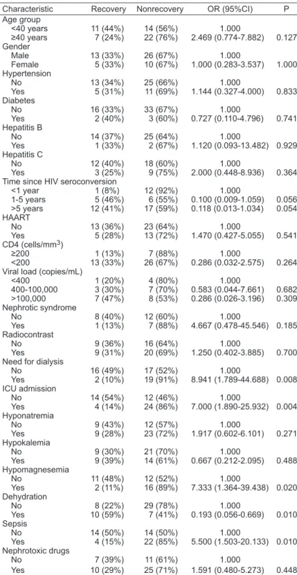
Risk factors for nonrecovery of renal function
Failure to recover renal function did
not correlate significantly with gender (P
> 0.999), age (P = 0.127), diabetes (P = 0.741), coinfection with hepatitis B (P = 0.929), coinfection with hepatitis C (P = 0.364), HAART use (P = 0.541), CD4 count (P = 0.264), proteinuria >3 g/day (P = 0.185), hyponatremia (P = 0.271), or hypokalemia (P = 0.488). Using univari-ate analysis (Table 2), we found that the nonrecovery of renal function correlated
significantly with ICU admission (P = 0.004),
the need for dialysis (P = 0.008), sepsis (P = 0.010), dehydration (P = 0.010), and hypomagnesemia (P = 0.020).
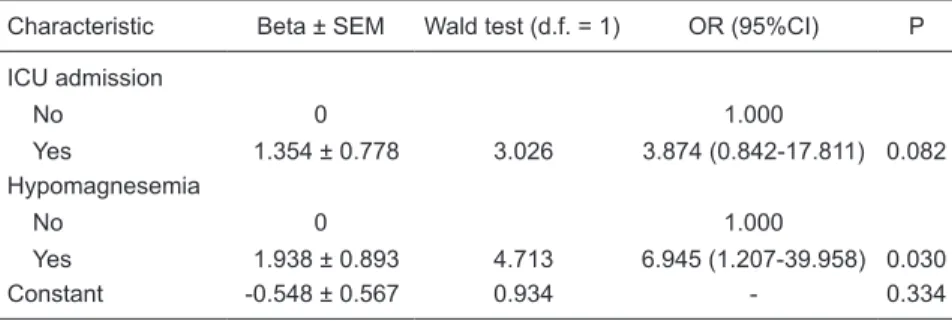
In the multivariate logistic regression analysis (Table 3), the independent risk factors for nonrecovery of renal function were hypomagnesemia (P = 0.03) and ICU admission (P = 0.082). The risk for nonrecovery of renal function was 3.8 times greater in patients admitted to the ICU than in those who did not require intensive care (P = 0.02). Interestingly, hypomagnesemia was also a risk factor for the nonrecovery of renal function. Of the 23 patients who
presented serum magnesium levels ≥1.7
mg/dL, 11 (48%) recovered renal function. However, of the 18 patients who presented serum magnesium levels <1.7 mg/dL, only 2 (11%) recovered renal function (P =0.030). The risk for the nonrecovery of renal function was 6.9 times greater in patients with hypomagnesemia than in those without.
Risk factors for mortality
No significant correlation was found in
the univariate analysis (Table 4) between mortality and the followingvariables: age (P =0.626), gender (P=0.839), CD4 count (P =0.232), HAART use (P=0.087), coinfec-tion with hepatitis B (P=0.731), coinfection with hepatitis C (P=0.921), diabetes (P= 0.274), proteinuria >3 g/day (P =0.124), hypokalemia (P= 0.569), and hyponatremia (P=0.529). However, we could
demon-strate that mortality correlated significantly
with the following variables: ICU admission (P=0.003), requiring dialysis (P=0.043), sepsis (P < 0.001), and hypomagnesemia (P=0.013).
Mortality was quite high (64%)
Table 2. Univariate analysis of nonrecovery of renal function in relation to socio-demographic and clinical characteristics.
Characteristic Recovery Nonrecovery OR (95%CI) P Age group
<40 years 11 (44%) 14 (56%) 1.000
≥40 years 7 (24%) 22 (76%) 2.469 (0.774-7.882) 0.127 Gender
Male 13 (33%) 26 (67%) 1.000
Female 5 (33%) 10 (67%) 1.000 (0.283-3.537) 1.000 Hypertension
No 13 (34%) 25 (66%) 1.000
Yes 5 (31%) 11 (69%) 1.144 (0.327-4.000) 0.833 Diabetes
No 16 (33%) 33 (67%) 1.000
Yes 2 (40%) 3 (60%) 0.727 (0.110-4.796) 0.741 Hepatitis B
No 14 (37%) 25 (64%) 1.000
Yes 1 (33%) 2 (67%) 1.120 (0.093-13.482) 0.929 Hepatitis C
No 12 (40%) 18 (60%) 1.000
Yes 3 (25%) 9 (75%) 2.000 (0.448-8.936) 0.364 Time since HIV seroconversion
<1 year 1 (8%) 12 (92%) 1.000
1-5 years 5 (46%) 6 (55%) 0.100 (0.009-1.059) 0.056 >5 years 12 (41%) 17 (59%) 0.118 (0.013-1.034) 0.054 HAART
No 13 (36%) 23 (64%) 1.000
Yes 5 (28%) 13 (72%) 1.470 (0.427-5.055) 0.541 CD4 (cells/mm3)
≥200 1 (13%) 7 (88%) 1.000
<200 13 (33%) 26 (67%) 0.286 (0.032-2.575) 0.264 Viral load (copies/mL)
<400 1 (20%) 4 (80%) 1.000
400-100,000 3 (30%) 7 (70%) 0.583 (0.044-7.661) 0.682 >100,000 7 (47%) 8 (53%) 0.286 (0.026-3.196) 0.309 Nephrotic syndrome
No 8 (40%) 12 (60%) 1.000
Yes 1 (13%) 7 (88%) 4.667 (0.478-45.546) 0.185 Radiocontrast
No 9 (36%) 16 (64%) 1.000
Yes 9 (31%) 20 (69%) 1.250 (0.402-3.885) 0.700 Need for dialysis
No 16 (49%) 17 (52%) 1.000
Yes 2 (10%) 19 (91%) 8.941 (1.789-44.688) 0.008 ICU admission
No 14 (54%) 12 (46%) 1.000
Yes 4 (14%) 24 (86%) 7.000 (1.890-25.932) 0.004 Hyponatremia
No 9 (43%) 12 (57%) 1.000
Yes 9 (28%) 23 (72%) 1.917 (0.602-6.101) 0.271 Hypokalemia
No 9 (30%) 21 (70%) 1.000
Yes 9 (39%) 14 (61%) 0.667 (0.212-2.095) 0.488 Hypomagnesemia
No 11 (48%) 12 (52%) 1.000
Yes 2 (11%) 16 (89%) 7.333 (1.364-39.438) 0.020 Dehydration
No 8 (22%) 29 (78%) 1.000
Yes 10 (59%) 7 (41%) 0.193 (0.056-0.669) 0.010 Sepsis
No 14 (50%) 14 (50%) 1.000
Yes 4 (15%) 22 (85%) 5.500 (1.503-20.133) 0.010 Nephrotoxic drugs
No 7 (39%) 11 (61%) 1.000
Yes 10 (29%) 25 (71%) 1.591 (0.480-5.273) 0.448
among the patients admitted to the ICU.
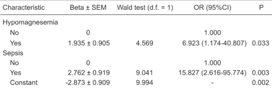
In multiple logistic regression analysis (Table 5), the independent risk factors for mortality were hypomagnesemia (P=0.033) and sepsis (P=0.003).
The most interesting finding in the present study was
the correlation between hypomagnesemia and mortality (P=0.033). Of the 18 patients who presented hypomag-nesemia, 11 (62%) died in the hospital, compared with only 5 (22%) of the 23 patients presenting normal serum
levels of magnesium. We found that the risk of death for
patients presenting serum magnesium levels <1.7 mg/dL was 6.92 times greater than that for those presenting serum
magnesium levels ≥1.7 mg/dL.
Discussion
The results of the present study emphasize the variety of clinical causes of AKI in hospitalized AIDS patients, the use of multiple nephrotoxic drugs for these patients and the impact that electrolyte disturbances have on the course of the disease.
Our study population was predominantly male. The majority of the patients evaluated had been HIV-infected for at least 5 years, presented low CD4 counts and were not receiving regular antiretroviral therapy. Noncompliance with treatment continues to be a challenge in the treatment of these patients.
Our findings are compatible with those of many other
studies in which renal dysfunction has been shown to occur predominantly in the more advanced stages of HIV infection (14,15). Sepsis is considered to be a major cause of AKI in AIDS patients (5,6,9). In a sample of patients with advanced HIV infection, approximately half of those presenting AKI also had sepsis, which causes prerenal azotemia by pro-moting systemic vasodilation and arterial hypotension (6). Sepsis was the leading cause of renal failure in our study population as well.
Sixty-five percent of the patients presenting AKI were
receiving potentially nephrotoxic drugs, contrast medium be-ing the most common. Contrast-induced AKI continues to be a leading cause of hospital-acquired renal failure (23). Vari-ous studies have demonstrated that diabetic nephropathy, volume depletion, dehydration, hypercholesterolemia, and
preexisting renal insufficiency are the most important risk
factors for the development of contrast-induced AKI (23). There are no data on whether AIDS can predispose to contrast-induced AKI. However, AIDS patients are typically under treatment with other potentially nephrotoxic drugs, as well as having sepsis, presenting a high risk for volume depletion (due to dehydration and diarrhea) and being frequently submitted to imaging studies involving the ad-ministration of contrast medium.
In view of these facts, it is not surprising that contrast medium was the most common potentially nephrotoxic drug employed in our cohort, and that vancomycin was the second most common. It should be borne in mind that vancomycin is used only in patients with severe infections,
which might explain the fact that a significant correlation
between vancomycin use and mortality, as well as between vancomycin use and nonrecovery of renal function, was found only in the univariate analysis.
In the present study, hyponatremia was the most com-mon electrolyte disturbance. Hyponatremia occurs in 36-56% of hospitalized AIDS patients and typically indicates inappropriate arginine vasopressin secretion or volume
depletion secondary to gastrointestinal fluid loss (15,24).
The second most common electrolyte disturbance de-tected in our study population was hypokalemia, which, since it can be caused by drugs such as gentamicin, amphotericin B, acyclovir, and tenofovir, is frequently observed in AIDS patients (18,20,25,26). In addition, the clinical symptoms of AIDS (diarrhea, malnutrition and anorexia) predispose to hypokalemia. It is also known that hypokalemia enhances the tubular damage resulting from ischemic injury (16).
Table 3. Final results of the multivariate analysis of sociodemographic and clinical charac-teristics associated with nonrecovery of renal function.
Characteristic Beta ± SEM Wald test (d.f. = 1) OR (95%CI) P
ICU admission
No 0 1.000
Yes 1.354 ± 0.778 3.026 3.874 (0.842-17.811) 0.082 Hypomagnesemia
No 0 1.000
Yes 1.938 ± 0.893 4.713 6.945 (1.207-39.958) 0.030 Constant -0.548 ± 0.567 0.934 - 0.334
Magnesium, AIDS and acute kidney injury 321
In contrast to what would be expected, hypokalemia did not correlate with the nonrecovery of renal function in the pres-ent study.
Hypomagnesemia is a common entity, occurring in up to 12% ofhospitalized pa-tients (27).The incidence can be as high as 65% in patients in intensive care settings, in which nutrition,diuretics, hypoalbuminemia, and aminoglycosides can play important roles (27,28). Gastrointestinal losses, renal losses (due to volume expansion, as well as to the use of pentamidine, amphoteri-cin and diuretic drugs), redistribution and hypokalemia are all potential causes of hypomagnesemia (27,28).
Hypomagnesemia is considered to be a common electrolyte disturbance in patients with AIDS (15). Since AIDS patients com-monly present diarrhea and malnutrition and are treated with drugs that can cause magnesium loss, they are at high risk for developing hypomagnesemia, which report-edly occurs in approximately 29% of such patients (15). Magnesium plays a basic role in various organ functions, and the kidney is the main regulator of magnesium (27,28). In mammals, the magnesium ion plays an important role, since the intracellular level of free magnesium regulates intermediary metabolism, DNA/RNA structure, DNA/RNA synthesis, cell growth, cell reproduction, membrane structure, potassium transport, calcium ion transport, signal transduction modulation, fat synthesis, and protein synthesis (27-29). Since the adenosine triphosphate-magnesium complex is bound and then hydrolyzed by enzymes, mag-nesium is a cofactor for most adenosine triphosphates (28,29).
It has been shown that hypomag-nesemia is a risk factor for AKI (22). de Araujo et al. (22)demonstrated that AZT
induces a decrease in glomerular filtration rate and in renal blood flow in rats with
hypomagnesemia. Hypomagnesemia also predisposes to AZT nephrotoxicity (21). Therefore, chronic administration of AZT can produce AKI in AIDS patients with hy-pomagnesemia. de Araujo et al. (22) also showed that magnesium supplementation protects against postischemic AKI. These
investigators found that renal blood flow in
ischemic rats receiving magnesium supple-mentation is comparable to that observed in
Table 4. Univariate analysis of mortality in relation to sociodemographic and clini-cal characteristics.
Characteristic Survival Death OR (95%CI) P Age group
<40 years 13 (52%) 12 (48%) 1.000
≥40 years 17 (59%) 12 (41%) 0.765 (0.260-2.247) 0.626 Gender
Male 22 (56%) 17 (44%) 1.000
Female 8 (53%) 7 (47%) 1.132 (0.343-3.743) 0.839 Hypertension
No 19 (50%) 19 (50%) 1.000
Yes 11 (69%) 5 (31%) 0.455 (0.132-1.561) 0.210 Diabetes
No 26 (53%) 23 (47%) 1.000
Yes 4 (80%) 1 (20%) 0.283 (0.029-2.714) 0.274 Hepatitis B
No 22 (56%) 17 (44%) 1.000
Yes 2 (67%) 1 (33%) 0.647 (0.054-7.746) 0.731 Hepatitis C
No 17 (57%) 13 (43%) 1.000
Yes 7 (58%) 5 (42%) 0.934 (0.241-3.625) 0.921 Time since HIV seroconversion
<1 year 3 (23%) 10 (77%) 1.000
1-5 years 6 (55%) 5 (46%) 0.250 (0.043-1.443) 0.121 >5 years 21 (72%) 8 (28%) 0.114 (0.025-0.526) 0.005 HAART
No 17 (47%) 19 (53%) 1.000
Yes 13 (72%) 5 (28%) 0.344 (0.101-1.167) 0.087 CD4 (cells/mm3)
≥200 6 (75%) 2 (25%) 1.000
<200 20 (51%) 19 (49%) 2.850 (0.511-15.901) 0.232 Viral load (copies/mL)
<400 4 (80%) 1 (20%) 1.000
400-100,000 6 (60%) 4 (40%) 2.667 (0.212-33.486) 0.447 >100,000 8 (53%) 7 (47%) 3.500 (0.313-39.153) 0.309 Nephrotic syndrome
No 16 (80%) 4 (20%) 1.000
Yes 4 (50%) 4 (50%) 4.000 (0.684-23.406) 0.124 Radiocontrast
No 16 (64%) 9 (36%) 1.000
Yes 14 (48%) 15 (52%) 1.905 (0.638-5.689) 0.248 Need for dialysis
No 22 (67%) 11 (33%) 1.000
Yes 8 (38%) 13 (62%) 3.250 (1.039-10.162) 0.043 ICU admission
No 20 (77%) 6 (23%) 1.000
Yes 10 (36%) 18 (64%) 6.000 (1.815-19.837) 0.003 Hyponatremia
No 13 (62%) 8 (38%) 1.000
Yes 17 (53%) 15 (47%) 1.434 (0.467-4.401) 0.529 Hypokalemia
No 18 (60%) 12 (40%) 1.000
Yes 12 (52%) 11 (48%) 1.375 (0.459-4.117) 0.569 Hypomagnesemia
No 18 (78%) 5 (22%) 1.000
Yes 7 (39%) 11 (61%) 5.657 (1.436-22.286) 0.013 Dehydration
No 19 (51%) 18 (49%) 1.000
Yes 11 (65%) 6 (35%) 0.576 (0.176-1.884) 0.361 Sepsis
No 24 (86%) 4 (14%) 1.000
Yes 6 (23%) 20 (77%) 20.000 (4.945-80.894) 0.000 Nephrotoxic drugs
No 11 (61%) 7 (39%) 1.000
Yes 18 (51%) 17 (49%) 1.484 (0.467-4.718) 0.503
References
1. Franceschini N, Napravnik S, Eron JJ Jr, Szczech LA, Finn
WF. Incidence and etiology of acute renal failure among
ambulatory HIV-infected patients. Kidney Int 2005; 67: 1526-1531.
2. D’Agati V, Appel GB. HIV infection and the kidney. J Am Soc Nephrol 1997; 8: 138-152.
3. Balow JE. Nephropathy in the context of HIV infection. Kid-ney Int 2005; 67: 1632-1633.
4. Dellow EL, Unwin RJ, Miller RF. Presentation, diagnosis, and
management of renal failure in patients with HIV infection.
AIDS Patient Care STDS 2000; 14: 71-77.
5. Wyatt CM, Arons RR, Klotman PE, Klotman ME. Acute renal
failure in hospitalized patients with HIV: risk factors and impact on in-hospital mortality. AIDS 2006; 20: 561-565. 6. Peraldi MN, Maslo C, Akposso K, Mougenot B, Rondeau E,
Sraer JD. Acute renal failure in the course of HIV infection: a single-institution retrospective study of ninety-two patients and sixty renal biopsies. Nephrol Dial Transplant 1999; 14:
nonischemic rats. It is known that hypomagnesemia leads to hypertension, atherogenesis and stroke, although the
mechanisms involved remain ill-defined (30).
It has also been shown that patients with
hyperten-sion, ischemic heart disease or stroke exhibit significant
magnesium depletion (31). Recent in vitro studies of aortic and cerebrovascular smooth muscle cells from humans, rats and dogs have demonstrated that reductions in serum magnesium levels to below those considered normal induce the expression of at least two proto-oncogenes (c-fos and
c-jun), as well as of the nuclear factor-kappa B (NF-κB)
transcription factor (30). Since it is known that mRNA
expression of c-fos, c-jun and NF-κB increases after renal
ischemia/reperfusion, these factors might also be involved
in the pro-inflammatory and oxidative renal processes
(32). Renal ischemia-induced hypomagnesemia might be associated with up-regulation of proto-oncogenes and of
NF-κB. In various cells, including peripheral blood mono -nuclear cells, magnesium is involved in myriad reactions and functions: enzymatic reactions, operation of channel, receptor and intracellular signaling molecules, as well as nucleic acid and protein conformation (27,33). Here, we
have demonstrated for the first time that hypomagnesemia
is a risk factor for mortality and for nonrecovery of renal function in AIDS patients.
The relationship between hy-pomagnesemia and mortality has been well described. Rubeiz et al. (34) reported that mortality rates were nearly twice as high in hypo-magnesemic patients as in patients presenting normal serum levels of magnesium. Soliman et al. (35) dem-onstrated that developing ionized hypomagnesemia during an ICU stay correlated with a poor progno-sis. However, there are no available reports showing that magnesium supplementation can improve mor-tality in hypomagnesemic patients in the ICU setting. In animal models,
magnesium deficiency has been
shown to increase production of
inflammatory cytokines (36).
As previously stated, sepsis was the leading cause of AKI in our study population. Salem et al. (37) showed that
progressive magnesium deficiency and hypomagnesemia
are strongly associated with increased mortality in experi-mental sepsis. In patients with sepsis or septic shock, the prevalence of hypomagnesemia is high, and sepsis has
been identified as an independent risk factor for developing
hypomagnesemia during an ICU stay (35). Moreover, stud-ies have suggested that low magnesium concentration may contribute to the pathogenesis of coronary atherosclerosis or acute thrombosis (38).
In order to classify the severity of AKI in hospitalized
patients, we used the definition of AKI established by the
Adult AIDS Clinical Trial Groups (39). This might constitute a limitation of the study, since it is possible that, by using
this definition, we missed some cases of AKI in which there
were smaller increases in serum creatinine.
In conclusion, hypomagnesemia in hospitalized AIDS patients is a risk factor for nonrecovery of renal function and for in-hospital mortality. Studies involving magnesium supplementation in AIDS patients are warranted in order to ascertain whether hypomagnesemia is a determinant or simply a marker of critical illness.
Table 5. Final results of the multivariate analysis of sociodemographic and clinical charac-teristics associated with mortality.
Characteristic Beta ± SEM Wald test (d.f. = 1) OR (95%CI) P
Hypomagnesemia
No 0 1.000
Yes 1.935 ± 0.905 4.569 6.923 (1.174-40.807) 0.033 Sepsis
No 0 1.000
Yes 2.762 ± 0.919 9.041 15.827 (2.616-95.774) 0.003 Constant -2.873 ± 0.909 9.994 - 0.002
Magnesium, AIDS and acute kidney injury 323
1578-1585.
7. Chertow GM, Soroko SH, Paganini EP, Cho KC, Himmelfarb J, Ikizler TA, et al. Mortality after acute renal failure: models
for prognostic stratification and risk adjustment. Kidney Int
2006; 70: 1120-1126.
8. Sandhu JS, Singla K, Sandhu P. HIV associated renal dis-ease. J Indian Acad Clim Med 2004; 5: 331-334.
9. Perazella MA. Acute renal failure in HIV-infected patients: a brief review of common causes. Am J Med Sci 2000; 319: 385-391.
10. Izzedine H, Launay-Vacher V, Deray G. Antiviral drug-in-duced nephrotoxicity. Am J Kidney Dis 2005; 45: 804-817. 11. Daugas E, Rougier JP, Hill G. HAART-related nephropathies
in HIV-infected patients. Kidney Int 2005; 67: 393-403. 12. Kimmel PL, Barisoni L, Kopp JB. Pathogenesis and
treat-ment of HIV-associated renal diseases: lessons from clinical and animal studies, molecular pathologic correlations, and genetic investigations. Ann Intern Med 2003; 139: 214-226.
13. Szczech LA. Renal diseases associated with human
immu-nodeficiency virus infection: epidemiology, clinical course,
and management. Clin Infect Dis 2001; 33: 115-119.
14. Weiner NJ, Goodman JW, Kimmel PL. The HIV-associated
renal diseases: current insight into pathogenesis and treat-ment. Kidney Int 2003; 63: 1618-1631.
15. Wyatt CM, Winston J. Renal disease in patients with HIV. Curr Infect Dis Rep 2006; 8: 76-81.
16. Menahem SA, Perry GJ, Dowling J, Thomson NM. Hypoka-laemia-induced acute renal failure. Nephrol Dial Transplant
1999; 14: 2216-2218.
17. Seguro AC, Shimizu MH, Monteiro JL, Rocha AS. Effect of potassium depletion on ischemic renal failure. Nephron
1989; 51: 350-354.
18. Goodhart GL, Handelsman S. Gentamicin and hypokalemia.
Ann Intern Med 1985; 103: 645-646.
19. Bernardo JF, Murakami S, Branch RA, Sabra R. Potassium depletion potentiates amphotericin-B-induced toxicity to renal tubules. Nephron 1995; 70: 235-241.
20. Sabra R, Branch RA. Amphotericin B nephrotoxicity. Drug Saf 1990; 5: 94-108.
21. Seguro AC, de Araujo M, Seguro FS, Rienzo M, Magaldi AJ, Campos SB. Effects of hypokalemia and hypomagnesemia on zidovudine (AZT) and didanosine (ddI) nephrotoxicity in rats. Clin Nephrol 2003; 59: 267-272.
22. de Araujo M, Andrade L, Coimbra TM, Rodrigues AC Jr, Seguro AC. Magnesium supplementation combined with N-acetylcysteine protects against postischemic acute renal failure. J Am Soc Nephrol 2005; 16: 3339-3349.
23. Marenzi G, Bartorelli AL. Recent advances in the prevention of radiocontrast-induced nephropathy. Curr Opin Crit Care
2004; 10: 505-509.
24. Upadhyay A, Jaber BL, Madias NE. Incidence and
preva-lence of hyponatremia. Am J Med 2006; 119: S30-S35. 25. Andrade L, Reboucas NA, Seguro AC. Down-regulation
of Na+ transporters and AQP2 is responsible for
acyclovir-induced polyuria and hypophosphatemia. Kidney Int 2004; 65: 175-183.
26. Libório AB, Andrade L, Pereira LV, Sanches TR, Shimizu MH, Seguro AC. Rosiglitazone reverses tenofovir-induced nephrotoxicity. Kidney Int 2008; 74: 910-918.
27. Agus ZS. Hypomagnesemia. J Am Soc Nephrol 1999; 10: 1616-1622.
28. Fawcett WJ, Haxby EJ, Male DA. Magnesium: physiology
and pharmacology. Br J Anaesth 1999; 83: 302-320. 29. Altura BM, Altura BT. Magnesium and cardiovascular
biol-ogy: an important link between cardiovascular risk factors and atherogenesis. Cell Mol Biol Res 1995; 41: 347-359.
30. Altura BM, Kostellow AB, Zhang A, Li W, Morrill GA, Gupta
RK, et al. Expression of the nuclear factor-kappaB and proto-oncogenes c-fos and c-jun are induced by low extra-cellular Mg2+ in aortic and cerebral vascular smooth muscle
cells: possible links to hypertension, atherogenesis, and stroke. Am J Hypertens 2003; 16: 701-707.
31. Altura BM, Altura BT, Gebrewold A, Ising H, Gunther T.
Mag-nesium deficiency and hypertension: correlation between magnesium-deficient diets and microcirculatory changes in situ. Science 1984; 223: 1315-1317.
32. Safirstein R, Price PM, Saggi SJ, Harris RC. Changes in
gene expression after temporary renal ischemia. Kidney Int
1990; 37: 1515-1521.
33. Shechter M, Sharir M, Labrador MJ, Forrester J, Silver B, Bairey Merz CN. Oral magnesium therapy improves en-dothelial function in patients with coronary artery disease.
Circulation 2000; 102: 2353-2358.
34. Rubeiz GJ, Thill-Baharozian M, Hardie D, Carlson RW.
Association of hypomagnesemia and mortality in acutely ill medical patients. Crit Care Med 1993; 21: 203-209. 35. Soliman HM, Mercan D, Lobo SS, Melot C, Vincent JL.
De-velopment of ionized hypomagnesemia is associated with higher mortality rates. Crit Care Med 2003; 31: 1082-1087.
36. Malpuech-Brugere C, Nowacki W, Daveau M, Gueux E, Linard C, Rock E, et al. Inflammatory response following acute magnesium deficiency in the rat. Biochim Biophys Acta 2000; 1501: 91-98.
37. Salem M, Kasinski N, Munoz R, Chernow B. Progressive
magnesium deficiency increases mortality from endotoxin
challenge: protective effects of acute magnesium replace-ment therapy. Crit Care Med 1995; 23: 108-118.
38. Liao F, Folsom AR, Brancati FL. Is low magnesium concen-tration a risk factor for coronary heart disease? The Athero-sclerosis Risk in Communities (ARIC) Study. Am Heart J
1998; 136: 480-490.