REVIEW ARTICLE
S149 Copyright ©2003 by Sociedade Brasileira de Pediatria
1. Assistant Professor, Clinical Pediatrics and Emergency Medicine, State University of New York at Buffallo. The Children’s Hospital of Buffalo. 2. MD, Intensive Care Unit, Children’s Hospital, Buffalo, NY.
3. Fellow in Pediatric Intensive Care, The Children’s Hospital of Buffalo, Buffalo, NY, USA.
Abstract
Objective: To review the current support and treatment strategies of the acute respiratory distress syndrome.
Sources of data: Original data from our research laboratory and from representative scientific articles on acute respiratory distress syndrome and acute lung Injury searched through Medline.
Summary of the findings: Despite advances in the understanding of the pathogenesis of acute respiratory distress syndrome, this syndrome still results in significant morbidity and mortality. Mechanical ventilation, the main therapeutic modality for acute respiratory distress syndrome, is no longer considered simply a support modality, but a therapy capable of influencing the course of the disease. New ventilation strategies, such as high-frequency oscillatory ventilation appear to be promising. This text reviews the current knowledge of acute respiratory distress syndrome management, including conventional and non-conventional ventilation, the use of surfactant, nitric oxide, modulators of inflammation, extracorporeal membrane oxygenation and prone position.
Conclusions: The last decade was marked by significant advances, such as the concept of protective ventilation for acute respiratory distress syndrome. The benefit of alternative strategies, such as high-frequency oscillatory ventilation, the use of surfactant and immunomodulators continue to be the target of study.
J Pediatr (Rio J) 2003;79(Suppl 2):S149-S60: Acute respiratory distress syndrome, mechanical
ventilation, high-frequency ventilation, surfactant, nitric oxide.
Management of the acute respiratory distress syndrome
Alexandre T. Rotta,1 Cláudia L. Kunrath,2 Budi Wiryawan3
Introduction
Acute respiratory distress syndrome (ARDS) is an entity marked by a significant inflammatory response to a local (pulmonary) or remote (systemic) insult which invariably results in hypoxemia and marked alterations to pulmonary
ARDS has an incidence of 13.5 cases per 100,000 people and that ALI affects 17.9 of every 100,000 people.2 Despite significant advances in general intensive care therapies, the dramatic alterations that are characteristic of ARDS are associated with an elevated mortality, varying between 35% and 71%.3-5
Despite having first been described several decades ago6 and being a significant causer of morbidity and mortality in pediatric intensive care units all over the world, ARDS has no specific pharmacological treatment. However, advances in the understanding of the pathogenesis and pathophysiology of ARDS over the years have resulted in the development of a series of support therapies capable of having an impact on the outcome of patients affected by this pathology (Table 1).
resources and a large capacity for integration among participating centers. The availability of clinical data specific to the pediatric ARDS population is even more limited due to the almost non-existence of controlled studies in this population. This being the case, many of the strategies employed for the management of pediatric ARDS and their indications have been adapted or inferred from studies of adult patients.
ARDS treatment strategies Control of the causative factor
While ARDS has no specific treatment, many of the factors causing and perpetuating the disease process can be treated or controlled. For example, patients with hypovolemic shock should be quickly identified and treated with rapid volumetric replacement, in order to minimize the impact on the evolution and maintenance of ARDS. Similarly, patients with infectious acute abdomen should be treated with antibiotics and early surgical intervention when indicated. Patients with septic shock or pneumonia that evolve to ARDS should be promptly treated with intravascular expansion and antibiotics, since the treatment of the infectious factor and hemodynamic control are fundamental to the success of managing the subsequent pulmonary pathology.
Controlled oxygen exposure
By definition, patients with ARDS exhibit significant hypoxemia (PaO2/FiO2 < 200).1 For this reason, oxygen is indicated for the management of the initial phase of the acute respiratory insufficiency. Severe hypoxemia in patients with ARDS is due to the intrapulmonary shunt, in which unventilated lung zones that result from edema, atelectasis or consolidation continue to receive blood supply, despite being incapable of participating in its oxygenation. Oxygen therapy, via mask, tent or non-invasive ventilation apparatus is capable of producing symptomatic improvement during the initial phase of acute respiratory failure. However, the rapid natural progression of ARDS with diminishing pulmonary compliance, increased exertion of respiratory muscles and subsequent exhaustion means that oxygen therapy only has value as a temporary symptom relief measure until mechanical ventilation is introduced. The great majority of patients that meet diagnostic criteria for ARDS cannot be managed exclusively with oxygen therapy, and will require mechanical ventilation. The health care professional who understands the pathophysiologic process of ARDS should recognize that a patient that meets diagnostic criteria and requires an accelerated escalation in oxygen therapy will need mechanical ventilation. Oxygen therapy should not delay the institution of ventilatory support, since intubation and initiation of mechanical ventilation for ARDS should be an elective decision made before the patient develops full-blown respiratory failure.
Table 1 - Therapeutic Strategies in ARDS
Control of the causative factor (sepsis, shock, etc.) Mechanical Ventilation
– Controlled oxygen exposure
– Avoidance of volutrauma (using reduced tidal volumes) – Avoidance of atelectrauma (using adequate PEEP) Careful fluid administration
Optimization of hemodynamics and tissue oxygen delivery Non-conventional ventilation
– High-frequency ventilation – Ventilação não invasive – Liquid ventilation Drug-based therapies – Surfactant – Nitric oxide
– Corticosteroids and other anti-inflammatory agents Extracorporeal membrane oxygenation (ECMO) Position therapy (proning)
Prevention and early diagnosis of intercurrent infections Analgesia and sedation
Nutritional support
Psychological support (patient and family)
The administration of oxygen, while simple, is not free from adverse effects. Continuous exposure to high concentrations of oxygen (FiO2 > 0.6) is capable of causing pulmonary injury, even in the absence of a pre-existing lesion.7 Pulmonary injury due to oxygen toxicity is the result of free radicals and reactive oxygen species that are spontaneously generated in hyperoxic environments or from the activation of neutrophils and alveolar macrophages.8,9 The normal lung deals with oxidative insults by means of a series of enzymes (superoxide dismutase, glutathione peroxidase, glutathione reductase, catalase) or antioxidants (vitamins C and E, albumin, etc.), and is capable of tolerating elevated oxygen concentrations for a number of days. However, an injured lung exposed to moderate concentrations of oxygen (which would not be harmful to a normal lung) can further aggravate pulmonary tissue damage even when the exposure is limited to just a few hours.8 This phenomenon occurs, presumably, due to an imbalance between oxidative stimuli and antioxidant protective mechanisms found in acute lung injury states.
Mechanical ventilation
Mechanical ventilation remains the primary support technique for ARDS and is indicated in the vast majority of cases.10 Nonetheless, the indications for mechanical ventilation in patients with ARDS are, to a certain extent, vague, based on clinical findings (dyspnea, tachypnea, use and fatigue of accessory muscles, diaphoresis, poor perfusion, etc.), laboratory findings (acidosis, hypoxemia, hypercapnia) and radiological findings (worsening alveolar infiltrates). An attempt at making the criteria for the institution of mechanical ventilation for ARDS more objective is the so-called “rule of 50s”, in which a PaO2 < 50 torr and a PaCO2 > 50 torr with a FiO2 of 50% characterize patients likely to require ventilatory support. These criteria, however, identify patients in extremely severe disease states with impending respiratory failure. One of the key points in the treatment of ARDS is the early identification of patients with respiratory involvement so that mechanical ventilation can be initiated before they reach an extreme state of respiratory failure.
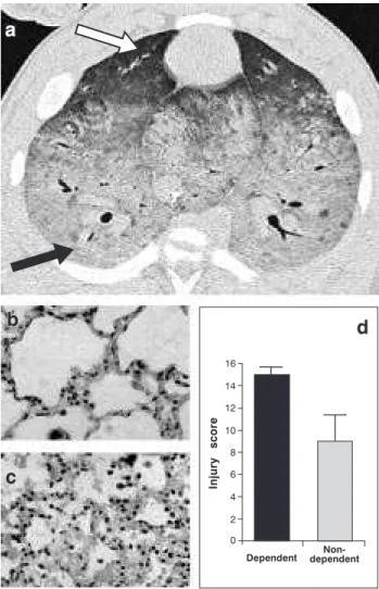
The heterogeneous distribution of lung disease in patients with ARDS makes mechanical ventilation a challenge to the intensive care specialist. In typical ARDS, gravitationally-dependent lung regions exhibit dense alveolar and interstitial inflammatory infiltrates, edema, cellular debris, atelectasis and consolidation, while non-dependant regions are relatively spared (Figure 1).11 In a healthy lung with homogeneous surface tension, tidal volume is evenly distributed among the various lung segments. In patients with ARDS, however, the tidal volume follows the path of least impediment, with a tendency to overdistend the more compliant alveoli (non-dependent) while failing to recruit the less compliant alveoli in the dependent areas. In addition to being heterogeneous, lung pathology in ARDS is also dynamic,12 as areas with relatively adequate compliance
can become poorly compliant in a matter of hours, as the syndrome evolves rapidly.
Mechanical ventilation for ARDS is much more than a mere support modality used to buy time until resolution of the lung disease process. We now know that the choice of ventilation strategy is capable of influencing the progression of the lung disease, with more favorable outcomes resulting from protective strategies. Similarly, non-protective ventilation strategies are associated with less favorable physiological outcomes and increased mortality.5,13-15
Figure 1 - a) Computerized axial tomography of an experimental ARDS model (swine) showing the heterogeneous distribution of lung disease. The gravitationally non-dependent lung region (white arrow) exhibits a relatively normal aspect, while the dependent lung region (black arrow) exhibits greater involvement. Histologic analysis of another animal model of ARDS (rabbits) also shows the heterogeneous distribution of this pathology, with less evidence of inflammation and tissue injury in the non-dependent region (b) in contrast with the dependent region (c). The use of an objective pulmonary injury score in this same model (d) confirms the heterogenous distribution of tissue injury
Injury
score
Dependent dependent Non-16
14
12
10
8
6
4
2
0
d
Tidal volume (Vt)
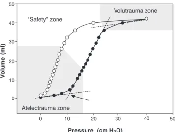
The use of an inadequately high Vt in experimental models is capable of promoting pulmonary injury even in healthy lungs.16 In experimental ARDS models, a Vt that has traditionally been considered adequate, such as 10 ml/kg, has been associated with progression and worsening of the pulmonary injury.17 This occurs because, in low pulmonary compliance states, the introduction of moderate or high Vt can lead to alveolar overdistension, marked by the upper inflection point on the volume-static pressure relationship curve (Figure 2), resulting in the so-called “volutrauma”. Based on this principle, Amato and colleagues have demonstrated significantly reduced 28-day mortality in ARDS patients treated with an open lung strategy consisting of a Vt of less than 6 ml/kg and PEEP set above the lower inflection point. However, two other studies, employing reduced Vt only18,19 failed to show any benefit from this strategy in patients with ARDS. More recently, a North American multi-center study involving 861 patients with ARDS15 showed a 22% reduction in mortality among patients treated with reduced Vt (6 ml/kg) in comparison with traditional Vt (12 ml/kg). The discrepancies between results of the various multi-center studies are related to significant methodologic variations, such as different Vt values employed for the intervention and control groups (Figure 3). Only studies with a sufficient difference in Vt between the reduced volume and the control groups5,15 yielded positive results.
To this date, no clinical studies have tested the hypothesis that reduced Vt would be beneficial in the pediatric population. However, considering that the recommendation to use reduced Vt has a strong physiological, experimental
and clinical support (in adults), pediatric patients with ARDS should be given mechanical ventilation with a Vt equal to or less than 6 ml/kg until data specific to this population become available.
Positive end-expiratory pressure (PEEP)
In ARDS, alveoli in the dependent lung regions exhibit greatly reduced compliance in comparison with non-dependent alveoli. As such, during every expiration the more dependent alveoli reach a critical closing volume, which results in alveolar collapse. This is followed by reopening of these collapsed alveoli during inspiration. The cyclical repetition of alveolar collapse and re-opening generates shearing forces capable of causing tissue damage (atelectrauma). The use PEEP is primarily aimed at avoiding the collapse of the less compliant alveoli at the end of expiration. Excessive use of PEEP increases the risk of pneumothorax, generates hyperinflation of certain pulmonary segments and can cause adverse hemodynamic effects by increasing intra-thoracic pressure and thus reducing venous return (pre-load). However, the application of inadequately low PEEP levels during mechanical ventilation provokes cyclic alveolar collapse and re-opening, resulting in atelectrauma.
The use of adequate levels of PEEP that target sufficient lung volume maintenance during is associated with favorable physiological outcomes.13,16,17 As has been mentioned above, Amato and colleagues5 have demonstrated a reduction in 28-day mortality in patients ventilated with a Vt lower than 6 ml/kg and PEEP level set above the lower inflection point. It is impossible to discern whether the observed effects5 are attributable to the limited Vt, the use
Figure 2 - Static pressure-volume relationship of the respiratory system in an animal model of ARDS (rabbits). The arrow indicates the lower inflection point
50
40
30
20
10
0
0
“Safety” zone
Volutrauma zone
Atelectrauma zone
10 20
Pressure (cm H O)2
V
olume
(ml)
of sufficient PEEP or both (Figure 3). However, strategies that apply sufficient PEEP while avoiding alveolar over distension can prevent the generation of pro-inflammatory mediators (biotrauma) that may adversely affect the progression of the pulmonary lesion,17,20 as well as damage remote organs if these substances were to enter into circulation.21 Despite the protective role of PEEP having been systematically documented in laboratory studies, the North American multi-center clinical trial of patients treated with a high pulmonary expiratory volume and low FiO2 compared with patients treated with a low pulmonary expiratory volume and high FiO2 was recently terminated due to futility after the inclusion of 550 patients.22
ARDS who develop significant hypercapnia when protective ventilation is initiated (elevated PEEP with limited Vt) are promptly started on high frequency oscillatory ventilation.
Ventilation mode
Modern conventional mechanical ventilators offer an increasing array of ventilation modes for use in patients with ARDS. Conceptually, however, most ventilation modes used in ARDS are similar in that they are cycled by time and limited by volume or pressure. A mode that is cycled by time and limited by volume implies that the cycle (inspiration and expiration) is controlled by time (inspiratory time and breath rate), and that during the inspiratory phase of the cycle a certain pre-determined volume is administered. A mode that is cycled by time and limited by pressure implies that the cycle (inspiration and expiration) is controlled by time (inspiratory time and breath rate), and that during the inspiratory phase of the cycle a certain pre-determined pressure is administered. In volume-limited ventilation, the Vt administered during each inspiration generates a certain airway pressure (which is measured and controlled in current ventilators). Similarly, in pressure-limited ventilation, the application of a specific pressure gradient between the ventilator and the airway results in the generation of a certain Vt that can be measured and controlled. Regardless of the ventilation mode used, it is important to emphasize that no one conventional ventilation mode has been shown to be clinically superior to another in the management of patients with ARDS, as long as the principles of protective ventilation are respected.
Considering that precise Vt control is a very important factor in ARDS support, time-cycled volume-limited modes are preferred by the most of intensive care specialists nowadays. In time-cycled volume-limited ventilation (controlled, assist-controlled, intermittent mandatory or intermittent mandatory with pressure support) the operator defines the exact Vt to be administered by each mandatory ventilator cycle. The pressure measurements generated by this set volume at the end of inspiration (dynamic) or after a pause (static or plateau pressure) are indicators of pulmonary compliance in ARDS. A peak inspiratory pressure which increases over time for a fixed volume generally indicates worsening compliance. In an analogous manner, a reduction in peak inspiratory pressure generally indicates an improvement in compliance. Volume-limited ventilation traditionally generates a triangular pressure waveform, in contrast with the rectangular waveform of pressure-limited ventilation (Figure 4). As the area under the pressure curve reflects mean airway pressure, volume-limited modes (triangular waveforms) generally have a slightly lower mean airway pressure than pressure-limited modes (rectangular waveform). Modern ventilators like the Servo 300, however, offer a mode known as pressure regulated volume control (PRVC), in which the shape of the pressure waveform of this volume-limited mode is similar to the rectangular format of the pressure-limited mode. As such, Figure 3 - Comparison of PEEP and tidal volume (Vt) among
different randomized controlled studies of reduced tidal volume strategies in ARDS
Amato5 Stewart19 Brochard18 ARDS Network15
0
5 5 5 5
0 0 0
10 PEEP (cm H O)2
VT (ml/kg) VT (ml/kg) VT (ml/kg) VT (ml/kg) PEEP (cm H O)2 PEEP (cm H O)2 PEEP (cm H O)2
10 10 10 10
10 10 10
20
15 15 15 15
20 20 20
In clinical practice, pediatric patients with ARDS should be ventilated with PEEP that is capable of maintaining adequate pulmonary volume at the end of expiration. This value is generally above 8 cm H2O and below 20 cm H2O, other than in exceptional cases. Positive end-expiratory pressure should be progressively increased (in 2 to 3 cm H2O increments) to optimize oxygenation (saturation between 90 and 95% with FiO2< 0.5) and pulmonary inflation checked with chest radiographs or computerized tomography. Patients with severe anasarca or other restrictive lesions of the chest (circumferential burns), as well as patients with excessive abdominal pressure, may require higher PEEP levels.
the use of PRVC has been gaining wide acceptance in the management of patients with ARDS.
Fluid administration
In caring for patients with ARDS, the intensive care specialist must ponder the quantity and quality of fluids that will be administered. For rapid intravascular expansion, the decision on the administration of colloids or crystalloids depends, to a certain extent, more on the personal convictions of the individual intensive care specialist than on established scientific facts. Those who prefer to give colloids justify the practice by the fact that these substances are capable of producing greater intravascular expansion per unit of volume, remain longer within the intravascular space and increase colloid osmotic pressure. Those who choose crystalloids do so because these are cheaper, more readily available, are capable of promoting intravascular expansion equivalent to colloids (when infused volumes are adjusted) and because they do not increase oncotic pressure in the pulmonary interstitium should they extravasate from the capillaries, as can occur with colloids. Controlled clinical studies are inconclusive on the superiority of colloids or crystalloids. Therefore, the choice of fluids for rapid intravascular expansion should be based on the patient’s needs at any given moment, taking into account the type of loss that has occurred, the urgency to resuscitate and the availability of fluids, in addition to plasma colloid osmotic pressure.
The amount of fluids administered to patients with ARDS is also the subject of debate. There is no question that patients in shock or with severe hypovolemia, both risk factors for ARDS, should be aggressively resuscitated, generally with infused volumes that exceed 60 ml/kg during the first hour, since this practice reduces
mortality and is not associated with an increased incidence of ARDS.25 Once hemodynamic stability is achieved in the patient with ARDS, the intensive care specialist should concentrate efforts on minimizing the capillary leak and pulmonary edema accumulation that occur in ARDS. Studies in animal models of acute lung injury indicate that the fluid accumulation in the lung can be attenuated by reducing left atrial pressure.26 This strategy of limiting fluid administration is also supported by some clinical studies of patients with ARDS.27,28 The North American study group involving 24 hospitals (ARDS Network) organized for the study of ARDS is currently conducting a controlled multi-center, randomized study of “conservative” versus “liberal” fluid administration. Until the results of this study become available, a sensible recommendation is to maintain intravascular volume at the lowest level that permits the maintenance of adequate systemic perfusion, assessed by renal and cardiac functions and by the acid-base balance.
Non-conventional ventilation
High frequency ventilation (HFV)
Mechanical ventilation techniques that employ supra-physiologic frequencies, generally between 60 and 900 cycles per minute, are collectively known as HFV. Various types of HFV are available, although only high frequency positive pressure ventilation (HFPPV), high frequency jet ventilation (HFJV) and high frequency oscillatory ventilation (HFOF) have gained significant penetration into clinical practice. Clinical studies of HFPPV and HFJV compared with conventional ventilation were disappointing and resulted in the virtual abandonment of these techniques for the management of patients with ARDS.29 The use of Figure 4 - Comparison of dynamic airway pressure
waveforms during pressure-controlled (a) and volume controlled (b) ventilation
a
b
Time
HFOV, however, is strongly supported by studies of experimental ARDS models.17,30,31 and has sufficient clinical evidence to justify its use under selected circumstances.32-34
In HFOV, tidal volumes that approximate dead space volume are actively pushed into and pulled out of the airway at a frequency of between 3 and 15 hertz (180 to 900 cycles per minute) by means of a piston or diaphragm. The proposed advantage of HFOV is that, due to the minute tidal volume of each cycle, the method is capable of ventilating patients with ARDS within a “Safety Zone” that avoids both alveolar overinflation during inspiration and cyclical closure and re-opening of the alveoli during expiration (Figure 2). Oxygenation and ventilation are controlled independently during HFOV. Controlling the mean airway pressure determines the state of pulmonary inflation and, consequently, oxygenation. Controlling the amplitude of oscillation indirectly determines the tidal volume of each cycle and, consequently, the efficacy of ventilation (CO2 elimination). As such, HFOV is ideal in situations when the patient with ARDS has worsening pulmonary compliance with hypoxemia, requiring a reduction in the Vt of conventional ventilation in order to avoid elevated peak inspiratory pressures, which leads to significant respiratory acidosis. The realization that HFOV can favorably influence the pulmonary inflammatory milieu in experimental models17,31,35 as well as reduce the incidence of chronic lung disease32,34 has been responsible for the enthusiasm about this method and for its increasingly early deployment in patients with ARDS. The use of HFOV in pediatric patients with ARDS requires deep sedation and neuromuscular relaxation, since spontaneous respiratory movements interfere with gas flow mechanics in this modality.
Non-invasive ventilation
The application of non-invasive positive pressure (CPAP or BiPAP) in patients with ARDS is capable of attenuating, albeit temporarily, the reduction in residual functional capacity responsible for the progressive hypoxemia that is characteristic of this pathology. The use of CPAP results in a transient improvement in oxygenation, yet it is not associated with reductions in the need for intubation, length of hospital stay or mortality of patients with ARDS.10 The use of CPAP for ARDS is also associated with an increased incidence of adverse effects.10 As such, the use of CPAP in the prophylaxis or treatment of patients with ARDS is not recommended.
Partial liquid ventilation
Partial liquid ventilation (PLV) is a technique that employs perfluorochemical substances capable of dissolving large quantities of oxygen and carbon dioxide.
In PLV, the lung is filled with a liquid perfluorocarbon via the endotracheal route so as to occupy the functional residual capacity, while volumes of gas are introduced through a conventional ventilator during each inspiratory cycle.36 The potential advantage of PLV in ARDS stems from the fact that when the lung is occupied by liquid it has a uniform surface tension, in contrast to the heterogeneous surface tension typical of ARDS. This occurs because the perfluorocarbon forms a liquid-liquid interface at the alveolar surface, in contrast to the liquid-gas interface found in conventional ventilation. A medical-grade perfluorocarbon called perflubron (C8-F17-Br1) has been successfully tested in the treatment of experimental acute lung injury. We now know that perflubron, as well as other perfluorocarbons that were considered biologically inert, have anti-inflammatory biological effects and protect cellular components against oxidative damage.37-41 However, the enthusiasm for PLV in the laboratory has not been repeated in the clinical arena. Controlled studies of children and adults with ARDS and acute lung injury have not demonstrated PLV to be superior to protective conventional ventilation.42 Further studies are necessary to test the impact of this method in specific clinical situations, such as progressive pulmonary recruitment (liquid PEEP) and intrapulmonary drug administration or viral vectors for genetic therapy. This treatment is not currently available for use outside of the research laboratory environment and cannot be recommended for the treatment of ARDS.
Drug-based therapies
Surfactant replacement
Nitric oxide
Nitric oxide is a potent vasodilator that can be administered via inhalation causing pulmonary vascular relaxation. Inhaled nitric oxide reaches the alveolus where it enters into direct contact with the pulmonary vasculature. During its migration through the wall of the blood vessel, nitric oxide causes direct relaxation of the muscular layer, before reaching the vascular lumen. Nitric oxide is then rapidly deactivated by binding with hemoglobin, resulting in the formation of methemoglobin and avoiding the undesirable effect of systemic vasodilation. The pulmonary vasodilatory effect of nitric oxide associated with the fact that the target vasculature is adjacent to the ventilated areas of the lung results in not only a decrease in pulmonary vascular resistance, but also in attenuation of ventilation-perfusion mismatch, thus improving oxygenation. The use of nitric oxide in newborns with pulmonary hypertension has achieved great clinical success, reducing morbidity and the need for extracorporeal support.45 However, its use in patients with ARDS has been disappointing. Despite producing a transient improvement in oxygenation, this benefit is of short duration and does not offer any objective gains. The use of nitric oxide in ARDS does not reduce mortality or the duration of mechanical ventilation46 and cannot therefore be routinely recommended in clinical practice. Nitric oxide can be used as a therapy of exception for temporary rescue of patients with hypoxemia that is refractory to more conventional interventions.
Corticosteroids
The fact that acute pulmonary damage in ARDS is primarily the result of an aggressive inflammatory process has lead intensive care specialists to consider anti-inflammatories in general, and corticosteroids in particular, as logical therapeutic alternatives. The use of corticosteroids, however, does not prevent the development of ARDS47 nor is it beneficial when employed during the initial phase of its clinical course.48 Corticosteroids appear to have some benefit when used during the later stages of the disease, which are marked by the reorganization of the acute inflammatory infiltration and fibrosing alveolitis.49 Currently, a multi-center randomized controlled North American study (ARDS Network) is being conducted to evaluate the efficacy of high doses of methylprednisolone during the later phases of ARDS. However, due to the fact that treatment with high doses of corticosteroids can increase the risk of secondary infections and other adverse effects, their routine use in ARDS treatment cannot yet be recommended. In our clinical practice, we reserve the use of corticosteroids as a rescue therapy in severe ARDS cases during the later phase of the disease (third or fourth week) when there is no progress in reducing the level of ventilatory support.
Other inflammation control agents
Despite having produced promising results in experimental models of acute lung injury, the use of non-steroidal drugs with anti-inflammatory effects, such as indomethacin, ibuprofen, procysteine, lisofylline and ketoconazole, have not been shown beneficial in the clinical arena.50 The use of these drugs for the treatment of patients with ARDS, therefore, cannot be recommended.
Extracorporeal membrane oxygenation (ECMO)
Extracorporeal membrane oxygenation consists of the use of a complex circuit of vascular cannulae, tubes, pumps, oxygenator, heat exchanger and monitoring systems used to provide respiratory support (in the case of veno-venous ECMO) or cardiorespiratory support (in the case of veno-arterial ECMO). To date, approximately 25,000 patients have undergone ECMO throughout the world with an overall survival rate of approximately 75%. The vast majority of these patients (17,000) were neonates with refractory pulmonary hypertension, while the experience in pediatric and adult cases of ECMO for treatment of ARDS is limited to approximately 3,000 cases (personal communication, ECMO Registry of the Extracorporeal Life Support Organization (ELSO), Ann Arbor, Michigan, November, 2002). Extracorporeal membrane oxygenation reduces the mortality of newborns with persistent pulmonary hypertension secondary to meconium aspiration syndrome (94% survival), but has yielded more modest results in older children with ARDS (52% survival). Clinical studies of the use of ECMO or an extracorporeal carbon dioxide elimination system with adults suffering from ARDS did not reveal any benefits in terms of reduced mortality.51 However, the outcome results for ECMO therapy in the international extracorporeal life support registry for pediatric patients with ARDS refractory to all other forms of treatment, and also in our personal practice, strongly suggest that this technique is of value in selected cases.
Positioning therapy
The simplicity and low cost of the use of prone positioning, associated with reports of improvements in oxygenation in 60 to 70% of patients with ARDS has made this therapeutic method popular. A number of different mechanisms have been suggested to explain this effect in patients placed in the prone position, such as an improvement in the ventilation-perfusion relationship,52 increased pulmonary volume at the end of expiration53 and regional ventilation changes associated with mechanical alterations of the thoracic wall.54 However, as has been demonstrated above, improvements in oxygenation do not necessarily translate to reduced mortality in ARDS.15 Recently, Gattinoni and colleagues55 reported the results of a multi-center, controlled study in which patients with ARDS were randomized to receive either conventional treatment (supine position) or treatment in the prone position for 6 or more hours per day for 10 days. In this study, despite causing an improvement in oxygenation, the use of the prone position did not result in a reduction in mortality.55 A number of different theories may explain these findings. The simplest is that the use of the prone position indeed does not prevent or attenuate the advance of pulmonary injury in patients with ARDS. On the other hand, despite including 304 patients, this study probably did not have sufficient statistical power to reveal differences between groups, since clinical studies of ARDS are marked by heterogeneous characteristics demanding large sample sizes. The patients randomized to the prone group assumed the position for approximately 7 hours per day (or just 30% of the time) and for a maximum of 10 days. It is possible that the limited duration of exposure to the prone position could explain the failure of this strategy. A multi-center study of pediatric patients with ARDS involving the use of the prone position for the greater part of the day and until resolution of the respiratory failure is in progress in tertiary ICUs in North America. Until concrete results are available, the recommendation to place patients with ARDS in the prone position in an attempt to improve oxygenation and allow exposure to lower concentrations of oxygen appears to have a reasonable theoretical foundation and few risks or costs associated with it.
Prevention and early diagnosis of intercurrent infections
As ARDS patients require invasive technology, such as vascular and urinary catheters, endotracheal intubation and mechanical ventilation for prolonged periods of time, they are often the target of secondary infections, especially pulmonary infections. Early diagnosis and precise treatment of these infections is extremely important, since secondary pneumonias act as an additional pro-inflammatory insult. Radiologic diagnosis of secondary pulmonary infections in patients with ARDS
is complicated by the fact that these patients exhibit pre-existing radiologic abnormalities. Clinical diagnosis also presents challenges, since symptoms such as fever, leukocytosis and increased tracheal secretions may already be part of the basic disease process. In clinical practice, early diagnosis may be achieved by integrating radiologic alterations, appearance and cellularity of tracheal secretions and routine cultures (tracheal aspirate, broncho-alveolar lavage and blood culture).
As with other nosocomial infections, prevention is the best method of reducing the risk of secondary pulmonary infections. Immunosuppressed or contagious patients should be isolated and the use of universal contact precautions and frequent hand washing are simple and highly effective measures. Criteria-based antibiotic therapy guided by the antibiogram of organisms isolated by cultures or on local epidemiological data also plays an important role in the prevention of secondary infections.
Analgesia and sedation
The comfort of patients with ARDS during their stay in the ICU should occupy a prominent position in the therapeutic strategy. Patients in the acute phase of the disease should receive infusions of medications to reduce the emotional stress and physical discomfort inherent to the pathology, as well as in anticipation of painful procedures. Our practice is to maintain patients with ARDS on continuous sedation and pain relief, with these needs being reevaluated on a daily basis. Infusions of midazolam (0.1 mg/kg/h) and fentanyl (2 µg/kg/h) are used in the majority of patients and doses are adjusted according to clinical requirements, with doses of 10 times higher than the original not being uncommon by the third week of the clinical course. Patients subjected to permissive hypercapnia or HFOV require the infusion of neuromuscular blocking agents, such as vecuronium (0.1 mg/kg/h). Patients with highly compromised pulmonary mechanics and during the acute phase of the disease also often require neuromuscular blocking agents.
Nutritional Support
References
1. Bernard GR, Artigas A, Brigham KL, Carlet J, Falke K, Hudson L, et al. The American-European Consensus Conference on ARDS. Definitions, mechanisms, relevant outcomes, and clinical trial coordination. Am J Respir Crit Care Med 1994;149:818-24. 2. Luhr OR, Antonsen K, Karlsson M, Aardal S, Thorsteinsson A, Frostell CG, et al. Incidence and mortality after acute respiratory failure and acute respiratory distress syndrome in Sweden, Denmark, and Iceland. The ARF Study Group. Am J Respir Crit Care Med 1999;159:1849-61.
3. Suchyta MR, Clemmer TP, Orme JF Jr, Morris AH, Elliott CG. Increased survival of ARDS patients with severe hypoxemia (ECMO criteria). Chest 1991;99:951-5.
4. Milberg JA, Davis DR, Steinberg KP, Hudson LD. Improved survival of patients with acute respiratory distress syndrome (ARDS): 1983-1993. JAMA 1995;273:306-9.
5. Amato MB, Barbas CS, Medeiros DM, Magaldi RB, Schettino GP, Lorenzi-Filho G, et al. Effect of a protective-ventilation strategy on mortality in the acute respiratory distress syndrome. N Engl J Med 1998;338:347-54.
6. Ashbaugh DG, Bigelow DB, Petty TL, Levine BE. Acute respiratory distress in adults. Lancet 1967;2:319-23.
7. Matalon S, Holm BA, Loewen GM, Baker RR, Notter RH. Sublethal hyperoxic injury to the alveolar epithelium and the pulmonary surfactant system. Exp Lung Res 1988;14 Suppl: 1021-33.
8. Nader-Djalal N, Knight PR 3rd, Thusu K, Davidson BA, Holm BA, Johnson KJ, et al. Reactive oxygen species contribute to oxygen-related lung injury after acid aspiration. Anesth Analg 1998;87:127-33.
9. Yusa T, Crapo JD, Freeman BA. Hyperoxia enhances lung and liver nuclear superoxide generation. Biochim Biophys Acta 1984;798:167-74.
10. Delclaux C, L’Her E, Alberti C, Mancebo J, Abroug F, Conti G, et al. Treatment of acute hypoxemic nonhypercapnic respiratory insufficiency with continuous positive airway pressure delivered by a face mask: A randomized controlled trial. JAMA 2000;284:2352-60.
11. Gattinoni L, Presenti A, Torresin A, Baglioni S, Rivolta M, Rossi F, et al. Adult respiratory distress syndrome profiles by computed tomography. J Thorac Imaging 1986;1:25-30.
12. Gattinoni L, Pesenti A, Bombino M, Baglioni S, Rivolta M, Rossi F, et al. Relationships between lung computed tomographic density, gas exchange, and PEEP in acute respiratory failure. Anesthesiology 1988;69:824-32.
13. Slutsky AS. Lung injury caused by mechanical ventilation. Chest. 1999;116(1 Suppl):9-15.
14. Ranieri VM, Suter PM, Tortorella C, De Tullio R, Dayer JM, Brienza A, et al. Effect of mechanical ventilation on inflammatory mediators in patients with acute respiratory distress syndrome: a randomized controlled trial. JAMA 1999;282:54-61.
15. The Acute Respiratory Distress Syndrome Network. Ventilation with lower tidal volumes as compared with traditional tidal volumes for acute lung injury and the acute respiratory distress syndrome. N Engl J Med 2000;342:1301-8.
16. Dreyfuss D, Soler P, Basset G, Saumon G. High inflation pressure pulmonary edema. Respective effects of high airway pressure, high tidal volume, and positive end-expiratory pressure. Am Rev Respir Dis 1988;137:1159-64.
17. Rotta AT, Gunnarsson B, Fuhrman BP, Hernan LJ, Steinhorn DM. Comparison of lung protective ventilation strategies in a rabbit model of acute lung injury. Crit Care Med 2001;29:2176-84. Psychological support
The psycho-social needs of the family and the patient with ARDS are extremely complex. Even in adequately sedated patients, factors such as anxiety over the uncertainty of the clinical outcome, the impossibility of speech due to the artificial airway, the occasional pain due to invasive procedures and the changes to the awake and sleep cycles, among others, cannot be neglected by the medical team. Attention must be afforded to explain to the patient (whenever possible) and the family all the diagnostic and therapeutic procedures and also the natural course and prognosis of the condition. It is common for adolescent patients and older children in the recovery phase of ARDS to exhibit delirium, depression or altered circadian patterns during prolonged hospitalization in an ICU environment. Such manifestations often require the involvement of a psychiatric consultant to monitor patients during recovery and after hospital discharge. The multidisciplinary medical team should always be alert to and available for the psychological needs of ARDS patients and their families, particularly because ICU hospital stays due to severe ARDS are prolonged and generally marked by oscillation between periods of frustration and optimism.
Monitoring the patient
18. Brochard L, Roudot-Thoraval F, Roupie E, Delclaux C, Chastre J, Fernandez-Mondejar E, et al. Tidal volume reduction for prevention of ventilator-induced lung injury in acute respiratory distress syndrome. The Multicenter Trial Group on Tidal Volume reduction in ARDS. Am J Respir Crit Care Med 1998;158:1831-8. 19. Stewart TE, Meade MO, Cook DJ, Granton JT, Hodder RV, Lapinsky SE, et al. Evaluation of a ventilation strategy to prevent barotrauma in patients at high risk for acute respiratory distress syndrome. Pressure- and Volume-Limited Ventilation Strategy Group. N Engl J Med 1998;338:355-61.
20. Ranieri VM, Giunta F, Suter PM, Slutsky AS. Mechanical ventilation as a mediator of multisystem organ failure in acute respiratory distress syndrome. JAMA 2000;284:43-4. 21. Tremblay L, Valenza F, Ribeiro SP, Li J, Slutsky AS. Injurious
ventilatory strategies increase cytokines and c-fos m-RNA expression in an isolated rat lung model. J Clin Invest 1997;99: 944-52.
22. The Acute Respiratory Distress Syndrome Network. Prospective, Randomized, Multi-Center Trial of Higher End-expiratory Lung Volume/Lower FiO2 versus Lower End-expiratory Lung Volume/ Higher FiO2 Ventilation in Acute Lung Injury and Acute Respiratory Distress Syndrome [site na internet] Disponível: http://hedwig.mgh.harvard.edu/ardsnet/ards04.html. Acessado: 7 de abril de 2003.
23. Carvalho CR, Barbas CS, Medeiros DM, Magaldi RB, Lorenzi Filho G, Kairalla RA, et al. Temporal hemodynamic effects of permissive hypercapnia associated with ideal PEEP in ARDS. Am J Respir Crit Care Med 1997;156:1458-66.
24. Feihl F, Perret C. Permissive hypercapnia. How permissive should we be? Am J Respir Crit Care Med 1994;150:1722-37. 25. Carcillo JA, Davis AL, Zaritsky A. Role of early fluid resuscitation
in pediatric septic shock. JAMA 1991;266:1242-5.
26. Prewitt RM, McCarthy J, Wood LD. Treatment of acute low pressure pulmonary edema in dogs: relative effects of hydrostatic and oncotic pressure, nitroprusside, and positive end-expiratory pressure. J Clin Invest 1981;67:409-18.
27. Mitchell JP, Schuller D, Calandrino FS, Schuster DP. Improved outcome based on fluid management in critically ill patients requiring pulmonary artery catheterization. Am Rev Respir Dis 1992;145:990-8.
28. Humphrey H, Hall J, Sznajder I, Silverstein M, Wood L. Improved survival in ARDS patients associated with a reduction in pulmonary capillary wedge pressure. Chest 1990;97:1176-80. 29. Wiswell TE, Graziani LJ, Kornhauser MS, Cullen J, Merton DA,
McKee L, et al. High-frequency jet ventilation in the early management of respiratory distress syndrome is associated with a greater risk for adverse outcomes. Pediatrics 1996;98:1035-43. 30. Froese AB. Role of lung volume in lung injury: HFO in the atelectasis-prone lung. Acta Anaesthesiol Scand Suppl 1989;90:126-30.
31. McCulloch PR, Forkert PG, Froese AB. Lung volume maintenance prevents lung injury during high frequency oscillatory ventilation in surfactant-deficient rabbits. Am Rev Respir Dis 1988;137: 1185-92.
32. Arnold JH, Hanson JH, Toro-Figuero LO, Gutierrez J, Berens RJ, Anglin DL. Prospective, randomized comparison of high-frequency oscillatory ventilation and conventional mechanical ventilation in pediatric respiratory failure. Crit Care Med 1994;22:1530-9.
33. Derdak S, Mehta S, Stewart TE, Smith T, Rogers M, Buchman TG, et al. High-frequency oscillatory ventilation for acute respiratory distress syndrome in adults: a randomized, controlled trial. Am J Respir Crit Care Med 2002;166:801-8.
34. Courtney SE, Durand DJ, Asselin JM, Hudak ML, Aschner JL, Shoemaker CT. High-frequency oscillatory ventilation versus conventional mechanical ventilation for very-low-birth-weight infants. N Engl J Med 2002;347:643-52.
35. Sugiura M, McCulloch PR, Wren S, Dawson RH, Froese AB. Ventilator pattern influences neutrophil influx and activation in atelectasis-prone rabbit lung. J Appl Physiol 1994;77:1355-65. 36. Fuhrman BP, Paczan PR, DeFrancisis M.
Perfluorocarbon-associated gas exchange. Crit Care Med 1991;19:712-22. 37. Rotta AT, Steinhorn DM. Partial liquid ventilation reduces
pulmonary neutrophil accumulation in an experimental model of systemic endotoxemia and acute lung injury. Crit Care Med 1998;26:1707-15.
38. Rotta AT, Gunnarsson B, Hernan LJ, Fuhrman BP, Steinhorn DM. Partial liquid ventilation with perflubron attenuates in vivo oxidative damage to proteins and lipids. Crit Care Med 2000; 28:202-8.
39. Rotta AT, Gunnarsson B, Hernan LJ, Fuhrman BP, Steinhorn DM. Partial liquid ventilation influences pulmonary histopathology in an animal model of acute lung injury. J Crit Care 1999;14:84-92.
40. Rotta AT, Gunnarsson B, Hernan LJ, Fuhrman BP, Steinhorn DM. Perflubron protects cell monolayers against direct in vitro oxidative injury. Crit Care Med 1999;27:A40.
41. Rotta AT, Gunnarsson B, Hernan LJ, Fuhrman BP, Steinhorn DM. Perfluorocarbon protects against fatty acid oxidation in a non-biological, in vitro model. Crit Care Med 1999;27:A133. 42. Hirschl RB, Croce M, Gore D, Wiedemann H, Davis K,
Zwischenberger J, et al. Prospective, randomized, controlled pilot study of partial liquid ventilation in adult acute respiratory distress syndrome. Am J Respir Crit Care Med 2002;165:781-7. 43. Anzueto A, Baughman RP, Guntupalli KK, Weg JG, Wiedemann HP, Raventos AA, et al. Aerosolized surfactant in adults with sepsis-induced acute respiratory distress syndrome. Exosurf Acute Respiratory Distress Syndrome Sepsis Study Group. N Engl J Med 1996;334:1417-21.
44. Willson DF, Zaritsky A, Bauman LA, Dockery K, James RL, Conrad D, et al. Instillation of calf lung surfactant extract (calfactant) is beneficial in pediatric acute hypoxemic respiratory failure. Members of the Mid-Atlantic Pediatric Critical Care Network. Crit Care Med 1999;27:188-95.
45. Clark RH, Kueser TJ, Walker MW, Southgate WM, Huckaby JL, Perez JA, et al. Low-dose nitric oxide therapy for persistent pulmonary hypertension of the newborn. Clinical Inhaled Nitric Oxide Research Group. N Engl J Med 2000;342:469-74. 46. Dellinger RP, Zimmerman JL, Taylor RW, Straube RC, Hauser
DL, Criner GJ, et al. Effects of inhaled nitric oxide in patients with acute respiratory distress syndrome: results of a randomized phase II trial. Inhaled Nitric Oxide in ARDS Study Group. Crit Care Med 1998;26:15-23.
47. Luce JM, Montgomery AB, Marks JD, Turner J, Metz CA, Murray JF. Ineffectiveness of high-dose methylprednisolone in preventing parenchymal lung injury and improving mortality in patients with septic shock. Am Rev Respir Dis 1988;138:62-8. 48. Bernard GR, Luce JM, Sprung CL, Rinaldo JE, Tate RM, Sibbald WJ, et al. High-dose corticosteroids in patients with the adult respiratory distress syndrome. N Engl J Med 1987;317:1565-70. 49. Meduri GU, Belenchia JM, Estes RJ, Wunderink RG, el Torky M, Leeper KV Jr. Fibroproliferative phase of ARDS. Clinical findings and effects of corticosteroids. Chest 1991;100:943-52. 50. ARDS N. Ketoconazole for early treatment of acute lung injury and acute respiratory distress syndrome: a randomized controlled trial. The ARDS Network. JAMA 2000;283:1995-2002. 51. Morris AH, Wallace CJ, Menlove RL, Clemmer TP, Orme JF Jr,
Corresponding author: Alexandre Tellechea Rotta Division of Pediatric Critical Care, The Children’s Hospital of Buffalo 219 Bryant Street
Buffalo, NY 14222, USA Tel.: (716) 878.7442 Fax: (716) 878.7101 E-mail: [email protected] 52. Pappert D, Rossaint R, Slama K, Gruning T, Falke KJ. Influence
of positioning on ventilation-perfusion relationships in severe adult respiratory distress syndrome. Chest 1994;106:1511-6. 53. Douglas WW, Rehder K, Beynen FM, Sessler AD, Marsh HM.
Improved oxygenation in patients with acute respiratory failure: the prone position. Am Rev Respir Dis 1977;115:559-66. 54. Pelosi P, Tubiolo D, Mascheroni D, Vicardi P, Crotti S, Valenza
F, et al. Effects of the prone position on respiratory mechanics and gas exchange during acute lung injury. Am J Respir Crit Care Med 1998;157:387-93.