REVIEW ARTICLE
S139 Copyright © 2003 by Sociedade Brasileira de Pediatria
1. Department of Pediatrics, School of Medicine, Universidade Federal do Rio de Janeiro. Specialist in Intensive Care. PICU, Hospital Copa D’Or, Rio de Janeiro, RJ, Brazil
2. Department of Pediatrics, School of Medicine, Universidade Estácio de Sá. Specialist in Intensive Care. Clinical Division, School of Medicine, Universidade Estácio de Sá, Rio de Janeiro, RJ, Brazil.
Abstract
Objective: The main concepts involved in the therapeutic management of intracranial hypertension are revisited, including pathophysiology, monitoring, the traditional approach, and also the presentation of recently proposed therapies.
Source of data: The main medical literature data bases (especially Medline and Lilacs) were searched for articles published in the last 10 years, and traditional text books and dissertations focusing the subject were consulted.
Summary of the findings: Intracranial hypertension may be associated with several cerebral neurologic lesions from traumatic, infectious or metabolic origin, and in severe cases may represent an important factor for morbidity and mortality. Increases in intracranial pressure interfere with cerebral blood flow; in order to maintain an adequate cerebral perfusion pressure, it is necessary to both reduce and control intracranial hypertension and combat low blood pressure from the very beginning of treatment. Adequate monitoring is essential, and the main points addressed in conventional management include elevation of the head of bed to 30 °C, central positioning of the head, optimization of hypocapnia, perfect metabolic and hydro-electrolyte balance, sedation, anti-convulsive therapy, liquor drainage, and the use of barbiturics, mannitol and furosemide. New therapies currently under investigation include the use of dexanabinol, hypertonic saline solutions, moderate hypothermia, decompressive craniectomy, optimization of cerebral perfusion pressure, and reduction in cerebral microvasculature pressure (Lund therapy).
Conclusions: The use of new therapies to effectively control intracranial hypertension, especially in cases that are refractory to the usual treatment, represent a promising scenario in the management of this problem.
J Pediatr (Rio J) 2003;79(Suppl 2):S139-S48: Intracranial hypertension, diagnosis, therapy.
New therapies for intracranial hypertension
Arnaldo Prata Barbosa,1 Sérgio Augusto Cabral2
Introduction
Many different types of brain damage, whether of a traumatic, infectious, metabolic or hypoxic origin, may result in an increase in intracranial pressure (ICP). In
these concepts that provides the primary objective of the present article.
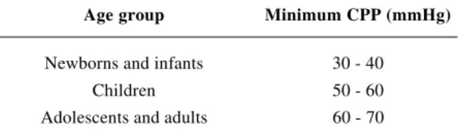
Intracranial pressure varies exponentially as a function of the volume of the contents of the skull. This volume consists of the sum of the volumes of the encephalon (80%), blood (10%), and cerebrospinal fluid (10%) compartments, and possibly of other components with a mass effect (Table 1). A number of different pathologies can cause intracranial hypertension as a result of an increase in the volume of these compartments (Table 2). With infants, the presence of fontanels and cranial sutures that are still open permits greater tolerance of ICP, but with children whose fontanels are already closed up and no longer have any capacity for expansion of the skull, small variations in the volume of one compartment will be balanced by changes to the volume of others sectors. Large variations, however, cannot be compensated for and will undoubtedly result in increased intracranial pressure. Increased ICP, in turn, is an important limitation to cerebral perfusion and can result in significantly reduced cerebral blood flow. As will be seen below, a number of different therapeutic measures are based upon an understanding of these effects. Normal ICP levels by age group and their maximum acceptable variations are presented in Table 3 and the minimum acceptable levels for cerebral perfusion pressure (CPP) can be found in Table 4.
Table 1 - Volume of the contents of the skull in cases of trauma
Volume of the encephalon
(80% of intracranial pressure - 75 to 85% of water) Volume increases with edema. Types of edema:
– Vasogenic (common surrounding tumors, rare in trauma) – Cytotoxic (common in trauma, secondary to cell lesion) – Interstitial (due to the elevated hydrostatic pressure)
Blood volume
(autoregulation maintains CBF constant)
Constant CBF depends on: – Cerebral perfusion pressure – Cerebral metabolic demand – PCO2 (pH)
– PO2 Fluid volume
(reabsortion blocked due to inflammation, edema or mass effect)
Volume of other contents (usually bruises)
Table 2 - Causes of intracranial hypertension
Increase in brain tissue volume
Generalized edema (trauma, toxins, metabolic disorders, hypoxia, infections)
Focal edema (localized trauma, perilesional edema)
Increase in cerebral blood volume
Obstruction of venous flow (thrombosis, inappropriate head position)
Hypercapnia, hypoxia Arterial hypertension
Autoregulation failure (trauma, tumor, ischemia, hypertension, hypotension)
Anesthetic drugs (halotan) Hyperemia (swelling)
Dysfunction of fluid dynamics
Hydrocephaly comunicante, obstruction of fluid circulation outside the ventricular system
Obstruction of the fluid circulation inside the ventricular system Anomaly of the coroid plexus
Reduction in fluid absorption (cerebral pseudotumor)
Mass effect Abcess Tumor Hemorrhage
Table 3 - Normal ICP levels by age group and their maximum acceptable variations
Age group ICP (mmHg)
Newborns and infants Up to 5
Children 6 - 15
Adolescents and adults < 15
Significant intracranial hypertension: 20 - 24 mmHg/30 minutes
25 - 29 mmHg/10 minutes > 30 mmHg/1 minute
Mild hypertension: 15 - 25 mmHg Moderate hypertension: 25 - 40 mmHg Severe hypertension: > 40 mmHg
Table 4 - Minimum acceptable levels for cerebral perfusion pressure (CPP) according to age group
Age group Minimum CPP (mmHg)
Newborns and infants 30 - 40
Children 50 - 60
Monitoring
Correct monitoring of patients with intracranial hypertension allows constant evaluation and maintenance of vital signs, with particular attention to developments such as hypotension, hypoxia, cerebral edema, hemorrhages, herniation and alterations to cerebral hemodynamics and cerebral metabolism of blood. The following are recommended: (1) basic monitoring: ECG, pulse oximetry, non-invasive arterial pressure, central venous pressure (CVP), temperature and urinary output and, ideally, invasive arterial pressure, capnography and pulmonary capillary pressure; (2) metabolic monitoring: gasometry and serum electrolytes, glycemia, serum osmolarity and urinary density and, particularly in intracranial hypertension cases resulting from trauma (3) cerebral monitoring: intracranial pressure, cerebral perfusion pressure, jugular venous bulb oximetry (SjvO2), cerebral oxygen extraction fraction (OEF) and, very often, continuous EEG.
The traditional approach
The first priority must always be to identify and correct life-threatening conditions before continuing with the rest of the neurological examination thus avoiding secondary cerebral lesions. This initial evaluation is known as the First evaluation. Next, with the patient stabilized, the Second evaluation can take place which aims to be a complete investigation with the objective of identifying all possible lesions and to direct treatment.
First evaluation
The first evaluation and initial resuscitation efforts should take place simultaneously, generally during the first 5 to 10 minutes. Vital signs should be continuously re-evaluated, every five minutes during the first evaluation and every fifteen thereafter, until the patient is stabilized, always bearing in mind, right from the start, the importance of avoiding maneuvers which might increase intracranial pressure. This first phase can be summed up by the algorithm presented in Table 5.
Second evaluation
During this phase, with the patient’s vital functions already established, a minutely detailed physical examination should be performed, as complete as possible a personal history should be elicited, initial laboratory tests should be performed and pertinent imaging studies requested (computerized tomography, nuclear magnetic resonance, Doppler transcranial) in order to assess the extent of lesions and make decisions about the need for surgical intervention and/or continued treatment within the ICU.
Controlling intracranial pressure
Patients with severe intracranial hypertension have a high risk of developing potentially fatal herniating syndromes which must be quickly recognized and require immediate action aimed at reducing ICP and maintaining it at acceptable levels. These “acceptable levels” are not precisely understood, particularly not with children, but it is believed that levels between 20 and 25 mmHg should be considered as an upper limit, above which further measures to reduce ICP should be instigated.1
For a long time ICP management was considered the foundation upon which the whole of the clinical care of patients with intracranial hypertension should be based. In recent years, however, emphasis has been given to other important parameters, in particular to the maintenance of optimum cerebral perfusion pressure2 and to attending to cerebral hemodynamics and metabolism.3 In any case, maintaining ICP at safe levels guarantees that CPP will remain at least at a minimally acceptable levels and respect for basic hemodynamic and metabolic parameters guides the majority of treatment protocols. The principal points to be followed are described below.
Table 5 - Initial care of patients with neurologic condition
N - Neck Consider the possibility of cervical trauma Do not move the patients’ neck until there is possibility of trauma
A - Airways Protect airways
Intubate the patient if Glasgow Coma Scale is < 9
B - Ventilation Avoid hypoxemia Keep normal PaCO2 C - Circulation Avoid and treat hypotension D - Drugs Consider exogenous intoxication
Check available antidotes
E - Epilepsy History and risk factors for seizures Signs: tongue biting, bruises
F - Fever Signs of infection of NCS: meningism, rash, bruises
G - Glasgow Glasgow coma scale
H - Herniation Check for herniation signs. If there are signs: – hyperventilation with Ambu bag: keep PaCO2
between 25-30 mmHg – manitol 1 g/kg – dexametasone
Specific surgical procedure: – drainage of bruises – decompressive craniotomy – ventricular derivation
Bed position
The head should be maintained in a neutral position (no lateral dislocation), with the head of the bed inclined at 30 degrees in order to encourage venous drainage through the jugulars, thus minimizing any influence on ICP. Angles greater than 30 degrees may, for a given AAP value, result in reduced CPP.4
Mechanical ventilation
The aim should be to maintain adequate oxygenation (SaO2 > 95%), for which PEEP may be employed at between 3 and 5 cm H2O without contributing to any significant increase in ICP. Any anemia should be corrected by concentrated erythrocyte transfusion, maintaining hemoglobin at around 10 g/dl, since more elevated levels will exponentially increase blood viscosity and reduce CBF. Levels of PaCO2 should be maintained between 30 and 40 mmHg, for which capnography is of great utility, improving control of PCO2 levels.
Empirical hyperventilation should be avoided, particularly in CET cases, during the first five days and particularly during the first 24 hours. Studies of the cerebral blood flow and cerebral metabolism [(a-vO2 diff), SjvO2, OEF] of patients who had been victims of severe CET demonstrate a significant flow reduction early on during the first few hours post-trauma with test result values close to those associated with ischemia. In these scenario the use of hyperventilation would only further aggravate ischemic lesions and at least one randomized clinical study has demonstrated worsened prognosis among patients given prophylactic hyperventilation.1 Empirical hyperventilation only has a role in cases where there are clinical signs of cerebral herniation (sudden anisocoria or mydriasis); when performed manually with a valve-bag it can rapidly reduce ICP and prevent herniation. Optimized hyperventilation can be use when cerebral oxygen supply and consumption are observed to have become uncoupled, resulting in e hyperemia or “luxury perfusion”, where CBF is abnormally elevated in relation to cerebral metabolic demand. Such states can be detected by blood oxygen saturation measurement at the jugular venous bulb (SjvO2 > 75%) and by calculating cerebral oxygen extraction (OEF < 24%).5
While there remains a risk or signs of intracranial hypertension, aspiration via the tracheal tube should be preceded (30 seconds) by the administration of intravenous lidocaine at 1% (1 mg/kg), in order to suppress the cough reflex, which can cause significant ICP increases. Supplementary sedation can also be employed such as a short action barbiturate like thiopental (3-5 mg/kg/dose) or a non-depolarizing neuromuscular blocking agent (vecuronium or rocuronium), as long as the patient is adequately sedated and on pain relief.
Hemodynamic, hydro-electrolytic and metabolic balance
The importance of instituting volumetric resuscitation and shock control in order to avoid secondary lesions from hypotension and hypoxia has been duly pointed out. Monitoring these patients’ CVP allows early detection of hypovolemic states even before AAP has altered, making correction and control possible. Invasive AAP monitoring, however, does permit the identification of episodes of hypotension. Hypotension, at any point during treatment, has been identified as one of the primary factors which effectively influence prognosis, worsening it. Studies have demonstrated that the record of a single episode of hypotension is associated with a doubling of the mortality rate and a significant increase in morbidity with severe head traumas.6 During the maintenance phase, therefore, it is important to be alert to any sign of hemodynamic instability, particularly hypotension episodes, which should be aggressively corrected with staged 20 ml/kg of saline every 10 to 20 minutes or albumin solutions at 5% in saline, with the objective of normalizing AP, improving peripheral perfusion and the reestablishment of diuresis. A small number of studies have demonstrated that the use of hypertonic solutions (NaCl at 3%) is better than normal saline for volumetric resuscitation. In these studies patients who received hypertonic solutions had better pressure response, lower volume requirements and required fewer interventions for ICP control, had fewer complications and better prognosis.7,8 If there is no response to volumetric expansion, the use of dopamine (5-15 µg/kg/min) is indicated, and dobutamine (5-15 µg/kg/min) can be added if there is echocardiographic evidence of ventricular dysfunction. Refractive hypotension cases should be tackled with such as noradrenaline (0,05-1 µg/kg/min) or phenyleprine (0,1-0,5 µg/kg/min), while average and ICP should be monitored in order to maintain CPP within minimum acceptable limits. In such situations a pulmonary artery catheter can be of great help in hemodynamic control and to inform treatment.
Hypertension, on the other hand, is normally considered to be a response to increased intracranial pressure and, therefore, essential to the maintenance of CPP. It should not be corrected while its cause is not completely clear. If the decision is taken to correct hypertension calcium channel blockers or beta-blockers should be used rather than vasodilators (such as sodium nitroprusside), in order to avoid sudden arterial hypotension and cerebral vasodilation (increased ICP), which would cause a drastic reduction in CPP.9
necessary with a parallel glucose infusion at 50%, starting with 0.5 ml/kg/hour (GIR of 4 mg/kg/min).10 Glucose is essential to cerebral metabolism and hypoglycemia should be avoided. However, its excess under anaerobiosis conditions, as may occur in certain, poorly perfused, encephalic areas, may cause elevated lactate levels, contributing to worsening of neuron damage. Serum sodium levels should be kept between 140 and 150 mEq/l. In cases where Na < 140 mEq/l, correct this with NaCl at 3% solution, 3 ml/kg per hour and continue with 0.5 ml/kg/hour until levels return to within the desired range.
Sedation, pain relief and neuromuscular blockade
Maintaining the patient correctly sedated and on pain relief allows greater tolerance of nursing procedures, reduces psycho-motor and muscular activity, which could, in the final analysis, increase ICP. Additionally, their use contributes to reduced metabolic cerebral oxygen consumption. Normally a continuous infusion of a benzodiazepine (midazolam 0,05 - 0,3 mg/kg/hour) is used in association with an effective analgesic (fentanyl 0.5 - 2 µg/kg/hour). Recently, one study has demonstrated similar effects when a continuous infusion of midazolam (0.05 - 0.15 mg/kg/hour) was used and when ketamine (3.5 - 6.5 mg/kg/hour), was used with no prejudice to ICP or CPP.11 Concerns about masking neurological status do not justify leaving the patient without adequate sedation and pain relief, particularly when it is possible to effect a neurological assessment using other parameters such as monitoring ICP, CPP, SjvO2, OEF and control CT. One sedation option which could make clinical neurological assessment easier would be the use of agents with ultra short half-lives, such as propofol, which has been used in the intensive care of adults but the license as a continued-use pediatric sedative is not yet approved. In cases of continuing severe intracranial hypertension the decision can be taken to use neuromuscular blocking agents. In this case the non-depolarizing type are indicated, preferably those with the least hemodynamic effects, such as vecuronium (attack dose 0.06 to 0.08 mg/kg, maintenance dose 0.02 to 0.03 mg/kg/hour) or atracurium (attack dose 0.3 to 0.5 mg/kg, maintenance dose 0.2 to 1 mg/kg/hour), although other agents may be employed, such as pancuronium (attack dose 0.06 to 0.08 mg/kg, maintenance dose 0.02 to 0.03 mg/kg/hour). When neuromuscular blocking agents are employed the EEG should be monitored to rule out convulsive states, in addition to, whenever possible, monitoring their use with a peripheral nerve stimulator.
Prevention and control of convulsive episodes
Convulsions can have a negative effect on prognosis, caused by the even greater increase to intracranial pressure, the increased cerebral metabolic demand, hypoxia and hypoventilation which are associated with it. For these
reasons the use of prophylactic diphenyl-hydantoin or carbamazepine until the crisis stabilizes is recommended. In CET cases particularly their use is not recommended after the first week.12 In the event of a convulsive episode the usual procedure should be followed, using a rapid acting benzodiazepine such as diazepam (0.2-0.3 mg/kg/dose), followed by maintenance treatment, at least initially, with diphenyl-hydantoin. The need for EEG to be performed for all patients on neuromuscular blocking agents cannot be over-emphasized.
Specific intracranial hypertension treatment
Significant intracranial hypertension (ICP > 20-24 mmHg for 30 minutes; 25-29 mmHg for 10 minutes or > 30 mmHg for 1 minute) can be tackled by a number of different methods, summarized below:
1. Hyperventilation - as has been mentioned above, hyperventilation can be used to rapidly reduce intracranial pressure if there are clinical signs of herniation or if ICP > 40 mmHg. This is because the encephalic vessels respond rapidly with vasoconstriction to hypocapnia, causing an immediate reduction in cerebral blood volume and therefore ICP. The procedure should be performed manually with a valve-bag, for no more than 2 minutes;
2. Mannitol and furosemide – there are two class I studies, one class II study and numerous class III studies supporting the use of mannitol for intracranial hypertension control.13 The dose is between 0.25 and 0.5 g/kg (1.25-2.5 ml/kg of a 20% solution) and should be administered continuously, but only in cases of significant intracranial hypertension. The administration of more than three doses daily should be avoided due to the risk of cerebral accumulation making edema more likely. Other important concerns are to avoid hypovolemia and serum osmolarity of > 320 mOsm/l, and to take into account the fact that its excretion in urine interferes with urine density assessments. Furosemide is also effective in reducing ICP acting to reduce cerebrospinal fluid production and causing excretion to favor water over solutes in the distal tubules of the kidney. It is more useful when employed in conjunction with mannitol, particularly if given 15 minutes later, at a dose of 1 mg/kg. In cases of elevated ICP with arterial hypotension the use of diuretics can worsen hypotension. In such cases, in order to maintain CPP, hypotension should be corrected as described above and the use of diuretics avoided;
3. Cerebrospinal fluid drainage – if the patient has a ventricular catheter for ICP monitoring, then this can be used to drain cerebrospinal fluid, opening the system for 1 minute. Continuous drainage may be employed after a neurosurgeon’s assessment;
a CT scan as soon as possible in order to rule out lesions with a mass effect, since effusions and further hemorrhages can occur as the cases evolves;
5. Barbiturates – the use of barbiturates at high dosages is effective in reducing ICP and it has been demonstrated that their use is capable of reducing mortality in cases of intracranial hypertension that are refractory to all conventionally measures.14 Its use as a prophylactic, however, is not indicated. Its beneficial effects are the result of a number of different mechanisms, such as altering the vascular tone, inhibiting lipid peroxidation, inhibiting oxygen free radical formation and suppressing the cerebral metabolism. The objective of the treatment with high dose barbiturates is to achieve an electroencephalographic stage known as burst suppression, which requires the use of an EEG for control. The primary side effects are hypotension and resuscitation toxicity. This is, therefore a treatment which requires a great deal of careful monitoring because of the risks of hemodynamic instability and cerebral oligemic hypoxia (SjvO2 < 55%, OEF > 42%). Normally the barbiturate used is pentobarbital (not available in Brazil), at an attack dose of 10-15 mg/kg in 1-2 hours, followed by maintenance doses of 1 mg/kg/hour, which can be increased to 2 to 3 mg/kg/hour, until the burst suppression pattern is obtained on the EEG. One alternative available in our locale would be thiopental, at an attack dosage of 4-6 mg/kg, maintaining at 1-5 mg/kg/hour.
6. Increasing average arterial pressure – with the aim of maintaining adequate CPP, a strategy of increasing AAP can be used in conjunction with the measures for reducing ICP. This can be done by (a) maintaining normovolemia, if necessary by infusing volume; (b) giving dopamine 5-15 µg/kg/min, especially if barbiturates have been used; (c) the use of vasopressors, such as noradrenaline (0.05-1 µg/kg/min) and phenyleprine (0.1-0.5 µg/kg/min).
7. Corticoids – there is a great deal of evidence to show that corticoids do not reduce ICP nor improve prognosis, at least not with severe CET patients, there use is not therefore indicated for these purposes.15
New treatments
Since, in the final analysis, intracranial hypertension results from the interaction of a number of different pathophysiological mechanisms which act by ischemia, neurotoxic cascades, edema or inflammation, a series of therapeutic measures aimed at the control of these events has been studied in clinical experiments. Below we will see the main innovations in this area.
Dexanabinol
Dexanabinol is a synthetic compound, a nonpsychotropic cannabinnoid, which functions as a non-competitive
antagonist to the NMDA (N-methyl-d-aspartate) receptors.16 These receptors are responsible for the neuroexcitotoxic reaction of glutamate, which is found in high concentrations in the cerebrospinal fluid of patients suffering from hypoxic-ischemic encephalopathy and head traumas, as is thought to be one of the main causes of neuronal lesions in these cases, resulting from neuron invasion by sodium and calcium and the consequent formation of edema, oxidative stress and cellular degeneration.17 Dexanabinol appears to function as a buffer for hydroxile and peroxide radicals, in addition to inhibiting tumor necrosis factor (TNF) production.18 The preliminary studies carried out with adults with CET and cerebral vascular accidents demonstrated that it can be beneficial during the first 6 hours after the primary event.19,20 One phase I study demonstrated that doses of up to 200 mg (for adults) are safe.21 A phase II study, with CET patients, tasting safety and efficacy at single doses of 48, 150 or 200 mg, showed a reduction in the time that ICP remained above 25 mmHg and systolic arterial pressure < 90 mmHg.22 Currently, phase III clinical trials are in progress with CET patients. The observed ICP reduction and the anti-hypotensive effects of dexanabinol, in conjunction with its anti-excitotoxic, antioxidant and antiinflammatory effects make the use of this drug particularly promising for the control of the multiple mechanisms which result in intracranial hypertension.
Hypertonic saline solutions
The use of hypertonic saline solutions for the treatment of intracranial hypertension is based on the hypothesis that the osmolar tissue load could be more important than the increase in cerebral blood volume and the permeability of the blood brain barrier to the development of the cerebral edema. In addition to their osmotic properties, hypertonic solutions have hemodynamic, vasoregulatory and immunomodulatory effects. The hemodynamic effects occur because of the expansion of the intravascular volume and the maintenance of average arterial pressure;23 the vasoregulatory effects are the result of an increase in vascular diameter due to plasma expansion, reduced endothelial cell edema and reduced secondary vascular resistance to the liberation of nitric oxide, while the immunoregulatory effects are associated with leukocyte activation inhibition.24 However, it is probable that the beneficial effects of hypertonic saline solutions result primarily from their osmotic effect on brain tissue, contributing to reduce edema and the water content of the brain.
adults with severe CET did not find any significant effects. In contrast, in a similar study with children, Simma et al. used a hypertonic saline solution at 1.7% during the first 72h post-CET, and demonstrated an inverse relationship between serum sodium levels and ICP.8 Retrospective studies of both adults and children, employing saline solutions at 3% in continuous infusions demonstrated ICP control at serum sodium levels of between 145 and 155 mEq/l.26-28 An uncontrolled prospective study used hypertonic saline at 3% (514 mEq/l), with infusion adjusted to achieve a serum sodium level that maintained ICP < 20 mmHg which was employed for an average of 7.6 days with 10 children with severe CET and intracranial hypertension refractory to conventional treatment. The authors found results that were effective for the control of ICP and to improve CPP, however, the average serum sodium levels that were reached were extremely elevated (170.7 mEq/l), as was plasma osmolarity (364.8 mOsm/l), although there were no associated complications.29
The main concerns in relation to the use of hypertonic saline solutions are due to the increased risk of subarachnoid hemorrhages, renal lesions, pontine myelinolisis (due to the sudden increase in sodium levels similar to what occurs when hyponatremia is corrected) and rebound effects once the infusion is withdrawn. Randomized clinical trials are awaited which could evaluate not just the efficacy and safety of the use of hypernatremia as a form of cerebral edema and intracranial hypertension control, but also its comparative effect to that of mannitol.
Moderate hypothermia
Hyperthermia is associated with increased cerebral oxygen consumption and should be combated. On the other hand, moderate hypothermia (32 to 34 °C) has been associated with beneficial effects and improved prognosis in some work done with both adult humans and laboratory animals. These studies have demonstrated that moderate hypothermia reduces endogenous antioxidant agents, reduces lipid peroxidation and has antiinflammatory effects demonstrated by reduced levels of interleukin-1-beta in cerebrospinal fluid and reduced plasma levels of interleukin-6 and prostanoids.30-32
Three randomized clinical trials have been conducted to date with adult CET victims, with conflicting results. Two of them studied small series of patients: Clifton et al studied 46 patients, comparing normal body temperature with moderate hypothermia started during the first 6 hours post-CET and maintained for 48h, without finding any differences between the groups;33 Marion et al studied 87 patients and found improvements on the Glasgow Prognosis Scale in the sub-group of patients with initial Glasgow scores of 5 to 7, but no differences for the sub-group with Glasgow scores between 3 and 4.30 In contrast, a multi-center study (11 centers), involving 392 patients with initial Glasgow scores of between 3 and 7, recruited during the first 48h post-trauma, did not only fail to find differences between the two
groups, but also concluded that complications such as bleeding, sepsis and pneumonia were more frequent among the group subjected to hypothermia.34 This study, however, has been criticized because the correct temperature was not achieved until 8.4 + 3hours post-trauma and the study group received significantly greater volume supply than the control group. Furthermore, two recent clinical studies of the use of moderate hypothermia with adult patients after cardiorespiratory arrest have demonstrated positive results.35,36
Moderate hypothermia, therefore, when employed should be begun during the first 6 hours after admission in patients who present with flaccidity or decorticate or decerebrate postures. In these cases the patient should be on mechanical ventilation and curare and sedated, and temperature maintained at around 33 degrees (using a thermal mattress for cooling) for 24 to 36 hours, then warming gradually at 1 degree every three hours until 37 degrees are reached.
Decompressive craniectomy
Traditionally used as the last resort for the control of refractory intracranial hypertension, recent studies have shown that decompressive craniectomy is effective at reducing ICP, although there are reports of hemorrhages and exacerbation of the cerebral edema after its use. The majority of studies performed to date involve children and results have been favorable.37,38 One retrospective study of 49 patients, however, was unable to demonstrate any advantages.39 The only randomized prospective study comparing early decompressive craniectomy with traditional treatment was performed by Taylor et al, who adopted the procedure as an early ICP control measure during the first 12 hours post-trauma in a group of children, obtaining not just a reduction in ICP very close to statistical significance (p = 0.057) during the first 48 hours post-craniectomy, but also better prognosis at 6 months (p = 0.046) in relation to the control group. The study involved a small group of patients (13 cases and 14 controls) and the analysis of prognosis was performed by telephone interview, but, in general, the results are promissing.10
Cerebral perfusion pressure optimization
measures to increase arterial pressure in addition to reducing ICP thus optimizing CPP. Some results have already been published.2
Reduction of cerebral microcirculation pressure (Lund Therapy)
Another approach proposed in literature for intracranial hypertension control in cases of head trauma is the Lund therapy or protocol, developed at the University of Lund, Sweden, which emphasizes the importance of reducing the pressure on the cerebral microcirculation, in order to minimize the formation of edema. The principle objectives of this approach are: (1) maintain normal colloid osmotic pressure, by means of albumin and concentrated erythrocyte infusion; (2) reduce capillary hydrostatic pressure by reducing systemic arterial pressure with metoprolol and clonidine; (3) reduce cerebral blood volume by means of precapillary vasoconstriction, achieved with low doses of thiopental and dihydroergotamine. Treatments which favor increased transcapillary filtration should be avoided, such as cerebrospinal fluid drainage, high dosage diuretic
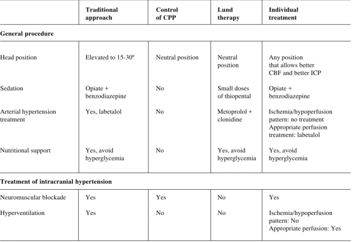
barbiturates and osmotic and CPP optimization. Decompressive craniectomy, which can also increase edema formation, is kept as a last resort. This protocol, very different from the approach traditionally employed in the majority of centers has been used for a number of years at Lund University and promising results have been published,40 clearly indicating the need for further study comparing the main approaches currently in vogue. Table 6 compares the various different approaches cited.
Other attempts
Other attempts to reduce refractory intracranial hypertension have been described in literature in isolated reports. Münch et al. reported favorable experiences with 23 adult patients using continuous cerebrospinal fluid drainage from a spinal tap in the lumbar region until ICP was controlled, as long as tomographic examinations revealed visible basal cisterns, so as to minimize the risk of transtentorial or tonsillar herniation.41 Förderreuther and Straube described success with venous indomethacin at 50 mg, in seven adult patients with refractory intracranial hypertension.42
Table 6 - Differences between several approaches proposed to treat intracranial hypertension in severe head trauma
Traditional Control Lund Individual
approach of CPP therapy treatment
General procedure
Head position Elevated to 15-30º Neutral position Neutral Any position
position that allows better CBF and better ICP
Sedation Opiate + No Small doses Opiate +
benzodiazepine of thiopental benzodiazepine
Arterial hypertension Yes, labetalol No Metoprolol + Ischemia/hypoperfusion
treatment clonidine pattern: no treatment
Appropriate perfusion treatment: labetalol
Nutritional support Yes, avoid No Yes, avoid Yes, avoid
hyperglycemia hyperglycemia hyperglycemia
Treatment of intracranial hypertension
Neuromuscular blockade Yes Yes No Yes
Hyperventilation Yes No No Ischemia/hypoperfusion
pattern: No
Table 6
Traditional Control Lund Individual
approach of CPP therapy treatment
Treatment of intracranial hypertension
Fluid drainage Yes Yes No Yes
Osmotic therapy Yes Yes No Hypoperfusion/edema
pattern: Yes Hyperemia/vascular dilation: No
Barbiturate-induced coma Yes No No Hypoperfusion/edema
pattern: No Hyperemia/vascular dilation: Yes
Control of cerebral perfusion pressure (CPP)
Objectives for CPP Not considered, > 70-80 mmHg > 50-60 Ischemia/hypoperfusion keep ICP < 20 mmHg (above lower limit (allowing pattern: increase CPP and normal AP of autoregulation) appropriate in order to improve CBF,
perfusion) especially if autoregulation is affected
Appropriate perfusion pattern: keep normal CPP
References
1. Brain Trauma Foundation, American Association of Neurological Surgeons, Joint Section on Neurotrauma and Critical Care. Hyperventilation. J Neurotrauma 2000;17:513-20.
2. Rosner MJ, Rosner SD, Johnson AH. Cerebral perfusion pressure: management protocol and clinical results. J Neurosurg 1995; 83:949-62.
3. Robertson CS, Cormio M. Cerebral metabolic management. New Horiz 1995;3:410-22.
4. Feldman Z, Kanter MJ, Robertson CS, Contant CF, Hayes C, Sheinberg MA, et al. Effect of head elevation on intracranial pressure, cerebral perfusion, and cerebral blood flow in head injured patients. J Neurosurg 1992;76:207-11.
5. Cruz J. Hemometabolismo cerebral: de medidas isoladas a medidas de monitorização terapêutica. Arq Neuropsiquiatr 1993; 51:1-7.
6. Chesnut RM, Marshall LF, Klauber MR, Blunt BA, Baldwin N, Eisenberg HM, et al. The role of secondary brain injury in determining outcome from severe head injury. J Trauma 1993;34:216-22.
7. Fisher B, Thomas D, Peterson B. Hypertonic saline lowers raised intracranial pressure in children after head trauma. J Neurosurg Anesthesiol 1992;4:4-10.
8. Simma B, Burger R, Falk M, Sacher P, Fanconi S. A prospective, randomized, and controlled study of fluid management in children with severe head injury: lactate Ringer’s solution versus hypertonic saline. Crit Care Med 1998;26:1265-70.
9. Tietjen CS, Hurn PD, Ulatowski JA, Kirsch JR. Treatment modalities for hypertensive patients with intracranial pathology: option and risks. Crit Care Med 1996;24:311-22.
10. Taylor A, Butt W, Rosenfeld J, Shann F, Ditchfield M, Lewis E, et al. A randomized trial of very early decompressive craniectomy in children with traumatic brain injury and sustained intracranial hypertension. Childs Nerv Syst 2001;17:154-62.
11. Bourgoin A, Albanese J, Wereszczynski N, Charbit M, Vialet R, Martin C. Safety of sedation with ketamine in severe head injury patients: comparison with sufentanil. Crit Care Med 2003;31: 711-17.
12. Brain Trauma Foundation, American Association of Neurological Surgeons, Joint Section on Neurotrauma and Critical Care. Role of antiseizure prophylaxis following head injury. J Neurotrauma 2000;17:549-53.
13. Brain Trauma Foundation, American Association of Neurological Surgeons, Joint Section on Neurotrauma and Critical Care. Use of mannitol. J Neurotrauma 2000;17:521-5.
14. Brain Trauma Foundation, American Association of Neurological Surgeons, Joint Section on Neurotrauma and Critical Care. Use of barbiturates in the control of intracranial hypertension. J Neurotrauma 2000;17:527-30.
15. Brain Trauma Foundation, American Association of Neurological Surgeons, Joint Section on Neurotrauma and Critical Care. Role of steroids. J Neurotrauma 2000;17:531-35.
16. Mechoulam R, Feigenbaum JJ, Lander N, Segal M, Jarbe TU, Hiltunen AJ, et al. Enantiomeric cannabinoids: stereospecificity of psychotropic activity. Experientia 1988;44:762-4.
18. Shohami E, Gallily R, Mechoulam R, Bass R, Ben-Hur T. Cytokine production in the brain following closed head injury: Dexanabinol (HU-211) is a novel TNF-alpha inhibitor and an effective neuroprotectant. J Neuroimmunol 1997;72:169-77. 19. Shohami E, Novikov M, Bass R. Long-term effect of HU-211, a
novel non-competitive NMDA antagonist, on motor and memory functions after closed head injury in the rat. Brain Res 1995; 674:55-62.
20. Belayev L, Busto R, Zhao W, Ginsberg MD. HU-211, a novel noncompetitive N-methil-D-aspartate antagonist, improves neurological deficit and reduces infarct volume after reversible focal cerebral ischemia in the rat. Stroke 1995;26:2313-19. 21. Brewster ME, Pop E, Foltz RL, Reuschel S, Griffith W, Amselem
S, et al. Clinical pharmacokinetics of escalating i.v. doses of dexanabinol (HU-211), a neuroprotectant agent, in normal volunteers. Int J Clin Pharmacol Ther 1997;35:361-5. 22. Knoller N, Levi L, Shoshan I, Reichenthal E, Razon N, et al.
Dexanabinol (HU-211) in the treatment of severe closed head injury: A randomised, placebo-controlled, phase II clinical trial. Crit Care Med 2002;30:548-54.
23. Holcroft JW, Vassar MJ, Turner JE, Derlet RW, Kramer GC. 3% NaCl and 7,5% NaCl/dextran 70 in the resuscitation of severely injury patients. Ann Surg 1987;206:279-88.
24. Hartl R, Medary MB, Ruge M, Arfors KE, Ghahremani F, Ghajar J. Hypertonic/hyperoncotic saline attenuates microcirculatory disturbances after traumatic brain injury. J Trauma 1997;42 Suppl 1:41-7.
25. Shackford SR, Bourguignon PR, Wald SL, Rogers FB, Osler TM, Clark DE. Hypertonic saline resuscitation of patients with head injury: A prospective, randomized clinical trial. J Trauma 1998;44:50-8.
26. Qureshi AI, Suarez JI, Bhardwaj A, Mirski M, Schnitzer MS, Hanley DF, et al. Use of hypertonic (3%) saline/acetate infusion in the treatment of cerebral edema: effect on intracranial pressure and lateral displacement of the brain. Crit Care Med 1998; 26:440-6.
27. Qureshi AI, Suarez JI, Castro A, Bhardwaj A. Use of hypertonic saline/acetate infusion in the treatment of cerebral edema in patients with head trauma: experience at a single center. J Trauma 1999;47:659-65.
28. Peterson B, Khanna S, Fisher B, Marshall L. Prolonged hypernatremia controls elevated intracranial pressure in head-injured pediatric patients. Crit Care Med 2000;28:1136-43. 29. Khanna S, Davis D, Peterson B, Fisher B, Tung H, O’Quigley J,
et al. Use of hypertonic saline in the treatment of severe refractory posttraumatic intracranial hypertension in pediatric traumatic brain injury. Crit Care Med 2000;28:1144-51.
30. Marion DW, Penrod LE, Kelsey SF, Obrist WD, Kochanek PM, Palmer AM, et al. Treatment of traumatic brain injury with moderate hypothermia. N Engl J Med 1997;336:540-6.
31. Globus MY, Alonso O, Dietrich WD, Busto R, Ginsberg MD. Glutamate release and free radical production following brain injury: Effects of post-traumatic hypothermia. J Neurochem 1995;65:1704-11.
32. Aibiki M, Maekawa S, Ogura S, Kinoshita Y, Kawai N, Yokono S. Effect of moderate hypothermia on systemic and internal jugular plasma IL-6 levels after traumatic brain injury in humans. J Neurotrauma 1999;16:225-32.
33. Clifton GL, Allen S, Barrodale P, Plenger P, Berry J, Koch S, et al. A phase II study of moderate hypothermia in severe brain injury. J Neurotrauma 1993;10:263-71.
34. Clifton GL, Miller ER, Choi SC, Levin HS, McCauley S, Smith KR Jr, et al. Lack of effect of induction of hypothermia after acute brain injury. N Engl J Med 2001;344:556-63.
35. Bernard SA, Gray TW, Buist MD, Jones BM, Silvester W, Gutteridge G, et al. Treatment of comatose survivors of out-of-hospital cardiac arrest with induced hypothermia. N Engl J Med 2002;346:557-63.
36. The Hypothermia After Cardiac Arrest Study Group: Mild therapeutic hypothermia to improve the neurologic outcome after cardiac arrest. N Eng J Med 2002;346:549-56.
37. Polin RS, Shaffrey ME, Bogaev CA, Tisdale N, Germanson T, Bocchicchio B, et al. Decompressive bifrontal craniectomy in the treatment of severe refractory posttraumatic cerebral edema. Neurosurgery 1997;41:84-92.
38. Guerra WK, Gaab MR, Dietz H, Mueller JU, Piek J, Fritsch MJ. Surgical decompression for traumatic brain swelling: Indications and results. J Neurosurg 1999;90:187-96.
39. Munch E, Horn P, Schurer L, Piepgras A, Paul T, Schmiedek P. Management of severe traumatic brain injury by decompressive craniectomy. Neurosurgery 2000;47(2):315-23.
40. Eker C, Asgeirsson B, Grande PO, Schalen W, Nordstrom CH. Improved outcome after severe head injury with a new therapy based on principles for brain volume regulation and preserved microcirculation. Crit Care Med 1998;26:1881-6.
41. Munch EC, Bauhuf C, Horn P, Roth HR, Schmiedek P, Vajkoczy P. Therapy of malignant intracranial hypertension by controlled lumbar cerebrospinal fluid drainage. Crit Care Med 2001;29: 976-81.
42. Förderreuther S, Straube A. Indomethacin reduces CSF pressure in intracranial hypertension. Neurology 2000;55:1043-5.
Corresponding author: Arnaldo Prata Barbosa Rua Siqueira Campos, 93/1004
CEP 22031-070 – Rio de Janeiro, RJ, Brazil Tel.: +55 (21) 2256.1894