ISSN 1806-3713 © 2017 Sociedade Brasileira de Pneumologia e Tisiologia
http://dx.doi.org/10.1590/S1806-37562016000000337
Pulmonary talcosis caused by intravenous
methadone injection
Dante Luiz Escuissato1, Rimarcs Gomes Ferreira2, João Adriano de Barros1, Edson Marchiori3
1. Universidade Federal do Paraná, Curitiba (PR) Brasil. 2. Universidade Federal de São Paulo, São Paulo (SP) Brasil. 3. Universidade Federal do Rio de Janeiro. Rio de Janeiro (RJ) Brasil.
TO THE EDITOR:
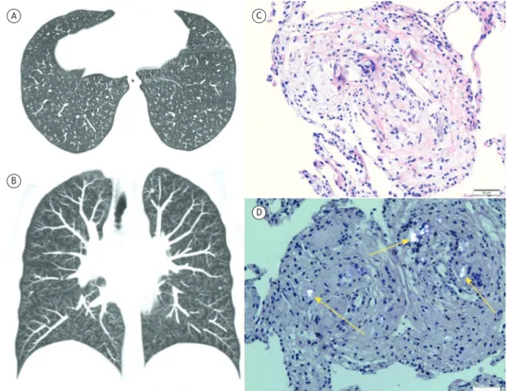
A 38-year-old woman presented to our pulmonology clinic with complaints of progressive dyspnea and dry cough for more than three months. She denied fever or weight loss. On physical examination, she presented as hypoxemic, with a room air oxygen saturation of 92% and an RR of 24 breaths/min. Pulmonary function tests showed that spirometric values were within normal limits, but there was a slight increase in residual volume (127% of predicted), as well as a reduction in DLCO (70% of predicted). Other laboratory test results were normal. A CT of the chest showed bilateral centrilobular nodules, most of them showing tree-in-bud appearance, scattered diffusely in the lung parenchyma (Figures 1A
and 1B). After the CT examination, lexible bronchoscopy
was then performed. Cultures of BALF were negative. Transbronchial biopsy performed in the left lower lobe showed multinucleated giant cell granulomas with
birefringent foreign material, compatible with talc (Figures 1C and 1D). The centrilobular nodules were determined histopathologically to be tiny foreign body particles lodged in the centrilobular arterioles and perivascular space.
Upon further discussion, the patient remembered that approximately one month before the onset of symptoms, she had self-administered an i.v. injection of a crushed methadone pill diluted in water, due to strong pain caused by trigeminal neuralgia. Based on the clinical history, CT
indings, and pulmonary biopsy results, the diagnosis of
pulmonary talcosis secondary to i.v. drug injection was made. Echocardiogram results were normal. No signs of pulmonary hypertension were seen. During 3 years of follow-up, the patient showed clinical stability with the persistence of dyspnea on exertion and dry cough. Findings of control CT examinations were unchanged.
Pulmonary talcosis is most commonly observed after inhalation of talc due to occupational exposure
Figure 1. Axial (in A) and coronal (in B) reformatted CT images showing numerous small bilateral centrilobular nodules, associated with the tree-in-bud pattern. In C, lung tissue biopsy demonstrating an interstitial granulomatous reaction to the talc particles with a giant-cell reaction (H&E; magniication, ×100), whereas, in D, under polarized light, birefringent crystals (arrows) are visible (H&E; magniication, ×100).
A C
B
D J Bras Pneumol. 2017;43(2):154-155
154
Escuissato DL, Ferreira RG, Barros JA, Marchiori E
(talc-induced pneumoconiosis), i.v. drug abuse (intravascular talcosis), and, occasionally, excessive use
of cosmetic talc. Clinical symptoms, imaging indings,
and histological presentations of pulmonary talcosis are essentially identical in these different etiologies.(1-4)
The most common form of pulmonary talcosis is caused by i.v. talc administration. Illegal street drugs commonly contain adulterants to increase their mass, and these adulterants commonly contain microscopic insoluble material. Another common source of such material is the injection of medications intended for oral use. The medications are typically crushed, mixed
with water, heated, and then injected i.v. The iller
materials (excipients) used in oral tablets include not only talc, but also other insoluble particles, such as cellulose, crospovidone, and starch, which can induce a foreign-body reaction in pulmonary arterioles. Heroin, cocaine, and methadone are the most commonly injected drugs.(1-5)However, other
medications, particularly analgesics and stimulants, are also used. Some authors(2)have suggested that
the term “intravascular talcosis” is a misnomer, since talc is only one of various possible materials used as excipients, and have proposed the term “excipient lung disease” to identify this condition.
Patients with talcosis can be asymptomatic or present with respiratory failure. The symptoms are
usually nonspeciic and can include dyspnea, cough,
fever, weight loss, chronic respiratory failure due to emphysema, and conditions related to pulmonary hypertension or fibrosis. Other complications of i.v. drug abuse resulting from the lack of a sterile technique include infections, such as endocarditis, septic embolism, HIV, and HCV. Physical examination
and laboratory test indings are usually unremarkable in patients with talcosis. A characteristic inding of
fundoscopy is the presence of crystals in retinal vessels. A history of i.v. drug abuse is an important clue to making the diagnosis; however, most i.v. drug abusers are reluctant to provide histories of exposure, and most diagnoses are made after lung biopsy.(1-4) The
i.v. administration of talc or other excipients results
in acute embolization of small vessels. Numerous tiny
particles become lodged in the pulmonary vessels and migrate to the pulmonary interstitium, where they cause a granulomatous foreign-body reaction, with
or without ibrosis. The granulomas can be visualized
under polarized light as birefringent needle-shaped
talc crystals in multinucleated giant cells.(1-3) The earlier CT indings of talcosis consist of a diffuse ine nodular pattern, which corresponds basically to
small centrilobular nodules and areas of ground-glass
attenuation in all lung zones. The centrilobular nodules
were determined histopathologically by tiny foreign body particles lodged in the centrilobular arterioles, and also in the perivascular space. The ground glass
opacities may represent the conluence of these
micronodules and/or microscopic granulomas below the resolution of HRCT.(1-3)
Periarteriolar, centrilobular micronodules can create a tree-in-bud pattern, mimicking bronchiolar disease. Centrilobular and panacinar emphysema patterns have been reported in i.v. drug users, with the lower-lobe panacinar pattern being predominant. Over time, talc micronodules may coalesce into perihilar conglomerate
masses, resembling progressive massive ibrosis
from silicosis or coal workers’ pneumoconiosis. The conglomerate masses in talcosis may contain high-at-tenuation material.(1,2,4,5)The differential diagnosis for
our patient (considering the presence of small bilateral centrilobular nodules, most with a tree-in-bud appear-ance) included arteriolar and bronchiolar diseases. The main conditions considered were infectious diseases (fungal, viral, and bacterial, particularly tuberculosis),
noninfectious bronchiolitis, cystic ibrosis, aspiration/
inhalation diseases, and peripheral pulmonary vascular diseases, such as pulmonary intravascular tumor embolism.There is no established treatment for talc granulomatosis. Patients must stop exposure and all tobacco use. Most authors believe that the use of
steroids and immunosuppressants has no beneit.
Associated pulmonary hypertension should be treated with vasodilators. Lung transplantation is considered to be a viable option for the treatment of talcosis. It is reserved as a last resort for patients with end-stage disease.(1)In our case, it was agreed that no treatment
was required due to the stable nature of the disease. In conclusion, the CT manifestations of intravascular talcosis consist of diffuse centrilobular nodules associated with a tree-in-bud pattern and ground-glass opacities, heterogeneous conglomerate masses containing areas of high attenuation, and panlobular emphysema in the lower lobes. The diagnosis should be considered in the setting of a history of i.v. drug abuse, but the
inal diagnosis is made after lung biopsy in most cases.
REFERENCES
1. Marchiori E, Lourenço S, Gasparetto TD, Zanetti G, Mano CM, Nobre
LF. Pulmonary talcosis: imaging indings. Lung. 2010;188(2):165-71.
https://doi.org/10.1007/s00408-010-9230-y
2. Nguyen VT, Chan ES, Chou SH, Godwin JD, Fligner CL, Schmidt RA, et al. Pulmonary effects of i.v. injection of crushed oral tablets:
“excipient lung disease”. AJR Am J Roentgenol.
2014;203(5):W506-15. https://doi.org/10.2214/AJR.14.12582
3. Grifith CC, Raval JS, Nichols L. Intravascular talcosis due to
intravenous drug use is an underrecognized cause of pulmonary
hypertension. Pulm Med. 2012;2012:617531. https://doi.
org/10.1155/2012/617531
4. Siddiqui MF, Saleem S, Badireddi S. Pulmonary talcosis with
intravenous drug abuse. Respir Care. 2013;58(10):e126-8. https://
doi.org/10.4187/respcare.02402
5. Almeida RR, Zanetti G, Souza AS Jr, Souza LS, Silva JL, Escuissato
DL, et al. Cocaine-induced pulmonary changes: HRCT indings. J Bras Pneumol. 2015;41(4):323-30.
https://doi.org/10.1590/S1806-37132015000000025