ABSTRACT
Objective: The halo sign consists of an area of ground-glass opacity surrounding pulmonary lesions on chest CT scans. We compared immunocompetent and immunosuppressed patients in terms of halo sign features and sought to identify those of greatest diagnostic value. Methods: This was a retrospective study of CT scans performed at any of seven centers between January of 2011 and May of 2015. Patients were classiied according to their immune status. Two thoracic radiologists reviewed the scans in order to determine the number of lesions, as well as their distribution, size, and contour, together with halo thickness and any other associated indings. Results: Of the 85 patients evaluated, 53 were immunocompetent and 32 were immunosuppressed. Of the 53 immunocompetent patients, 34 (64%) were diagnosed with primary neoplasm. Of the 32 immunosuppressed patients, 25 (78%) were diagnosed with aspergillosis. Multiple and randomly distributed lesions were more common in the immunosuppressed patients than in the immunocompetent patients (p < 0.001 for both). Halo thickness was found to be greater in the immunosuppressed patients (p < 0.05). Conclusions: Etiologies of the halo sign differ markedly between immunocompetent and immunosuppressed patients. Although thicker halos are more likely to occur in patients with infectious diseases, the number and distribution of lesions should also be taken into account when evaluating patients presenting with the halo sign.
Keywords: Tomography, X-ray computed; Aspergillosis; Lung neoplasms.
The halo sign: HRCT indings in 85 patients
Giordano Rafael Tronco Alves1, Edson Marchiori1, Klaus Irion2,Carlos Schuler Nin3, Guilherme Watte3, Alessandro Comarú Pasqualotto3, Luiz Carlos Severo3, Bruno Hochhegger1,3
Correspondence to:
Giordano Rafael Tronco Alves. Avenida Roraima, 1000, CEP 97105-900, Santa Maria, RS, Brasil. Tel.: 55 55 9729-1990. E-mail: [email protected]
Financial support: None. INTRODUCTION
The CT halo sign consists of an area of ground-glass opacity surrounding a pulmonary nodule or mass.(1) The halo sign was irst described in 1985 by Kuhlman et al., who reviewed chest CT scans of nine patients with acute leukemia who developed invasive pulmonary aspergillo-sis. (2) Since then—and despite its infrequency—the halo sign has been reported in association with a variety of conditions.(3,4) Its pathophysiology usually involves one of three mechanisms: hemorrhage, inlammation, or neoplastic growth.(5)
Most of the available information about the halo sign has been derived from patients with pre-existing conditions. However, there are limited data on its potential usefulness in predicting the inal diagnosis (or diagnostic group, e.g., infection or malignancy). Therefore, the objective of the present study was to determine the diagnostic value of the halo sign by exploring associations between CT measurements and immunological status in a cohort of patients presenting with the CT halo sign.
METHODS
This was a multicenter retrospective study of CT images obtained between January of 2010 and May of 2014 from patients presenting at any of seven tertiary care centers in southern Brazil, which is an endemic
area for granulomatous diseases. Scans were selected by searching the picture archiving and communication systems (PACSs) of all participating institutions using the terms “halo” and “halo sign”, as well as the following combinations of terms: “ground-glass” + “nodule”; “nodule” + “surround”; “nodule” + “periphery”; and “ground-glass” + “periphery”. The research protocol was approved by the local research ethics committee (Protocol no. 243.155). Given the retrospective nature of the study, informed consent was waived.
A general radiologist reviewed the iles retrieved from the PACSs in order to conirm the presence of the halo sign. Medical records were reviewed in order to determine patient immune status. For the purpose of the present study, patients were considered immunosuppressed in the presence of AIDS; any form of congenital immunodeiciency; or a recent (≤ two-month) history of chemotherapy, radiation therapy, or decreased white blood cell count (lymphopenia [absolute lymphocyte count ≤ 1.0 × 10⁹ L] or neutropenia [absolute neutrophil count ≤ 1.5 × 10⁹ L]).(6,7) All other patients were considered to be immunocompetent. If a inal diagnosis had not been reached by the time of medical record review, patients were followed until a deinitive diagnosis was made, with serological, histological, or microbiological conirmation. Histological sampling varied across the participating institutions, including transthoracic needle biopsy,
1. Programa de Pós-Graduação em Medicina (Radiologia), Universidade Federal do Rio de Janeiro, Rio de Janeiro (RJ) Brasil.
2. Radiology Department, Liverpool Heart and Chest Hospital, Liverpool, United Kingdom.
3. Universidade Federal de Ciências da Saúde de Porto Alegre, Porto Alegre (RS) Brasil.
Submitted: 28 February 2016. Accepted: 21 July 2016.
video-assisted thoracotomy, and open thoracotomy. Although serological and microbiological tests were performed in accordance with the corresponding (updated) guidelines, their performance was dependent on patient clinical background.
All CT scans were performed with multidetector scanners with at least 16 rows of detectors, the acquisition parameters being as follows: slice thickness, ≤ 1.25 mm; rotation time, 0.5 s; voltage, 120 kV; and electric current, 150-400 mA. Automatic exposure control was enabled. No contrast medium was used. For each examination, the number of lesions, as well as their outline (regular vs. irregular), size, and distribution, together with any other associated indings, were recorded. The criteria for CT indings were those deined in the Fleischner Society Glossary of Terms.(1) A nodule was deined as a rounded or irregular opacity that was well or poorly deined and ≤ 3 cm in diameter. Mediastinal and hilar lymph nodes range in size from sub-CT resolution to 10 mm. A cavity was deined as a gas-illed space, seen as a lucency or low-attenuation area within a pulmonary consolidation, mass, or nodule. The tree-in-bud pattern refers to centrilobular branching structures that resemble a budding tree. Ground-glass opacities were deined as hazy areas of increased opacity or attenuation with no obscuration of the underlying vessels. Consolidation was deined as homogeneous opaciication of the parenchyma with obscuration of the underlying vessels. Abnormalities were classiied as being located in the upper lobes, located in the lower lobes, or randomly distributed.
The CT scans were independently reviewed in random order by two chest radiologists who had more than 10 years of experience and who were blinded to patient clinical information. Subsequently, the two aforementioned radiologists and a third chest radiologist (with more than 40 years of experience) together reviewed the scans in order to make a inal consensus decision. The diameters of the nodules and halos were measured at their widest points on axial CT scans with lung window settings.
Microsoft Excel was used for data storage and descriptive analysis, and the Statistical Package for the Social Sciences, version 14.0 (SPSS Inc., Chicago, IL, USA), was used for correlations. The chi-square test was used for qualitative variables. The Kolmog-orov-Smirnov test was used in order to determine whether quantitative data were normally distributed. For parametric variables, the Student’s t-test was used. For nonparametric variables, the Mann-Whitney U test was used. All tests were two-tailed, and a signiicance level of 0.05 was used throughout.
RESULTS
Of the 20,210 CT examinations retrieved from the PACSs of the participating centers, 85 cases (0.42%) were selected for inclusion in the present study. Of those 85 patients, 46 were male. In addition, 32 were classiied as being immunosuppressed at the time of
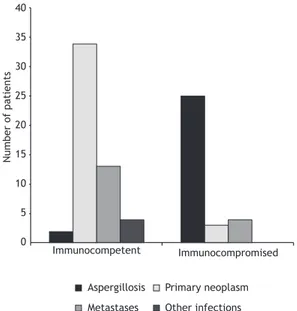
CT examination, and 53 were immunocompetent. Of those, 34 (64%) were diagnosed with a primary malignancy; of those, 24 had histopathologically conirmed adenocarcinoma. Of the 32 patients who were classiied as being immunosuppressed, 25 (78%) had indings suggestive of invasive aspergillosis (positive results on direct mycological examination or histological indings). All but one of the immunosuppressed patients presenting with invasive aspergillosis were found to have neutropenia. In addition, 7 of those 25 were diagnosed with proven invasive fungal disease (on the basis of specimen culture results and radiological indings), and 18 were diagnosed with probable invasive aspergillosis (on the basis of positive microbiological studies and radiological indings).(8) Other causes of the CT halo sign included metastases, lymphoproliferative diseases, tuberculosis, plasmacytoma, staphylococcal pneumonia, actinomycosis, cryptococcosis, and histiocytosis. Table 1 and Figure 1 show the frequencies of these diagnoses in the two study groups.
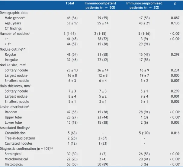
The number and distribution of lesions on CT scans were found to vary signiicantly according to patient immune status, with immunosuppressed patients tend-ing to exhibit multiple and randomly distributed lesions (Table 2). In cases of multiple lesions, halo thickness tended to be greater (≥ 9 mm) in immunosuppressed patients (p < 0.05; Figure 2); the same was not true for single lesions (p = 0.299).
DISCUSSION
The present retrospective study revealed ten dif-ferent etiologies of the CT halo sign in 85 individuals. Pathological indings of tumor cells, inlammatory iniltrates, and, most commonly, alveolar hemorrhage have been reported to account for the ground-glass opacities surrounding pulmonary lesions on CT
Other infections Metastases
Primary neoplasm Aspergillosis
Number of patients
Immunocompetent Immunocompromised 40
35
30
25
20
15
10
5
0
Table 1. Etiology of the CT halo sign, by patient immune status.a
Variable Immunocompetent patients
(n = 53)
Immunocompromised patients (n = 32)
Primary neoplasm 34 (64)
-Invasive aspergillosis - 25 (78)
Metastases 13 (25.0) 2 (6.3)
Lymphoproliferative diseases - 3 (9.4)
Tuberculosis 2 (3.8)
-Plasmacytoma - 2 (6.3)
Staphylococcal pneumonia 1 (1.8)
-Actinomycosis 1 (1.8)
-Cryptococcosis 1 (1.8)
-Histiocytosis 1 (1.8)
-aData presented as n (%).
Table 2. Demographic data and CT indings, by patient immune status.a
Total Immunocompetent
patients (n = 53)
Immunocompromised patients (n = 32)
p
Demographic data
Male genderb 46 (54) 29 (55) 17 (53) 0.887
Age, years 53 ± 17 55 ± 14 48 ± 21 0.135
CT indings
Number of nodulesc 3 (1-16) 2 (1-15) 5 (1-16) < 0.001
1b 41 (48) 38 (72) 3 (9) < 0.001
> 1b 44 (52) 15 (28) 29 (91)
Nodule outlineb,*
Regular 46 (54) 31 (58) 15 (47) 0.298
Irregular 39 (46) 22 (42) 17 (53)
Nodule size, mm†
Solitary nodule 25 ± 13 26 ± 14 16 ± 9 0.231
Largest nodule 16 ± 8 12 ± 8 19 ± 7 0.805
Smallest nodule 6 ± 3 6 ± 4 5 ± 2 0.007
Halo thickness, mm†
Solitary nodule 7 ± 3 7 ± 3 5 ± 1 0.299
Largest nodule 8 ± 4 5 ± 2 9 ± 4 0.001
Smallest nodule 5 ± 1 3 ± 1 5 ± 1 0.002
Lesion distributionb
Random 47 (55) 15 (28) 28 (91) < 0.001
Upper lobe 23 (27) 23 (44) 1 (3) < 0.001
Lower lobe 15 (18) 15 (28) 2 (6) 0.003
Associated indingsb
Consolidation 5 (63) - 5 (100) 0.016
Tree-in-bud pattern 2 (25) 2 (67)
-Cavitated nodules 1 (12) 1 (33)
-Diagnostic conirmation (n = 105)b,‡
Serological 30 (30) 4 (7) 26 (53) < 0.001 Microbiological 22 (20) 2 (4) 20 (41) < 0.001 Histological 53 (50) 50 (89) 3 (6) < 0.001
aData presented as mean ± SD, except where otherwise indicated. bData presented as n (%). cData presented as
median (range). *For nodules presenting with a peripheral halo sign. †For patients with multiple lesions, data for the largest and smallest lesions are displayed separately. ‡The number of diagnostic conirmations exceeds the number of cases because some diagnoses were conirmed by more than one method.
scans. (4,9) Although the halo sign has been causally associated with numerous other conditions, its presence is generally more helpful than challenging. (9) Studies involving immunocompromised patients have conirmed
present study was the irst to address the corre-lations between the halo sign and patient immune status. In addition, our study demonstrated that thicker halos are associated with infectious diseases, whereas thinner halos are associated with neoplastic diseases. In agreement with the results of previous studies, the results of the present study showed that adenocarcinoma and pulmonary aspergillosis were the conditions that were most commonly associated with the halo sign in immunocompetent and immu-nosuppressed patients, respectively.(4,12) In cases of multiple pulmonary nodules, halo thickness tended to be greater in immunocompromised patients than in immunocompetent patients. This inding is consistent with those of a study investigating characteristics of the reversed halo sign, in which greater rim thickness was associated with invasive fungal infection.(13) The same cannot be concluded for solitary nodules, and this is possibly due to a statistically insuficient number of single lesions among the immunosuppressed patients (i.e., only three).
According to Gao et al., the relationships between ground-glass lung nodules and adjacent blood vessels can aid in establishing a inal diagnosis based on the presence and degree of vascular distortion.(14) This is particularly relevant for immunocompetent patients presenting with the CT halo sign. In the present study, a neoplastic etiology was found to be more common in the immunocompetent patients than in the immunosuppressed patients. However, given the reduced halo thickness in the former and the variety of possible pathophysiological mechanisms underlying the halo sign, further studies are needed in order to
determine whether this correlation can be extrapolated to smaller areas of ground-glass opacities.
Our study has some limitations. First, the retro-spective nature of the study increases the likelihood that medical record inaccuracies affected our results. Second, the relatively small sample size did not allow us to perform an analysis of covariance in order to determine the combined contribution of halo sign features in predicting the inal diagnosis. Third, dynamic changes observed in the long-term evaluation of the appearance of the halo sign, particularly in cases of infectious disease, might have affected some of our results. Finally, the deinition of immunosuppression used in our study might be in disagreement with the constant improvements in the toxicity of chemotherapy and radiation therapy; however, that deinition was adopted for research purposes only, and many other clinical parameters should be taken into account in a more practical scenario.
In summary, our indings are consistent with available data on the etiologies of the CT halo sign in immuno-competent and immunosuppressed patients. Given the differences in radiological presentation between these two groups of patients, appropriate assessment of halo sign features can be useful in clinical investigation. A diagnosis of primary neoplasm appears to be common among immunocompetent patients, as does a diagnosis of invasive aspergillosis among immunosuppressed patients. Thicker halos are associated with infectious diseases, whereas thinner halos are associated with neoplastic diseases; the number and distribution of lesions should also be taken into account, given that they can predict the inal diagnosis.
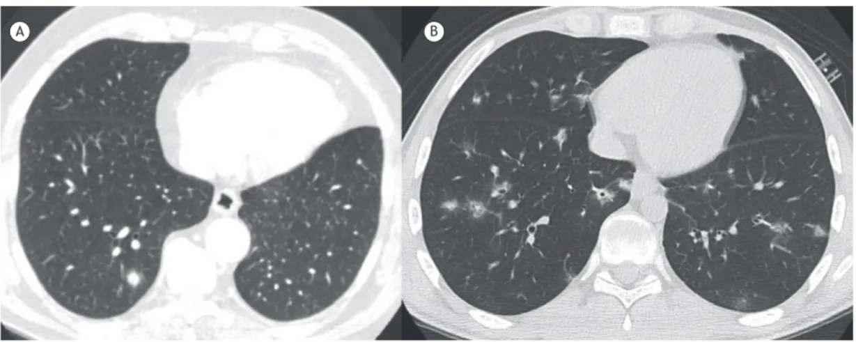
A B
Figure 2. In A, axial CT scan of the chest of an asymptomatic, immunocompetent 54-year-old male patient, showing a
right lower lobe pulmonary nodule surrounded by areas of ground-glass opacity (the CT halo sign); the inal diagnosis was primary adenocarcinoma. In B, axial CT scan of the chest of an immunosuppressed 19-year-old male patient, showing multiple, randomly distributed pulmonary nodules surrounded by ground-glass opacities (the CT halo sign); the inal diagnosis was aspergillosis.
REFERENCES
1. Hansell DM, Bankier AA, MacMahon H, McLoud TC, Müller NL, Remy J. Fleischner Society: glossary of terms for thoracic imaging. Radiology. 2008;246(3):697-722. http://dx.doi.org/10.1148/ radiol.2462070712
2. Kuhlman JE, Fishman EK, Siegelman SS. Invasive pulmonary
aspergillosis in acute leukemia: characteristic indings on
radiology.157.3.3864189
3. Lee YR, Choi YW, Lee KJ, Jeon SC, Park CK, Heo JN. CT halo sign: the spectrum of pulmonary diseases. Br J Radiol. 2005;78(933):862-5. http://dx.doi.org/10.1259/bjr/77712845
4. Kim Y, Lee KS, Jung KJ, Han J, Kim JS, Suh JS. Halo sign on high
resolution CT: indings in spectrum of pulmonary diseases with
pathologic correlation. J Comput Assist Tomogr. 1999;23(4):622-6. 5. Parrón M, Torres I, Pardo M, Morales C, Navarro M,
Martínez-Schmizcraft M. The halo sign in computed tomography images:
differential diagnosis and correlation with pathology indings [Article
in Spanish]. Arch Bronconeumol. 2008;44(7):386-92. http://dx.doi. org/10.1016/S0300-2896(08)70453-8
6. Ng WL, Chu CM, Wu AK, Cheng VC, Yuen KY. Lymphopenia at presentation is associated with increased risk of infections in patients with systemic lupus erythematosus. QJM. 2006;99(1):37-47. http:// dx.doi.org/10.1093/qjmed/hci155
7. Valent P. Low blood counts: immune mediated, idiopathic, or myelodysplasia. Hematology Am Soc Hematol Educ Program. 2012;2012:485-91. http://dx.doi.org/10.1182/ asheducation-2012.1.485
8. De Pauw B, Walsh TJ, Donnelly JP, Stevens DA, Edwards JE,
Calandra T, et al. Revised deinitions of invasive fungal disease from
the European Organization for Research and Treatment of Cancer/ Invasive Fungal Infections Cooperative Group and the National Institute of Allergy and Infectious Diseases Mycoses Study Group
(EORTC/MSG) Consensus Group. Clin Infect Dis. 2008;46(12):1813-21. http://dx.doi.org/10.1086/588660
9. Pinto PS. The CT Halo Sign. Radiology. 2004;230(1):109-10. http:// dx.doi.org/10.1148/radiol.2301020649
10. Escuissato DL, Gasparetto EL, Marchiori E, Rocha Gde M, Inoue C, Pasquini R, et al. Pulmonary infections after bone marrow
transplantation: high-resolution CT indings in 111 patients. AJR
Am J Roentgenol. 2005;185(3):608-15. http://dx.doi.org/10.2214/ ajr.185.3.01850608
11. Blum U, Windfuhr M, Buitrago-Tellez C, Sigmund G, Herbst EW, Langer M. Invasive pulmonary aspergillosis. MRI, CT, and plain
radiographic indings and their contribution for early diagnosis. Chest.
1994;106(4):1156-61. http://dx.doi.org/10.1378/chest.106.4.1156 12. Gaeta M, Blandino A, Scribano E, Minutoli F, Volta S, Pandolfo I.
Computed tomography halo sign in pulmonary nodules: frequency and diagnostic value. J Thorac Imaging. 1999;14(2):109-13. 13. Marchiori E, Marom EM, Zanetti G, Hochhegger B, Irion KL, Godoy
MC. Reversed halo sign in invasive fungal infections: criteria for differentiation from organizing pneumonia. Chest. 2012;142(6):1469-73. http://dx.doi.org/10.1378/chest.12-0114