www.bjorl.org.br
Brazilian Journal of
OtOrhinOlaryngOlOgy
1808-8694/$ - see front matter © 2014 associação Brasileira de Otorrinolaringologia e Cirurgia Cérvico-Facial. Published by Elsevier Editora ltda. all rights reserved.
DOi: 10.5935/1808-8694.20140004
ORIGINAL ARTICLE
Influence of obesity on the correlation between
laryngopharyngeal reflux and obstructive sleep apnea
Marcos Marques Rodrigues
a,b,*, Ralph Silveira Dibbern
c, Victor José Barbosa Santos
d,
Luis Augusto Passeri
e,fa Associação Brasileira de Otorrinolaringologia e Cirurgia Cérvico-Facial, Brazilian Association of Otorhinolaryngology and
Cervi-cofacial Surgery (ABORLCCF/AMB), São Paulo, SP, Brazil
b School of Medicine, Centro Universitário de Araraquara (UNIARA), Araraquara, SP, Brazil c School of Medicine, Universidade de São Paulo (USP), Ribeirão Preto, SP, Brazil
d School of Medicine, Universidade Estadual Paulista (UNESP), Botucatu, SP, Brazil e Southwestern Medical Center, Texas University, Dallas, Texas, USA
f Department of Surgery, School of Medical Sciences, Universidade Estadual de Campinas (UNICAMP), Campinas, SP, Brazil
Received 14 June 2012; accepted 9 September 2013
KEYWORDS Sleep apnea obstructive; Obesity; Laryngitis
PALAVRAS-CHAVE Apneia obstrutiva do sono;
Obesidade; Laringite
Abstract
Introduction: The obstructive sleep apnea (OSA) is caused by recurrent episodes of partial or total obstruction of the upper airway lasting more than 10 seconds during sleep. Laryngopharyngeal
re-lux (LPR) is a variant of the disease Gastroesophageal Rere-lux that affects the larynx and pharynx.
Objectives: Evaluate the inluence of obesity on the relationship between RFL and OSAS in
patients with OSA.
Materials and methods: An observational retrospective cross. We reviewed care protocol for
patients with OSA that includes validated questionnaires for RFL as Sympton Relux Index (RSI) and Relux Finding Score (RSI), and polysomnography nasolaringoibroscopia.
Results: 105 patients were divided into obese group (39 patients) and non-obese patients (66 patients). In the evaluation of the mean RSI group of non-obese was similar between patients with mild OSA (11.96) and moderate (11.43). In the obese group the mean RSI was 6.7 in pa-tients with mild OSA and 11.53 in papa-tients with moderate to severe OSA (p < 0.05).
Discussion: The subgroup of patients with OSA and RFL have several factors that promote in
-lammation of the upper airway. Patients with OSA should be screened and treated as the RFL
increasing the quality of life.
Conclusion: The RFL are positively correlated and OSAS in obese patients.
© 2014 Associação Brasileira de Otorrinolaringologia e Cirurgia Cérvico-Facial. Published by Elsevier
Editora Ltda. All rights reserved.
Inluência da obesidade na correlação entre reluxo faringolaríngeo e apneia obstrutiva do sono
Resumo
Introdução: A apneia obstrutiva do sono (AOS) é causada por episódios recorrentes de obstrução total
ou parcial da via aérea superior com duração superior a 10 segundos durante o sono. Reluxo faringo
-laríngeo (RFL) é uma variante da doença do reluxo gastroesofágico que afeta a laringe e a faringe.
Please cite this article as: Rodrigues MM, Dibbern RS, Santos VJ, Passeri LA. Inluence of obesity on the correlation between laryngopha
-ryngeal relux and obstructive sleep apnea. Braz J Otorhinolaryngol. 2014;80:5-10.
* Corresponding author.
Introduction
Obstructive sleep apnea (OSA) is caused by recurrent ep-isodes of partial or complete obstruction of the upper
airways (UAs) lasting more than 10 seconds during sleep, causing a decrease in oxyhemoglobin saturation and an in -crease in adrenergic discharge. A thorough upper airway assessment is very important to evaluate the obstruction
points. In 1999, the American Academy of Sleep Medicine1
established the diagnostic criteria for OSA, which included presence of excessive daytime sleepiness not explained by
other factors; presence of at least two of the following:
nocturnal choking, recurrent arousals, unrefreshing sleep, daytime fatigue, and decrease in concentration; and pres
-ence of apnea-hypopnea index (AHI) score greater than ive events per hour, a mandatory criterion.
The physiopathogeny of OSA has not yet been fully
elu-cidated. During respiratory events, there is a decrease in oxygen saturation, which leads to barorelex activation, triggering a generalized activation of the sympathetic au -tonomic nervous system. There is an adrenergic discharge leading to peaks of tachycardia and hypertension. This process is repeated several times during sleep in apneic
patients and, in the long-term, it leads to high sensitivity of the peripheral chemorelex, with exaggerated response even in normoxia, barorelex dysfunction, increased ad
-renergic discharge, cardiovascular dysfunction in the long term, systemic inlammation, and metabolic dysregulation
with insulin resistance and type II diabetes mellitus.2 All
these alterations result in chronic inlammation of the en
-tire upper airway, leading to the appearance of the varied
symptoms in these patients.
Studies have demonstrated the diversity of OSA
prev-alence, as it can affect children, young adults, and the
elderly. It is associated with different risk factors such as
anatomical abnormalities, diseases, and habits. An epide
-miological study performed in São Paulo, Brazil showed that the prevalence of OSA was 32.8% of the adult population of
the city. The risk factors associated with the development
of the syndrome were male gender, body mass index (BMI)
> 25 kg/m2, low socioeconomic status, age, and menopausal status.3 Snoring has a prevalence of 19.1% among men and
7.9% among women in the population aged 30 to 60 years.4
Approximately 20% of the adult population complains of snoring, a igure that increases to 60% when considering
males older than 40 years of age.5
Laryngopharyngeal relux (LPR) is a variant of the gas
-troesophageal relux disease (GERD) that affects the larynx and pharynx. In most cases, it is secondary to retrograde low of gastric contents into the laryngopharynx, resulting
in a series of laryngeal signs and symptoms.6 It has become one of the most common conditions in otorhinolaryngology.
It is diagnosed in approximately 10% of patients with otorhi
-nolaryngological symptoms and in at least 50% of patients
with voice-related complaints.7 Obesity has also been
rec-ognized as an important factor, which is increasingly more
prevalent in the world’s population.
LPR tends to occur during the day and has no clear asso
-ciation with obesity, in contrast to GERD.
Belafsky et al. validated two questionnaires for the
sys-tematic evaluation of the LPR-related complaints. The irst, called the Relux Symptom Index (RSI), assesses LPR symp
-toms. The scale consists of nine items scored 0 to 5, with a minimum score of 0 (asymptomatic) and maximum of 45.8 An RSI index ≥ 13 is considered to be suggestive of LPR.
The other scale developed by Belafsky et al. is the Relux Finding Score(RFS) of laryngoscopic indings suggestive of LPR. An LPR score > 7 is considered positive for LPR.8
OSA and LPR are two conditions that cause chronic upper airway inlammation. Symptoms such as hoarseness, pha
-ryngeal globus, dysphagia, and choking are observed in both diseases. The association between OSA and LPR has been discussed in the literature in recent years, but the correla -tion between the two diseases has not reached a consensus. Gastroesophageal motility is decreased during sleep.
Intrae-sophageal pressure is decreased in patients with OSA, but
Kuribayashi et al. demonstrated that the upper esophageal sphincter and the gastroesophageal junction increase their
tonus, decreasing LPR events.9
The presence of LPR or GERD is associated with an in
-creased number of awakenings, regardless of the presence of OSA. Patients with mild to moderate OSA experience greater inluence from LPR on the number of awakenings than patients with severe OSA. Few studies have evaluated the inluence of obesity on the association between OSA and LPR. Thus, this assessment is relevant, since the incidence of obesity has
Objetivos: Avaliar a inluência da obesidade na relação entre RFL e AOS em pacientes com SAOS.
Materiais e métodos: Estudo observacional transversal retrospectivo. Foram revisados protoco
-los de atendimento de pacientes com AOS que incluem questionários validados para RFL como Relux Sympton Index (RSI) e Relux Finding Score (RSI), nasolaringoibroscopia e polissonograia.
Resultados: Cento e cinco pacientes foram divididos em grupo de obesos (39 pacientes) e não obesos (66 pacientes). Na avaliação das médias do RSI o grupo de não obesos foi semelhante
entre pacientes com AOS leve (11,96) e moderada (11,43). No grupo de obesos a média do RSI foi de 6,7 em pacientes com AOS leve e de 11,53 em pacientes com AOS moderada a grave (p < 0,05).
Discussão: O subgrupo de pacientes com AOS e RFL apresenta vários fatores que promovem a
inlamação da via aérea superior. Pacientes com AOS devem ser pesquisados e tratados quanto a RFL, aumentando a qualidade de vida.
Conclusão: O RFL e a AOS se correlacionam positivamente em pacientes obesos.
© 2014 Associação Brasileira de Otorrinolaringologia e Cirurgia Cérvico-Facial. Publicado por Elsevier
been consistently increasing and has an important inluence
on the natural course of OSA.
Objectives
To evaluate the inluence of obesity on the association be
-tween LPR and OSA in a population of patients with OSA.
Materials and methods
This study was approved by the Ethics and Research
Commit-tee under protocol 113/08, and registered at the Clinical Tri
-als Registry under ID NCT00883025. This was a retrospective, cross-sectional, and observational study, which evaluated
patients with OSA who attended the otorhinolaryngology ser-vice by reviewing treatment protocols from medical records.
The evaluation protocol included anamnesis; the
appli-cation of Epworth Sleepiness Scale, snoring scale, Friedman classiication, and validated questionnaires for LPR (RSI and RFS); and complete otorhinolaryngological examination
with nasal endoscopy and polysomnography.
The Friedman classiication evaluates palatine tonsils, modiied Mallampati score, and BMI, and comprises four stages (I, II, III, and IV).10
Patients underwent nasal endoscopy with a 3.2-mm Ma
-chida ENT PIII nasal endoscope. The level of upper airway obstruction was evaluated using the Fujita classiication.11
Patients were referred for type I polysomnography per
-formed in a sleep laboratory by a trained technician. Moni
-toring included the following channels: eye movements, leg movements, nasal airlow, chest movement, electroenceph
-alogram (EEG), electrocardiogram (ECG), heart rate, and oxygen saturation. Patients were divided regarding disease severity, according to the criteria of the task force of the American Academy of Sleep Medicine.1
Statistical analysis
A study model for continuous variables with t-test was used for data analysis. Levene’s t-test was used for equality of vari-ances and the Student’s t-test was used for equality of means.
The following statistical hypotheses were analyzed in
the statistical model used:
• Null hypothesis - The severity of OSA by AHI does not correlate with LPR assessed by RSI.
• Alternative hypothesis – The severity of OSA by AHI is positively correlated with LPR assessed by RSI. The signiicance level used in the analysis was 0.05 for type α error and 0.2 for type β error. The Statistical Package for Social Sciences (SSPS) software for Mac, release 20.0,
was used for the statistical analysis.
Inclusion and exclusion criteria
Inclusion
1. Patients belonging to the OSA Outpatient Clinic cohort;
2. Patients with complete protocol;
3. Patients aged 18 to 80 years;
4. Patients of both genders.
Exclusion
1. Patients with upper airway tumors and/or polyps;
2. Patients with craniofacial deformities such as craniosyn -ostosis and cranio-dys-ostosis;
3. Previous history of airway and/or abdominal surgery;
4. Incomplete protocol and/or no analysis of key study
vari-ables such as RSI, RFS, and polysomnography.
Results
The number of patients in the cohort described in “Mate
-rials and methods”totaled 343. One hundred and ive pa
-tients who had LPR evaluation data assessed by the RSI, RFS scales, and polysomnography were included.
The sample was divided by BMI using a cutoff of 30 kg/m2
for the obese group, with 39 patients (37.1%); the non-obese group had 66 patients (62.9%). Gender distribution showed 57 (53.3%) males and 50 (46.7%) females.
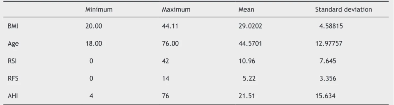
Table 1 shows the description of continuous variables evaluated in this study.
Table 1 Description of continuous variables.
Minimum Maximum Mean Standard deviation
BMI 20.00 44.11 29.0202 4.58815
Age 18.00 76.00 44.5701 12.97757
RSI 0 42 10.96 7.645
RFS 0 14 5.22 3.356
AHI 4 76 21.51 15.634
For the purposes of paired analysis using the t-test, and in order to maximize the statistical power, the AHI was divided into two groups with a cutoff of 15 events per hour. Mild OSA patients totaled 51 (48.5%) patients, and 53 (51.5%) had mod -erate and severe OSA. Both categories were correlated with
the continuous variables RSI and RFS using the t-test. Table 2
shows the results of pairing in the group of non-obese patients. This study was designed to assess obesity alone as a
confound-ing factor, with balanced samples and obesity deined by BMI.
When analyzing non-obese patients, Table 2 evidences
the association between apnea severity and the indices
val-idated in LPR assessment (RSI and RFS). In this subgroup, there was no statistical signiicance. In relation to the same analysis in Table 3, in the group of obese patients, there was no statistical signiicance between RSI and the OSA severity, as shown by the test of equality of means, with p < 0.05. The association between RFS and OSA severity was not sig
-niicant. This association is depicted in Fig. 1.
Figure 1 Box-plot between Relux Symptom Index (RSI) and Apnea-hyponea Index (AHI) in obese patients. Mild obstructive sleep apnea (OSA), AHI < 15 events/hour; moderate/severe OSA, AHI ≥ 15 events/hour.
Mild OSA
25
20
15
10
5
0
MODERATE/SEVERE OSA
OSA severity
Re
lu
x
Sy
m
pt
on
In
de
x
Table 2t-test for equality of means in non-obese patients.
Mean Standard deviation p
RSI Mild OSA 11.96 8.470 0.333a
Moderate-severe OSA 11.43 8.891
RFS Mild OSA 6.00 3.651 0.587a
Moderate-severe OSA 5.35 3.673
OSA, obstructive sleep apnea; RSI, Reflux Symptom Index; RFS, Reflux Finding Score.
a Non-significant.
Table 3t-test for equality of means in obese patients.
Mean Standard deviation p
RSI Mild OSA 6.70 5.498 0.054a
Moderate-severe OSA 11.30 6.097
RFS Mild OSA 4.43 2.507 0.648b
Moderate-severe OSA 4.69 3.199
OSA, obstructive sleep apnea; RSI, Reflux Symptom Index; RFS, Reflux Finding Score.
a Significant.
Discussion
The importance of OSA and LPR in otolaryngology has con -siderably increased in recent years; they are among the main complaints in the specialty clinics. These are chronic
diseases that affect the airways, causing chronic mucosal inlammation and decreased quality of life. According to Wise et al., GERD is more commonly found in patients with OSA, but the direct correlation was not signiicant.12
In a recent systematic review, Karkos et al. conclud
-ed that the association between LPR and OSA has been previously described in the literature, but the studies
had low levels of evidence with few controlled studies.13
Several factors may generate biases in this analysis,
since these are multifactorial diseases that are
influ-enced by anatomic upper airway abnormalities, weight,
and comorbidities.
Obesity is closely related to OSA, and it is considered as
an independent predictor.10 According to Halum et al., the
correlation with GERD is also quite clear, but the association with LPR alone is not.14 Xiao et al. studied the correlation
between LPR and OSA using multichannel pH monitoring and found no signiicant correlation, but listed BMI as a con -founding variable that could affect the results of association
and prediction of LPR in OSA.15
In the present study, when evaluating the correlation in patients with BMI < 30 kg/m2, i.e. not obese, no increase in
RSI and RFS in patients with moderate to severe OSA (AHI > 15) were observed. Mean values were similar; demonstrat
-ing that, in this subgroup of patients, LPR and OSA are inde -pendent factors.
When evaluating the group of obese patients, a change in trend was observed, as the RSI of obese patients was sig
-niicantly higher in patients with moderate to severe OSA,
with almost twice the mean RSI. The same correlation was
not observed in the evaluation of RFS.
Obesity has a major influence on the natural course
of OSA, and is directly associated with disease severi -ty.16 In the present series, obese patients and patients
with OSA had higher incidence of LPR. This subgroup of patients, therefore, has several factors (OSA, LPR, and obesity) that increase upper airway inflammation, in
-creasing edema and causing greater laxity of the mucosa and musculature, with morbidity worsening in both dis -eases. The aforementioned factors act together to pro-mote inflammatory changes that increase the symptoms
of upper airway irritation, which justifies the increase
of RSI in obese patients and patients with moderate to severe OSA.
Obesity leads to increased intra-abdominal pressure; this mechanism has important physiopathological implications in
LPR and OSA. In the latter, it causes increased respiratory effort, resulting in decreased thoracic range of movement. Stomach clearance is decreased and the incidence of relux
episodes increases.
In the present study, no positive correlation was observed between the severity of OSA by AHI and LPR laryngeal alterations measured by RFS. Obesity did not influence this variable behavior in both groups, as RFS
maintained similar means within the normal range in patients with mild OSA and in those with moderate to severe OSA.
Symptoms of LPR and OSA are mistaken for symptoms of chronic airway inlammation. In obese patients, these symp -toms become more intense in patients with moderate and severe OSA. This correlation was not sustained in the
laryn-goscopic evaluation, and the possibility of a false positive
result in this review cannot be ignored.
During the evaluation of patients with OSA, the coexis
-tence of LPR should be considered, which must be treated,
promoting increased quality of life and reducing airway
in-lammation, as well as attenuating the deleterious effects
on the upper airways.
Conclusion
LPR, OSA, and obesity are positively correlated. Obese pa -tients with moderate to severe OSA have more severe symp-toms than non-obese patients. The study demonstrated the
importance of diagnosis and treatment of LPR in patients
with OSA and obesity.
Conlicts of interest
The authors declare no conlicts of interest.
References
1. Sleep-related breathing disorders in adults: recommendations
for syndrome deinition and measurement techniques in cli -nical research. The report of an American Academy of Sleep
Medicine Task Force. Sleep. 1999;22:667-89.
2. Caples SM, Gami AS, Somers VK. Obstructive sleep apnea. Ann Intern Med. 2005;142:187-97
3. Tuik S, Santos-Silva R, Taddei JA, Bittencourt LRA. Obstructive sleep apnea syndrome in the São Paulo Epidemiologic Sleep Study. Sleep Med. 2010;11:441-6.
4. Jennum P, Sjol A. Epidemiology of snoring and obstructive sleep apnea in a Danish population, age 30-60. Sleep Res.
1992;1:240-4.
5. Hoffstein V, Mateika JH, Mateika S. Snoring and sleep architec
-ture. Am Rev Respir Dis. 1991;143:92-6.
6. Ali MS. Laryngopharyngeal relux: diagnosis and treatment
of a controversial disease. Curr Opin Allergy Clin Immunol.
2008;8:28-33.
7. Remacle M, Lawson G. Diagnosis and management of laryn
-gopharyngeal relux disease. Curr Opin Otolaryngol Head Neck
Surg. 2006;14:143-9.
8. Belafsky PC, Postma GN, Koufman JA. The validity and re
-liability of the Relux Finding Score (RFS). Laryngoscope.
2001;111:1313-7.
9. Kuribayashi S, Massey BT, Hafeezullah M, Perera L, Hussaini SQ, Tatro L, et al. Upper esophageal sphincter and gastro -esophageal junction pressure changes act to prevent
gas-troesophageal and esophagopharyngeal relux during apneic
episodes in patients with obstructive sleep apnea. Chest. 2010;137:769-76.
10. Rodrigues MM, Dibbern RS, Goulart CW, Palma RA. Correlation between the Friedman classiication and the Apnea-Hypopnea Index in a population with OSAHS. Braz J Otorhinolaryngol.
2010;76:557-60.
11. Fujita S, Conway W, Zorick F, Roth T. Surgical correction of
anatomic abnormalities in obstructive sleep apnea
12. Wise SK, Wise JC, DelGaudio JM. Gastroesophageal relux and laryngopharyngeal relux in patients with sleep-disordered bre
-athing. Otolaryngol Head Neck Surg. 2006;135:253-7.
13. Karkos PD, Leong SC, Benton J, Sastry A, Assimakopoulos DA, Issing WJ. Relux and sleeping disorders: a systematic review.
J Laryngol Otol. 2009;123:372-4.
14. Halum SL, Postma GN, Johnston C, Belafsky PC, Koufman JA. Patients with isolated laryngopharyngeal relux are not obese.
Laryngoscope. 2005;115:1042-5
15. Xiao YL, Liu FQ, Li J, Lv JT, Lin JK, Wen WP, et al. Gas
-troesophageal and laryngopharyngeal reflux profiles in
patients with obstructive sleep apnea/hypopnea syndro-me as determined by combined multichannel
intralumi-nal impedance-pH monitoring. Neurogastroenterol Motil. 2012;24(6):e258-65.
16. Berger G, Berger R, Oksenberg A. Progression of snoring and
obstructive sleep apnoea: the role of increasing weight