Guidelines for the assessment and acceptance of
potential brain-dead organ donors
INTRODUCTION
Organ transplantation is often the only therapeutic option for patients with end-stage failure of diferent vital organs. However, there is a worrisome disproportion between the demand for organ transplants and the number of transplants that are actually performed in Brazil and other countries.
he Brazilian medical authorities are actively seeking to minimize the discrepancy between the supply and demand for organs. Many of the problems on the supply side derive from laws in the recognition of brain death, interaction with potential donor relatives, clinical maintenance of brain-dead donors and determination of contraindications. Although the corrective measures seem obvious, this problem does not receive due attention in most intensive care units (ICU) in Brazil, as evidenced by the almost absolute absence of systematics for the care of potential multiple-organ donors. his matter is beyond the mere technical sphere; it is a humanitarian and civic issue that concerns all actors involved in the maintenance of brain-dead potential donors, among whom intensivists should play a leadership role. he lack of robust evidence on the subject strongly points to the relevance of formal orientations (even when merely consensus-based in many respects) to have a minimum uniformity in the performance of protocols for the assessment and acceptance of brain-dead potential donors. Given the weakness of the available evidence, it is important to note that divergences in the recommendations formulated by the Conselho Federal de Medicina (CFM) are indeed a possibility. In any case, the CFM recommendations should be followed.
Glauco Adrieno Westphal, Valter Duro Garcia, Rafael Lisboa de Souza, Cristiano Augusto Franke, Kalinca Daberkow Vieira, Viviane Renata Zaclikevis Birckholz, Miriam Cristine Machado, Eliana Régia Barbosa de Almeida, Fernando Osni Machado, Luiz Antônio da Costa Sardinha, Raquel Wanzuita, Carlos Eduardo Soares Silvado, Gerson Costa, Vera Braatz, Milton Caldeira Filho, Rodrigo Furtado, Luana Alves Tannous, André Gustavo Neves de Albuquerque, Edson Abdala, Anderson Ricardo Roman Gonçalves, Lúcio Filgueiras Pacheco-Moreira, Fernando Suparregui Dias, Rogério Fernandes, Frederico Di Giovanni, Frederico Bruzzi de Carvalho, Alfredo Fiorelli, Cassiano Teixeira, Cristiano Feijó, Spencer Marcantonio Camargo, Neymar Elias de Oliveira, André Ibrahim David, Rafael Augusto Dantas Prinz, Laura Brasil Herranz, Joel de Andrade and Associação de Medicina Intensiva Brasileira, Associação Brasileira de Transplante de Órgãos
The present guideline is jointly formulated by the Associação de Medicina Intensiva Brasileira (AMIB) and the Associação Brasileira de Transplante de Órgãos (ABTO) with the support of the Central de Notificação, Captação, Distribuição de Órgãos e Tecidos para Transplantes do Estado de Santa Catarina (CNCDO/SC).
Organ transplantation is the only alternative for many patients with terminal diseases. he increasing disproportion between the high demand for organ transplants and the low rate of transplants actually performed is worrisome. Some of the
Conflicts of interest: None.
Final version: February 2016
Corresponding author:
Glauco Adrieno Westphal Centro Hospitalar Unimed Rua Orestes Guimarães, 905
Zip code: 89204-060 - Joinville (SC), Brazil E-mail: [email protected]
Responsible editor: Alexandre Biasi Cavalcanti
Diretrizes para avaliação e validação do potencial doador de
órgãos em morte encefálica
ABSTRACT causes of this disproportion are errors
in the identiication of potential organ donors and in the determination of contraindications by the attending staf. herefore, the aim of the present document is to provide guidelines for intensive care multi-professional stafs for the recognition, assessment and acceptance of potential organ donors.
he present guidelines discuss essential aspects of the protocol for the assessment and acceptance of brain-dead potential donors and seek to provide grounds for the diagnosis of brain death and determination of the eligibility of potential multiple-organ donors.
he aim of the present guidelines is to contribute to intensive care and institutional transplant coordination through uniform care standards for brain-dead donors to optimize organ transplantation both quantitatively and quantitatively based on measures that are applicable to Brazilian society.
METHODS
Preliminary questions were formulated based on an extensive literature review conducted by an ad hoc Writing and Planning Committee composed of doctors from the
Associação de Medicina Intensiva Brasileira (AMIB) and the
Associação Brasileira de Transplante de Órgãos (ABTO). he preliminary questions were sent to all authors as a starting point for suggestions, replacements and the formulation of new questions. he questions were revised by the Executive Committee and then sent to the authors for text development.
he primary sources consulted were located in the following databases: Scientiic Electronic Library Online (SciELO), Embase and MEDLINE®
, using the PubMed search engine. he search was performed to answer questions styled according to the PICO (Population, Intervention, Comparison, Outcome) method. he following search terms selected from the MeSH (Medical Subject Headings) database were used: (potential brain-death donor OR brain brain-death diagnosis OR brain brain-death deinitions OR brain death criteria AND consensus), (potential brain-death donor OR brain death diagnosis AND clinical test OR ancillary test), (organ donor OR potential brain-death donor AND mechanical ventilation OR strategies of ventilation OR apnea test), (potential brain-death donor OR brain death diagnosis AND lazarus phenomenon OR lazarus sign), (potential brain-death donor OR brain brain-death diagnosis AND sedation OR sedatives OR drugs OR neuromuscular blockers OR anesthetics OR neurologic depressors), (potential brain-death donor OR expanded criteria donors), (potential brain-death donor OR clinical evaluation AND expanded criteria donors OR expanding the donor pool), (potential brain-death donor OR organ donation AND increased infectious risk donors OR infectious disease OR virus transmisson OR bacterial transmission
OR fungal transmission), (potential brain-death donor OR organ donation AND tumoral disease OR cancer AND neoplasia), (potential brain-death donor OR organ donation AND older adults OR aging), (organ donor OR lung transplantation AND contraindications AND expanded criteria donors), (organ donor OR kidney transplantation AND contraindications AND expanded criteria donors), (organ donor OR liver transplantation AND contraindications AND expanded criteria donors OR expanding the donor pool), (brain-death OR organ donor AND renal donation), (renal function AND brain-death organ donation), (brain-death organ donor AND kidney transplantation), (organ transplantation OR donor kidneys OR management donor kidneys), (transplantability AND liver OR hepatic AND donor), (cadaveric donor AND timing AND liver transplantation), (expanding the donor pool AND liver OR marginal donor liver AND outcome OR extended criteria donor AND MELD), (deceased cardiac donor AND cardiac transplantation AND contraindications AND expanded criteria donors OR expanding the donor pool).
Given the nature of the present document, laws, bills, decrees, ordinances and resolutions were also considered bibliographical references. he located references were subjected to critical analysis and categorized according to strength of the evidence and the recommendations formulated by the Guidelines Project of the Associação Médica Brasileira (AMB) and CFM (Table 1).
Topics were divided into four subgroups: (1) concepts and screening for potential donors, (2) diagnosis of brain death, (3) criteria for potential donor selection and (4) organ-speciic contraindications.
Texts written based on the formulated questions were organized by the Writing and Planning Committee, reviewed by the Executive Committee and returned to the authors for revision. Once the inal text was produced, it was distributed to all participants and discussed at two plenary meetings held in May and October 2015.
Table 1 - Method for grading the quality of evidence and defining the strength of the recommendations
Quality of scientific evidence per study type*
A Experimental or observational studies with greater consistency
1A: Systematic reviews (with homogeneity) of randomized controlled trials 1B: Individual randomized controlled trials with narrow confidence intervals 1C: “All or none” randomized controlled trials
B Experimental or observational studies with lower consistency 2A: Systematic reviews (with homogeneity) of cohort studies
2B: Individual cohort study (including low quality randomized controlled trials) 2C: Outcomes research; ecological studies
3A: Systematic reviews (with homogeneity) of case-control studies 3B: Individual case-control studies
C Case series (and poor quality cohort and case-control studies)
D Expert opinion without explicit critical appraisal, or based on physiology, bench research or “first principles”
Strength of the recommendations according to GRADE**
Strong Must be followed (we recommend)
Weak Perhaps should be followed (we suggest)
Unspecific There are neither advantages nor disadvantages
Sources: * Associação Médica Brasileira - AMB, Conselho Federal de Medicina - CFM. Projeto Diretrizes. São Paulo: AMB/CFM; 2001. Available at http://www.projetodiretrizes.org.br/projeto_ diretrizes/texto_introdutorio.pdf; ** Guyatt GH, Oxman AD, Vist GE, Kunz R, Falck-Ytter Y, Alonso-Coello P, Schünemann HJ; GRADE Working Group. GRADE: an emerging consensus on rating quality of evidence and strength of recommendations. BMJ. 2008;336(7650):924-6. GRADE: Grading of Recommendation, Assessment, Development and Evaluation.
the undesirable efects. In a weak recommendation for a given intervention, the desired efects are likely to outweigh the undesirable efects, but the group making the recommendation is not completely conident, either because some of the evidence is low quality or because additional studies are needed. In the case of an unspeciic recommendation, its beneits and disadvantages are balanced and thus must be evaluated on a case-by-case basis. Strong recommendations should be understood as “we recommend” and weak recommendations as “we suggest”.
PART 1: CONCEPTS AND SCREENING FOR POTENTIAL DONORS
1. How to define brain death?
Comment: Brain death is deined as the irreversible loss of all functions of the brain, including the brainstem (D).(1-6) Brain death is equivalent to death, despite the
maintenance of a heartbeat and spinal cord function (D).(1) he diagnosis of brain death demands proof of
irreversible loss of consciousness, brainstem relexes and the ability to breath (D).(5)
Recommendation: Brain death is deined as the irreversible loss of brain functions (including the brainstem) manifested by unresponsive coma, absence of brainstem relexes and apnea (D). Strong Recommendation.
2. What criteria and terminology define a patient as likely to become an organ and tissue donor?
Comment: he most common causes of brain death are traumatic brain injury (TBI) and stroke, which account for more than 90% of potential organ donors. Other causes include brain tumors, central nervous system (CNS) infections and post cardiac arrest anoxic brain injury (B).(7,8) Historically, TBI was the main cause of
brain death in Brazil; however, this situation is changing in some of the country’s states.
Before progressing to brain death, many patients exhibit a state known as “imminent brain death” from which they might pass to the status of possible organ donors (D).(9) his condition should be clearly deined
and recognized in critically ill patient care services, and it should be emphasized that these patients are not brain dead and thus must receive the required intensive care until irreversibility is conirmed.
In 2008, a group of specialists from the World Health Organization (WHO) and he Transplantation Society (TTS) harmonized the terminology for the donation-transplantation process. After three meetings, the new glossary was presented as a WHO recommendation in 2010 (D).(10) his harmonization was necessary because
he terminology recommended by WHO and TTS is as follows:
- Possible donor: patient with a devastating brain injury or lesion sustained with mechanical ventilation (D).(10)
- Potential donor: a person whose clinical condition is suspected to fulill brain death criteria, i.e., a patient is considered a potential donor the moment the brain death protocol is started. - Eligible donor: the diagnosis of brain death is
conirmed, and there are no previously known contraindications to donation.
- Actual donor: a person in whom an operative incision was made with the intent of organ recovery.
- Utilized donor: an actual donor from whom at least one organ was transplanted.
Recommendation: Individuals with severe brain injury or brain death must be classiied according to the terminology formulated by the WHO (D). Weak Recommendation.
3. Should there be a strategy for the systematic search for possible donors or brain-dead individuals? What clinical criteria are considered essential for such a systematic search? What protocol features are considered essential? Who should apply the protocol?
Comment: he hird WHO Global Consultation on Organ Donation and Transplantation called on each country to strive to achieve self-suiciency in organ transplants (D).(11) he resulting document also instructed
countries to maximize donation through the application of adequate protocols for end-of-life care. he WHO call agrees with the principle asserting that decision-making on end-of-life care should be based on an assessment of the patient’s best interests, which go beyond his or her physical needs to encompass broader issues such as social, ethical and moral aspects, including the desire to donate organs. Successful donation programs essentially depend on the identiication and notiication of all potential donors. Equally important is the recognition of the occurrence of brain death as soon as possible to institute efective maintenance measures and reduce the risk of family refusal (D).(11,12)
Until 2010, more than half of the individuals who died in a state of brain death in Brazil were not identiied, considering an underestimated rate of 70 brain death cases per 1 million inhabitants per year. Since then, slightly
more than 70% of cases have been identiied, i.e., three out of 10 cases of brain death in Brazil are not identiied (C).(13) Similar results have been reported in Europe
and Canada (C)(14) (D).(15) he ACCORD Consortium
(Achieving Comprehensive Coordination in Organ Donation throughout the European Union) found that 35% of the patients who died by devastating brain injury at European hospitals were not reported as such, and thus the possibility of organ donation could not even be considered (D).(14)
herefore, a systematic search of individuals with brain death is crucial to correct identiication laws (D).(16,17) he
process of identiication of possible and potential donors ideally should include the following features:
1. Determination of sites where possible donors are usually found. All hospital sectors with patients under invasive mechanical ventilation, especially critically ill patient care services.
2. Accurate knowledge of the deined criteria for possible and potential donors. Identiication and notiication of the In-hospital Comissão Intra-hospitalar de Doação de Órgãos e Tecidos para
Transplante (CIHDOTT) and the Central de
Captação, Notiicação e Distribuição de Órgãos e Tecidos do Estado (CNCDO) of all possible donors fulilling the following criteria: under mechanical ventilation, with devastating irreversible brain injury of known origin, score of 3 on the Glasgow Coma Scale and absence of one or more brainstem relexes (D);(12,18) and all individuals who fulill the
brain death criteria formulated by the CFM and described in question 4.
3. Establishment of the minimum frequency of the active search, which is twice per day (D).(16,17)
4. Identiication of ICU team leaders and transplant coordinators presenting the conditions required to systematize the identiication of brain-dead individuals. In institutions with established transplant coordination, an active search must be performed by transplant coordinators (doctors or nurses). In all other institutions, an active search should be conducted by professionals with broad experience in the management of neurocritical care patients (D).(16,17) Active participation of
Recommendation: Daily rounds should focus on the identiication of brain-dead individuals and possible donors with devastating irreversible brain injury, score of 3 on the Glasgow Coma Scale and absence of one or more brainstem relexes (D). Strong Recommendation.
Recommendation: Daily rounds conducted to identify possible donors should be performed systematically by transplant coordinators and/or the professionals in charge of units that provide care to critically ill patients, in all sectors with mechanical ventilators (D). Strong Recommendation.
Recommendation: If there are no clinical contraindications, discontinue sedation for some time every day and assess possible donors with devastating irreversible brain injury during this period (D). Weak Recommendation.
Recommendation: As a suggestion, rounds should be performed at least twice per day (D). Weak Recommendation.
Recommendation: Notify all potential donors to CIHDOTT, Organização de Procura de Órgãos (OPO) or your state’s CNCDO (D). Strong Recommendation.
Recommendation: Notify all possible donors with devastating irreversible brain injury, score of 3 on the Glasgow Coma Scale and absence of one or more brainstem relexes to CIHDOTT, OPO or your state’s CNCDO (D). Weak Recommendation.
PART 2: DIAGNOSIS OF BRAIN DEATH
4. What are the clinical criteria for the diagnosis of brain death?
Comment: To comply with law no. 9,434, the CFM passed resolution no. 1,480, which “establishes criteria to characterize Brain Death” (D).(18,19) his resolution deines
criteria, procedures and steps for the determination of brain death. On legal and ethical grounds, the diagnosis of brain death should rigorously comply with the stipulations in both legal documents.
Article 4 of CFM resolution no. 1,480 establishes that “he clinical parameters required for determination of brain death are: unresponsive coma with absence of supraspinal motor activity, absence of brainstem relexes and apnea”. he appendix titled “Declaration of Brain Death” describes the “elements on neurological examination” that - in the absence of irreversible causes of coma - are conirmatory of brain death, which is incompatible with life.
Clinical conirmation of brain death requires the following:
1. he presence of unresponsive coma due to a well-deined cause, in the absence of spontaneous movements and of supraspinal motor responses to stimuli applied to the area of distribution of the cranial nerves on both sides of the body (D).(2,18-20)
Important: Some brain-dead individuals might exhibit spinal relexes, which do not suice to exclude the diagnosis of brain death (D).(18,20) (See question 10).
2. Absence of brainstem relexes (D):(2,18-20)
Pupillary relex: absence of contraction of the iris sphincter muscle in response to light on both eyes, resulting in medium-sized or dilated ixed pupils.
Corneal relex: absence of response to stimulation of the cornea by touching.
Oculocephalic relex: absence of eye movements upon rotating the head to the sides.
Vestibulo-ocular response: absence of eye movements on the caloric test.
Cough relex: absence of the cough relex.
3. Absence of respiratory eforts conirmed by the apnea test (D).(2,18,19,21)
Recommendation: Clinical diagnosis of brain death requires the presence of unresponsive coma of known cause, absence of all brainstem relexes (pupillary, corneal, oculocephalic, vestibulo-ocular and cough relex) and apnea (D). Strong Recommendation.
5. How to clinically assess coma in a patient with suspected brain death?
Comment: Prior to the assessment of coma in the strict sense, some prerequisites should be fulilled to rule out coma by reversible causes (see question 10): (1) presence of irreversible brain injury of known etiology able to cause the condition; (2) absence of evidence of exogenous intoxication or use of CNS depressants; (3) absence of severe hydroelectrolytic or acid-base abnormalities that are not due to the condition that caused coma but that might be the cause of coma; (4) core temperature ideally ≥ 35ºC (core blood, rectal, bladder or esophageal temperature); and (5) mean arterial pressure (MAP) ≥ 60mmHg or systolic arterial pressure (SAP) ≥ 100mmHg. Brain-dead individuals must exhibit unresponsive coma, with an absence of supraspinal activity, an absence of brainstem relexes and apnea (D).(2,3,6,22)
stimuli, such as supraorbital and/or temporomandibular joint and/or nail bed pressure. he individual should not exhibit evidence of supraspinal motor responses to painful stimuli, but spinal relexes might be present.
Recommendation: Rule out reversible causes of coma and verify the absence of supraspinal motor responses to standardized painful stimuli (supraorbital and/or temporomandibular joint and/or nail bed pressure) (D). Strong Recommendation.
6. How should the brainstem reflexes be tested?
Comment: he brainstem relexes should be assessed as follows (D):(2,3,6,20,22)
Pupillary relex: documented absence of response to light in both eyes, usually with ixed medium-sized or dilated pupils. Preexisting pupil abnormalities or prior surgery might interfere with the assessment.
Corneal relex: absence of response to stimulation of the cornea by touching it with a non-traumatic device (e.g., 1 drop of 0.9% saline and cotton).
Oculocephalic relex: presence of cervical spine injury must be ruled out irst. While holding the patient’s eyes open, the head is briskly turned from side to side; when the relex is absent, the eyes rotate to the same side as the head and do not move within the orbits.
Vestibulo-ocular relex: irst conirm that the tympanum is intact and the external auditory meatus is clear. Performance of this test is not recommended in the presence of signs of basilar skull fracture. he patient’s head is kept in a neutral position and elevated to 30º; 50mL of ice water (caloric test) are irrigated into the external auditory meatus; when the relex is absent, the eyes do not move after a 1-minute observation. Each ear should be separately tested with a 5-minute interval. Smaller water volumes should be used for children under 2 years of age.
Cough relex: absence of cough during gentle stimulation of the tracheal carina by inserting an aspiration cannula through the orotracheal tube.
Recommendation: Once the presence of unresponsive coma is established, all relexes involving the cranial nerves should be tested (pupillary, corneal, oculocephalic, vestibulo-ocular and cough relex), and the presence of apnea should be assessed according to a standardized technique (D). Strong Recommendation.
7. How should the apnea test be performed?
Comment: To perform the apnea test, the patient must have normal blood pressure, normal body luid content and temperature, satisfactory oxygenation and a partial
pressure of carbon dioxide (PaCO2) of 40 - 45mmHg. he test assesses the absence of ventilatory drive in the presence of carbon dioxide (CO2) retention. he minimum PaCO2 level should be ≥ 60mmHg according to American guidelines and ≥ 50mmHg according to British recommendations, while the Canadian guidelines recommend a PaCO2 ≥ 60mmHg and ≥ 20mmHg increase over the baseline level (D).(6,22,23) he post-test
PaCO2 level recommended in Brazil is ≥ 55mmHg (D).(19)
he test should be performed as follows (D):(3,6,23,24)
- Keep SAP ≥ 100mmHg.
- Pre-oxygenate by ventilation with a fraction of inspired oxygen (FiO2) of 100% for 10 minutes. - Adjust the ventilator frequency to attain
normocapnia (40 - 45mmHg).
- Collect an arterial blood sample for arterial blood gas testing.
- Disconnect the ventilator.
- Introduce a catheter through the tracheal tube up to the carina and administer oxygen at a low rate of 6L/minute.
- Observe the patient for respiratory movements for 8 to 10 minutes.
- Stop the test if SAP < 90mmHg, oxygen saturation (SatO2) < 85% or cardiac arrhythmia develops. - Collect a new blood sample for arterial blood gas
testing.
- Reconnect the ventilator, reduce FiO2 (suicient to maintain SaO2 > 90%) and reset the ventilation parameters to the pretest levels. Alveolar recruitment maneuvers might be needed after performance of the apnea test.(25)
Alternative options for patients who cannot tolerate being disconnected from the ventilator include the following:
1. Connect a T-piece coupled to a continuous positive airway pressure (CPAP) valve to the orotracheal tube and ventilate at a CPAP of 10cmH2O and an oxygen low rate of 12L/minute (B).(26)
2. Perform the apnea test using noninvasive ventilation equipment that permits a supplemental oxygen low; set CPAP to 10cmH2O and the oxygen low rate to 10 - 12L/minute. he apnea test should not be performed when the ventilator cannot provide the desired oxygen low rate when operating in CPAP mode because it will cause hypoxemia (D).(25,27,28)
he apnea test results should be interpreted cautiously in the case of patients with severe lung disease who were CO2 retainers (previous hypercapnia) (D).(23) A PaCO
2 of
55 - 60mmHg may not suice as a respiratory stimulus when the baseline PaCO2 is slightly lower. In such a situation, consider variations greater than 20mmHg over the baseline PaCO2 in addition to PaCO2 ≥ 55mmHg (D).(26)
Recommendation: he apnea test must not last longer than 10 minutes and should be monitored by a doctor at the bedside. he test is considered to be positive for brain death when spontaneous respiratory eforts are absent with PaCO2 ≥ 55mmHg (D). Strong Recommendation.
Recommendation: When the test is stopped before 10 minutes have elapsed, the results should be interpreted as follows:
- PaCO2 ≥ 55mmHg: compatible with brain death (D).Strong Recommendation.
- PaCO2 < 55mmHg: inconclusive (test must be repeated) (D).Strong Recommendation.
Recommendation: Set the ventilatory parameters to attain a PaCO2 of 40 - 45mmHg. For patients with severe lung disease, higher baseline PaCO2levels are acceptable (D). Weak Recommendation.
Recommendation: he apnea test can be performed using three diferent techniques.
1. he patient is disconnected from the ventilator and administered oxygen at 6 L/minute (D). Weak Recommendation.
2. he patient is disconnected from the ventilator and administered oxygen at 6L/minute with CPAP at 10cmH2O (B). Weak Recommendation.
3. Using a speciic noninvasive ventilation device, CPAP is set to 10cmH2O, and the oxygen low rate is set to 10 - 12L/minute (D). Weak Recommendation.
8. Who should perform the clinical tests for brain death and what is the minimum interval between clinical tests?
Comment: Article 3 of law no. 9,434 (February 4, 1997), also known as the Transplant Bill, stipulates that “Postmortem removal of tissues, organs or parts of a human body for transplantation or treatment should be preceded by a diagnosis of brain death established and recorded by two doctors not belonging to the removal and transplantation teams, based on clinical and technological criteria described in resolutions of the Conselho Federal de Medicina” (D).(19,29)
According to decree no. 2,268, from June 30, 1997, a diagnosis of brain death should be conirmed by at least two doctors, one of whom is a specialist in neurology (D).(29) he order of examination is irrelevant, i.e., it does
not matter whether the neurologist performs the irst or the second examination. A later CFM ruling established that a diagnosis of brain death may be performed by a neurosurgeon or a pediatric neurologist instead of a specialist in neurology.
he minimum interval between the two clinical assessments required for the determination of brain death varies according to the age of the individual as follows (D):(19) 48 hours for infants aged 7 days to under 2 months
old; 24 hours for infants aged 2 months to under 1 year old; 12 hours for infants aged 1 to under 2 years old; and 6 hours for individuals over 2 years old.
he interval between clinical assessments varies considerably at the global level (D).(30,31) Although CFM
resolution no. 1,480 regulates the duration of this interval, the time interval deined for the diagnosis of brain death per age range is arbitrary (D).(3)
Recommendation: Until laws establishing new criteria for the determination of brain death are passed, the clinical tests for the conirmation of brain death should be performed by at least two diferent doctors, one of whom should be a neurologist or neurosurgeon, at intervals established according to the age range (D). Strong Recommendation.
9. Do spinal reflexes exclude a diagnosis of brain death? What is their frequency? Can neuromuscular blockers be used to inhibit spinal reflexes?
Comment: he essential criteria for the determination of brain death are complete unresponsiveness, an absence of brainstem relexes and permanent apnea (D).(2)
However, a variety of relex movements was observed in brain-dead individuals, such as plantar lexion and extension responses, muscle stretch relexes, abdominal relexes and inger jerks (D).(32) Because these are spinal
relexes, their presence does not exclude a diagnosis of brain death.
and at times also trunk lexion during the apnea test or passive head movement) have been reported (B).(33)
While several hypotheses have been proposed to account for the occurrence of these relex movements, the underlying mechanisms are not yet fully understood. According to one hypothesis, the relex movements represent hypoxia- and hypercapnia-induced activity of cervical cord neurons (D).(32) Alternatively, they might be
due to disinhibition of movement generators of the spinal cord. Another hypothesis is that mechanical compression/ decompression of the spinal root or cervical spinal cord by neck lexion/extension can generate movement (B).(33)
he same intact spinal cord that exhibits spinal relexes to noxious stimuli is also capable of inducing the release of catecholamines through the adrenergic loop, with deleterious systemic consequences when the uncontrolled sympathetic outlow is not prevented or treated (D).(34)
Use of neuromuscular blocking agents can be considered in the pre- and transoperative management of brain-dead organ donors to prevent spinal relexes in response to perioperative stimulation and once a deinitive diagnosis of brain death is established (D).(34)
Recommendation: Do not exclude a diagnosis of brain death due to the occurrence of spinal relexes (B). Strong recommendation.
Recommendation: Spinal relexes can be inhibited through the use of muscle relaxants once the diagnosis of brain death is established (D). Weak Recommendation.
10. Which reversible causes of unresponsive coma should be excluded?
Comment: he irst step to excluding reversible causes of unresponsive coma is to objectively determine the etiology of coma based on the patient’s medical history, physical examination, neuroimaging and laboratory tests. Once the etiology of coma is established, ive conditions that are frequently mentioned in the literature as brain death mimics must be proactively investigated and excluded, namely (1) use of CNS depressants; (2) severe metabolic disorders; (3) severe hypothermia; (4) severe hypotension; and (5) drugs or diseases that cause motor paralysis.
Use of CNS depressants is discussed in full detail in question 11. Severe metabolic disorders, demonstrated by laboratory values that deviate markedly from the normal range and include blood glucose, electrolytes (sodium, phosphorus and calcium), acid-base abnormalities and kidney and liver failure, may cause coma, although there is no evidence that these disorders abolish brainstem relexes.
Judicious clinical judgment is needed to establish a causal link between coma and existing metabolic disorders. IMPORTANT: Metabolic disorders that develop after the setting of coma cannot be considered as the cause and do not interfere with the determination of brain death (D).(3,35)
Severe hypothermia may also mimic brain death because the pupillary light relex is lost at a body temperature ranging from 28 - 32ºC, as well as other brainstem relexes when the body temperature falls below 28ºC (C).(36) Hypothermia must be corrected ideally
to ≥ 35ºC (core blood, rectal, bladder or esophageal temperature) (D)(3,36-38) before initiating the neurological
examination of patients with suspected brain death (D). Severe hypotension, independently of its etiology, might cause coma. Ideally the clinical examination for brain death determination should be performed with MAP ≥ 60mmHg or SAP ≥ 100mmHg. he correction of hypotension with luid infusion and/or vasopressors is adequate (D).(3)
he locked-in syndrome, high spinal cord injury and the efects of neuromuscular blocking agents and paralyzing toxins should be considered in the diferential diagnosis of motor unresponsiveness (C).(39) However,
none of these conditions fulill the criteria for starting the brain death protocol.
Recommendation: Exclude the action of CNS depressants and neuromuscular blocking agents, MAP < 60mmHg or SAP < 100mmHg or body temperature < 35ºC to establish a diagnosis of brain death (D). Strong Recommendation.
Recommendation: Electrolyte abnormalities that develop after the setting of unresponsive coma do not interfere with the determination of brain death (D). Strong Recommendation.
11. How to exclude and evaluate the use of central nervous depressants during assessment of patients with suspected brain death?
Comment: Brain dead individuals almost always exhibit hypotension, hypothermia, a low metabolic rate and reduced tissue perfusion, resulting in impaired and unpredictable drug metabolism and/or elimination (D).(40) In addition, many CNS depressants have
be assessed based on the patient’s medical records and, whenever possible, measurement of the drug serum levels. For cases with a known previous history of intoxication, the administration of doses larger than the recommended ones or prolonged administration via continuous infusion, and the drug serum level cannot be measured. Wijdicks et al. suggest waiting a period equivalent to 4 to 5 times the drug half-life before beginning to evaluate brain death in patients with normal liver and kidney function who are not subjected to therapeutic hypothermia, which might delay drug elimination (D).(41) However, it should be noted that
this is a private suggestion with a more robust degree of evidence and attuned to the reality of countries in which auxiliary conirmatory tests are not mandatory. Because the law in Brazil requires the performance of conirmatory tests, we might safely set an interval equal to 4 to 5 times the drug half-life for patients without liver and kidney failure and not subjected to therapeutic hypothermia, and preferentially choose tests that are capable of assessing cerebral blood low, which is not afected by CNS depressants. When intravenous barbiturates are used, cerebral blood low imaging is mandatory because this type of drug strongly afects the brain metabolism and electrical activity. In patients with unresponsive coma who previously used drugs with potential CNS depressant activity but unlikely to cause unresponsive and arelexic coma at usual therapeutic doses (for example, enteral phenobarbital, phenytoin, clonidine, dexmedetomidine and morphine), these drugs cannot be considered as the cause of coma, and the start of the protocol for brain death diagnosis should not be delayed. By contrast, in patients with kidney or liver failure or who are subjected to therapeutic hypothermia, the pharmacokinetics of drugs might become considerably altered, with consequent prolongation of their efects. Under such circumstances, the interval between drug discontinuation and the start of the diagnostic protocol should be assessed on a case-by-case basis considering the severity of the liver and/or kidney dysfunction, the drug serum level and cerebral blood low imaging, the latter being mandatory in this case. Electroencephalograms should be avoided in cases with a history of CNS depressant use, induced hypothermia, metabolic disorders or impaired metabolism and excretion of CNS depressants because these conditions might interfere with the test to the point of showing electrical inactivity in patients with preserved cerebral blood low and consequently resulting in a false-positive diagnosis of brain death (D).(40-42) Serum levels below the therapeutic
range allow the exclusion of CNS depressants as the cause of coma; however, this test is not usually available
in Brazilian emergency services and thus is seldom used
(D).(40,41) While antagonists of benzodiazepines and
opioids, such as lumazenil and naloxone, respectively, may be used, they are only able to reveal the presence of brain activity that has been previously masked by CNS depressants, and they do not exclude potential efects of the latter when the patient remains in arelexic coma following their administration (C).(13) Attention should
also be paid to neuromuscular blocking agents, even when administered at their usual doses, because they cause paralysis and apnea and thus confound the neurological examination; the orientations given for CNS depressants also apply in this case (Table 2).
Recommendation: Rule out action of CNS depressants prior to the determination of brain death as follows (D). Strong Recommendation:
a. Unresponsive arelexic coma must not be attributed to CNS depressants without the potential to cause arelexic coma when administered at their usual therapeutic doses (e.g., enteral phenobarbital, phenytoin, clonidine, dexmedetomidine and morphine).
b. Wait a period equivalent to four to ive times the half-life of CNS depressants after their discontinuation, provided these agents were administered in continuous infusion and at their usual therapeutic doses; cerebral blood low imaging is advisable. In the case of intravenous barbiturates, cerebral low imaging is mandatory. c. For patients with liver or kidney failure or after
induced therapeutic hypothermia, estimate the interval between drug discontinuation and initiation of the brain death protocol, taking into account the severity of liver and/or kidney dysfunction. he serum levels of CNS depressants may be considered, and cerebral blood low imaging is mandatory as an auxiliary test. Recommendation: Any decision that has been made based on the use of CNS depressants should be recorded in complete detail with due justiication of the patient’s medical records (D). Weak recommendation.
12. Which auxiliary tests can be used for the diagnosis of brain death? Are there circumstances that require certain tests?
Comment: A technically scientiic diagnosis of brain death is established based on the clinical examination (D).(3) However, when a neurological examination cannot
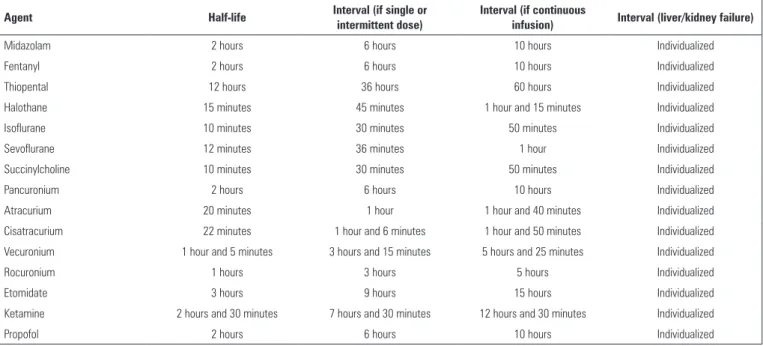
Table 2 - Main central nervous system depressants and interval from discontinuation to the start of the determination of brain death
Agent Half-life Interval (if single or
intermittent dose)
Interval (if continuous
infusion) Interval (liver/kidney failure)
Midazolam 2 hours 6 hours 10 hours Individualized
Fentanyl 2 hours 6 hours 10 hours Individualized
Thiopental 12 hours 36 hours 60 hours Individualized
Halothane 15 minutes 45 minutes 1 hour and 15 minutes Individualized
Isoflurane 10 minutes 30 minutes 50 minutes Individualized
Sevoflurane 12 minutes 36 minutes 1 hour Individualized
Succinylcholine 10 minutes 30 minutes 50 minutes Individualized
Pancuronium 2 hours 6 hours 10 hours Individualized
Atracurium 20 minutes 1 hour 1 hour and 40 minutes Individualized
Cisatracurium 22 minutes 1 hour and 6 minutes 1 hour and 50 minutes Individualized
Vecuronium 1 hour and 5 minutes 3 hours and 15 minutes 5 hours and 25 minutes Individualized
Rocuronium 1 hours 3 hours 5 hours Individualized
Etomidate 3 hours 9 hours 15 hours Individualized
Ketamine 2 hours and 30 minutes 7 hours and 30 minutes 12 hours and 30 minutes Individualized
Propofol 2 hours 6 hours 10 hours Individualized
Half-life - half-life time; intermittent dose - less than 4 doses/24 hours; continuous infusion - continuous infusion or more than 3 intermittent doses/24 hours. - If intermittent administration: interval of three times the half-life. Use of blood flow imaging test preferred.
- If continuous infusion administration: interval of five times the half-life. Use of blood flow imaging test preferred.
- In liver and/or kidney failure: determine the interval on a case-by-case basis, considering the severity of the abnormalities, discussing the case with the intensivist and with the on-call doctor of the Organ Procurement Organization/Center for Notification, Procurement and Allocation of Organs. In these cases, the blood flow imaging test is mandatory.
- In the case of an intravenous barbiturate, always perform the blood flow imaging test.
- The cause of unresponsive and areflexic coma should not be related to CNS depressants that are unlikely to cause areflexic coma when used at usual therapeutic doses. Examples: enteral phenobarbital, phenytoin, clonidine, dexmedetomidine, morphine.
apnea test not possible due to hypoxemia) or potential interference by confounding factors such as hypothermia, metabolic disorders and CNS depressant use, or due to legal reasons as in Brazil, an imaging test demonstrating an absence of cerebral blood low, electrical activity or metabolic and cephalic activity is required (D).(3,23,45) he
ideal auxiliary test should have adequate sensitivity but mainly 100% speciicity, which indicates that there will be no cases of patients presenting any evidence of brain or brainstem activity during the clinical examination for which the imaging test shows an absence of cerebral blood low or of electrical or metabolic activity (false positive). Safety and the immediate availability of the imaging test are also desirable characteristics. SAP ≥ 100mmHg and MAP ≥ 60mmHg are required for all imaging tests to avoid false-positive results (D).(46,47)
Several tests are accepted internationally for the determination of brain death (D).(3,32,45-48) Cerebral
angiography, which investigates the presence of blood low in the intracranial portion of the internal carotid and vertebral arteries, is considered the reference standard for the test comparison. However, it has some disadvantages, such as a need to move the patient outside the ICU, the use of potentially nephrotoxic contrast
agents and arterial puncture (D).(45,46) Transcranial
Doppler ultrasonography investigates the presence of blood low in the intracranial internal carotid, middle cerebral, vertebral and basilar arteries. Although this test should be performed by a professional with a high level of training, it has the following advantages: bedside availability, noninvasive and does not require the use of contrast medium (D).(45,46) Cerebral scintigraphy is
also a widely accepted cerebral blood low test; it assesses brain perfusion based on parenchymal uptake of the radionuclide technetium. his test does not require the use of iodinated contrast, is easy to interpret and exhibits high concordance with cerebral angiography (D).(45,46) As
of no cerebral blood low provides deinitive conirmation
(D).(45,46) Encephalography is a widely used imaging
test that investigates the presence of electrical activity. Its advantages are performance at the bedside, no requirement for contrast medium and wide availability. Its main disadvantage is that it might demonstrate an absence of electrical activity in the presence of confounding factors, namely, severe metabolic disorders, hypothermia and CNS depressant efects (D).(45,46) In this case, cerebral
blood low imaging must be performed. In this context, it is worth underscoring that continuous administration of barbiturates has a cumulative efect in which electrical activity might remain absent on electroencephalography for several hours after discontinuation (D).(3,23,45-48)
Several other tests have been assessed, including computed tomography angiography (CTA), a previously validated cerebral blood low test that is currently used in some countries and gaining increasing acceptance worldwide. his test is attractive because it is available at most medium- and large-sized hospitals, is easy to perform, does not require invasive puncture and employs a lower amount of iodinated contrast medium compared to conventional angiography. While most studies have assessed individuals with a conirmed diagnosis of brain death only (B),(49-51) Dupas et al. included a control
group, i.e., coma patients still exhibiting evidence of brain electrical activity, and found that the method had 100% speciicity (B).(52) here are no uniform international
radiological criteria for the interpretation of CTA; however, the Société Française de Radiologie published guidelines based on a so-called “four point scale” that assesses opaciication of the middle cerebral arteries and internal cerebral veins with 85% sensitivity (D).(53) Two
recent systematic reviews concluded that CTA might be used as an auxiliary to clinical examination for the diagnosis of brain death (B).(54,55)
Evoked potentials, which assess brain electrical activity, are of limited use because they investigate speciic neural pathways, even when including somatosensory and auditory evoked potentials, which assess the electrical response to stimulation of the median and vestibulocochlear nerve, respectively. he principle underlying the evoked potentials is alien to the concept of and rationale for the integral and global assessment of the brain function required for an accurate diagnosis of brain death. he test might evidence the absence of electrical signals in lesions proximal to the investigated pathways, even though other areas might be preserved from an anatomical and functional perspective. Few studies have investigated
evoked potentials in coma patients with severe brain injury but not fulilling the clinical criteria for brain death, which does not allow an accurate assessment of the speciicity of the method. Similarly to electroencephalograms, evoked potentials can also provide false-positive results in the presence of hypothermia, metabolic disorders or the use of CNS depressants (B).(56-58)
Intracranial pressure monitoring is indicated based on physiological reasons, but it has been assessed in only a few observational studies with a small number of patients. When the intracranial pressure remains above SAP continuously for at least 20 minutes, the test is considered to be positive. One further limitation of this method is the technical diiculty inherent to the available measurement techniques, which provide values with reduced accuracy. herefore, intracranial pressure monitoring should not be used for the diagnosis of brain death (C).(59-61)
Jugular venous oxygen saturation monitoring has a physiological rationale, namely, the drop in the oxygen extraction rate that occurs at the time of brain death. his parameter was assessed in a single prospective observational study that evaluated a central venous/jugular oxygen saturation ratio < 1 as predictor of brain death in a sample of 118 individuals with a clinical diagnosis of brain death and 152 head injury survivors. he test had 96.6% sensitivity and 99.3% speciicity for the diagnosis of brain death. Electroencephalography was the imaging method used as a reference for comparison. Jugular venous oxygen saturation monitoring is limited by technical diiculties related to the position of the catheter, and the results are inluenced by the PaO2 level. herefore, it should not be used for the diagnosis of brain death (B).(62)
Recommendation: he preferred imaging tests for the diagnosis of brain death are cerebral angiography, transcranial Doppler ultrasonography, cerebral scintigraphy and electroencephalogram (D). Strong Recommendation.
Recommendation: Intracranial pressure and jugular venous oxygen saturation should not be used as imaging tests for the diagnosis of brain death. (C). Strong Recommendation.
Recommendation: In cases with severe metabolic disorders, hypothermia and the use of CNS depressants, cerebral blood low tests are indicated: cerebral angiography, transcranial Doppler ultrasonography and cerebral scintigraphy (D). Strong Recommendation.
protocol for interpretation of the results, such as the
Société Française de Radiologie’s “four point scale” (B). Weak Recommendation.
Recommendation: In patients with some degree of skull opening, such as children under 1 year of age, individuals with open head injuries or after an extensive craniotomy, electroencephalography might be preferred, but only residual blood low has been demonstrated using other methods (D). Strong Recommendation.
13. In situations such as severe facial trauma, otorrhagia, eye agenesis and high cervical spine injury, which preclude the performance of a portion of the clinical examination, is it possible to establish a diagnosis of brain death?
Comment: In Brazil, the determination of brain death is based on a conirmed irreversible loss of all brainstem functions, as established on the clinical examination and by the apnea test, whereas an auxiliary test might be performed as an additional safety measure and to provide documented proof of the patient’s status. Any hindrance to the performance of some part of the brainstem function assessment might raise doubts regarding the diagnosis of brain death and, concomitantly, represent a situation of ethical-legal non-compliance with the stipulations of law no. 9,434 and CFM resolution 1,480 (D).(18,19)
here are no data in the literature or derived from daily clinical practice to contraindicate continuation of the process of brain death determination when one of the brainstem relexes cannot be evaluated, provided all other indings on the clinical examination are compatible with brain death (D).
he results of 18 years of experience since it was passed show that resolution no. 1,480 requires modiications (D).(19) In 2011, the CFM Brain Death Technical Board
passed a new resolution on brain death determination that has not yet been enforced. Article 3, paragraph 4 of this new resolution establishes that “in the presence of congenital or acquired structural abnormalities hindering the assessment of the relexes mentioned in the heading of this article, and provided all other indings on clinical examination conirm the status of brain death, due justiication for the aforementioned impossibility should be recorded in the [patient’s] medical records and [the process of brain death] determination should continue”. Only once the new resolution is in force will there will be legal and ethical grounds to continue the process of brain death determination under the aforementioned circumstances.
Recommendation: According to CFM resolution no. 1,480 and law no. 9,434, the examination of brain death cannot continue when it is not possible to assess all brainstem relexes (D). Strong Recommendation.
14. Who is responsible for filling and signing the death certificate? What time of death should be recorded in the death certificate? Can therapeutic support be discontinued after a diagnosis of brain death is established?
Comment: Only doctors can issue a death certiicate (D),(63) with the exception of cases of natural death in a
place where no doctor is available, as stipulated in law 12,842/2013 (D),(63) which regulates the practice of
medicine.
he death certiicate should be illed by the doctor who conirmed the occurrence of death, according to a CFM resolution (D).(64) In the case of brain-dead individuals, a
brain death certiicate is irst issued,(18) and when natural
death occurs, the death certiicate is issued by the doctor who cared for the patient or by a substitute or on-call doctor if the former is not available (D).(64,65) “Death
Veriication Services are institutions with the aim to determine that death has actually occurred, as well as its cause - when death was natural and there is no suspicion of violence - in cases of death without previous medical care, or in cases in which medical care was provided but death occurred due to an ill-deined condition”. herefore, wherever a Death Veriication Service is available, it may be called in when the doctor is unable to correlate death to the clinical condition of the patient, as recorded in his/ her medical records or institutional medical forms (D).(65)
In cases of unnatural death, also known as death due to external causes (homicide, accidents, suicide and suspicious deaths), at places where there is a Instituto Médico Legal (IML) unit, the death certiicate is issued by the coroner (D),(64,65) and the attending doctor should
complete the Cadaver Referral to the IML Form (Guia de Encaminhamento de Cadáver ao IML). In places where no IML unit is available, the death certiicate is issued by a local doctor who is appointed as the ad hoc coroner by legal or police authorities (D).(64,65)
he date and time of death recorded in the death certiicate should be those corresponding to the determination of brain death, according to CFM resolution no. 1,826/2007 (D).(66)
all support procedures that artiicially sustain the function of vital organs. In this case, discontinuation of life support does not characterize orthothanasia, euthanasia or any threat to life because the subject is not a terminal patient but a cadaver. Resolution no. 1,827/2007 states that “discontinuation of therapeutic support procedures after brain death was determined in a non-donor is legal and ethical” (D).(66) Here the term “non-donor” encompasses
not only cases due to family refusal but also to medical contraindication and/or to administrative/logistic problems. Doctors should communicate the patient’s death to the family or legal representatives in a clear and detailed manner and enter in the patient’s medical records the date and time of the communication, as well as the names of the individuals who were present. Maintenance of therapeutic support for brain-dead non-donors may be considered in the case of pregnant women with a living fetus, in which case the corresponding decisions should be made by an obstetrician.
Recommendation: In cases of death due to natural causes, the death certiicate must be completed and signed by the doctor who provided care to the patient and determined the presence of brain death or by a substitute (D). Strong Recommendation.
Recommendation: In cases of death due to unnatural causes, the death certiicate must be completed and signed by a coroner, while the attending physician, a substitute or an on-call doctor must provide all the relevant information related to the case in point (D). Strong Recommendation.
Recommendation: he date and time of death recorded in the death certiicate should be those corresponding to the last procedure for the determination of brain death (D). Strong Recommendation.
Recommendation: All therapeutic support should be discontinued after brain death is determined in a non-donor and this information has been communicated to the family with an explanation for the patient’s death (D). Strong Recommendation.
PART 3: CRITERIA FOR POTENTIAL DONOR SELECTION
15. How should the clinical and laboratory assessment of potential organ and tissue donors be organized and performed?
Comment: Any risk of the transmission of infectious or neoplastic diseases through organ or tissue transplantation should be completely eliminated. he risks associated with the procedure should always be considered in relation to the
high risk of the death of patients on the transplant waiting list. he increasing number of individuals on waiting lists and of needed transplant organs has led transplantation teams to use organs from donors with expanded criteria, with satisfactory outcomes, but with a higher potential for complications such as disease transmission. All procedures needed to gather the clinical and laboratory data to determine a minimum risk to the recipient of the organs and tissues used for transplantation should be performed. he time available to assess potentially deceased donors is usually quite short, particularly in the case of solid organ donors.(17,67,68) Consequently,
well-structured approaches are required to be applied by health care providers with direct participation in the donation-transplantation process. To ensure the safety and quality of the donation-transplantation process, which includes the clinical assessment of potential donors and performance of auxiliary tests, it is advisable to appoint a duly trained professional to oversee the entire process (D).(17)
he assessment comprises the following steps: (1) clinical history (analysis of the patient’s medical records and interviews with relatives), (2) physical examination including anthropometric measurements, (3) auxiliary tests and (4) inventory during organ removal surgery.
1. Clinical history: aims at ruling out transmissible (infectious and neoplastic) diseases in the donor, in addition to determining the functional status of the organs to be harvested and transplanted. For this purpose, the donor’s clinical history should be carefully reviewed to guide selection of the necessary auxiliary tests (D).(68)
A careful review of the potential donor’s medical records allows information to be gathered concerning the cause of death, current disease, past pathological history, treatments administered and intercurrent events (D).(68)
A detailed clinical history serves to conirm the donor’s past medical history (with an emphasis on neoplastic and infectious diseases), social habits (diet, alcohol and/or illegal drug use, smoking), sexual behavior, occurrence of menstrual irregularity after pregnancy (choriocarcinoma), admission/stay at institutions (arrests/psychiatric hospitals), origin and geographical provenance (D).(68)
2. Physical examination and anthropometric
geographic characteristics, masses/enlarged lymph nodes, skin neoplasms and scars derived from past surgical interventions.
he anthropometric variables to be assessed are as follows:
- Body weight and height: all donors
- Pediatric kidney donor: > 15kg, separate removal; < 15kg, en bloc removal of the kidneys.
- Liver donor: approximately 10 - 20% variation, with a donor weight and graft weight to recipient weight ratio of 1% - the latter especially in the case of children.
- Pancreas donor: acceptable when the body weight is between 30 and 90kg.
- Heart donor: < 20% lower weight.
- Chest circumference at the level of the nipple: lung donors.
3. Auxiliary tests: allow monitoring of clinical parameters during donor maintenance to detect organ dysfunctions and transmissible diseases and provide guidance to prioritize possible recipients included in the waiting list according to their blood type.
- Periodic biochemical testing every 24 hours to attain normal physiological parameters and ensure adequate functioning of the transplant organs (D).(69,70)
- Particular tests should be performed according to the organs to be transplanted: Heart donor - creatine kinase MB isoenzyme (CK-MB) and/ or troponin every 24 hours, electrocardiogram and echocardiogram; cardiac catheterization may be considered for donors >45 years old. Liver donor - aspartate aminotransferase (AST), alanine aminotransferase (ALT) and bilirubin at least every 24 hours. Kidney donor - urea and creatinine (Cr) every 24 hours and urinalysis. Pancreas donor - amylase and blood glucose every 24 hours. Lung donor - arterial blood gases with FiO2 at 100% and chest radiography (D).(68,69,71)
- he presence of transmissible diseases should be eliminated by performing serologic tests for Chagas disease, anti-toxoplasma antibodies, Venereal Disease Research Laboratory (VDRL, when positive, the luorescent treponemal antibody absorption test (FTA-ABS) should be performed), anti-human immunodeiciency virus (HIV) antibodies, anti-human T lymphotropic virus (HTLV) 1 and 2 antibodies, surface antigen of hepatitis B virus (HBsAg), hepatitis
B core antibodies (anti-HBc), hepatitis B surface antibodies (anti-HBs), hepatitis C antibodies HCV), cytomegalovirus antibodies (anti-CMV), Epstein-Barr virus antibodies (anti-EBV) and serologic tests for malaria in endemic areas (North Brazil).
- Two blood cultures and one urine culture should be performed for all potential donors. Cultures with samples collected from other body sites should be performed in the case of suspected infection; the results must be supplied to the transplantation teams/transplant centers (D).(71,72)
- Tumor markers: see question 21.
4. Surgical inventory during organ removal: the chest and abdominal organs should be examined during removal surgery to detect potentially hidden tumors or pathological lymph nodes. he kidneys and liver should be carefully examined due to the high numbers of tumors found in kidneys after removal (D).(73,74)
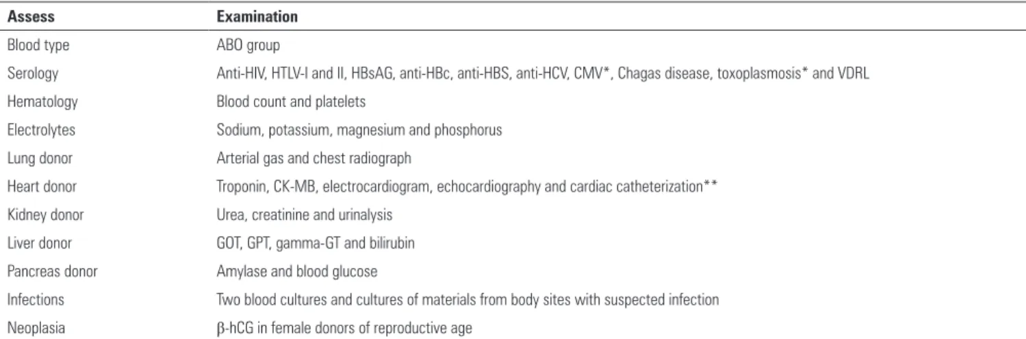
Recommendation: Perform a complete clinical history, including a past pathological history and careful physical examination, order auxiliary tests (Table 3) and perform a surgical inventory during organ removal (D). Strong Recommendation.
Recommendation: Enter all indings on the complete clinical assessment in the medical records of the potential donor (D). Strong Recommendation.
16. Which organs and tissues harvested from brain-dead donors can be donated?
Comment: Brain-dead deceased donors are the main source of transplant organs and tissues (D).(75) According to
the Brazilian Ministry of Health ordinance no. 2,600, from September 21, 2006 (D),(76) the organs that can be donated
and individually used for transplantation are the heart, lungs, kidneys, liver, pancreas and intestine (B).(69,76-78)
Multi-organ transplantation can also be performed, which involves joint donation and transplantation of the liver, pancreas, stomach, duodenum and small intestine into a single recipient; other possibilities are kidney-pancreas and liver-kidney transplantation. Tissues that can be donated for transplantation are the cornea, sclera, skin, bone, cartilage, tendon, meniscus, muscle fascia, heart valves, pericardium and blood vessels (B).(69,76-78) Hematopoietic
stem cells retrieved from the bone marrow, peripheral blood and the umbilical cord/placenta can be donated by living donors. here are reports in the literature of limb (C),(79,80) face (C),(81) larynx and trachea (C)(82) transplants,
Table 3 - Examinations to be solicited for the evaluation of the potential donor.
Assess Examination
Blood type ABO group
Serology Anti-HIV, HTLV-I and II, HBsAG, anti-HBc, anti-HBS, anti-HCV, CMV*, Chagas disease, toxoplasmosis* and VDRL
Hematology Blood count and platelets
Electrolytes Sodium, potassium, magnesium and phosphorus
Lung donor Arterial gas and chest radiograph
Heart donor Troponin, CK-MB, electrocardiogram, echocardiography and cardiac catheterization**
Kidney donor Urea, creatinine and urinalysis
Liver donor GOT, GPT, gamma-GT and bilirubin
Pancreas donor Amylase and blood glucose
Infections Two blood cultures and cultures of materials from body sites with suspected infection
Neoplasia β-hCG in female donors of reproductive age
HTLV - human T-lymphotropic virus; HBsAG - hepatitis B virus surface antigen; anti-HBc - hepatitis B core antibody; anti-HBS - antibodies against hepatitis B surface antigen; anti-HCV - antibodies against hepatitis C virus; CMV - cytomegalovirus; VDRL - Venereal Disease Research Laboratory; CK-MB - creatine kinase MB isoenzyme; GOT - glutamic-oxalacetic transaminase; GPT - glutamic-pyruvic transaminase; gamma-GT - gamma-glutamyltransferase; β-hCG - beta-human chorionic gonadotropin. * Results may be obtained after transplantation. ** For patients older than 45 years.
Recommendation: Organs that can be donated by brain-dead deceased donors include the heart, lungs, kidneys, liver, pancreas and intestine (B). Strong Recommendation.
Recommendation: Tissues that can be donated by deceased donors are the corneas, sclera, skin, bone, cartilage, tendon, meniscus, muscle fascia, heart valves and blood vessels (B). Strong Recommendation.
17. What characterizes the expanded criteria donor?
Comment: Several terms are used to designate donors that barely meet the selection criteria, such as suboptimal, unit, high-risk, marginal, borderline and expanded criteria donors. he terms “high risk”, “marginal” and “expanded criteria” donors are the most widely used (D).(83) here is
no universal deinition of marginal or expanded criteria donors (ECDs). However, the presence of some conditions associated with shortened survival, reduced graft function or the risk of disease transmission has been used to characterize organs as of “marginal” quality (D).(83-85)
he characteristics of marginal donors are as follows: (D):(83-85)
- Relative to graft function: higher short-term morbidity (delayed graft function or primary graft nonfunction) and shorter graft survival. hese events might be associated with the donor’s age, past pathological history, anthropometric measurements, cause of death, previous function of the organ to be donated, anatomical abnormalities, intoxications and poisonings, hemodynamic instability, prolonged ischemia time and donation after circulatory death (D).(86-93)
- Relative to disease transmission: infections and neoplasias.
he use of marginal donors is only justiied when the life expectancy after transplantation is higher compared with conventional clinical treatment. Under borderline circumstances, the decision to transplant organs is made by the transplantation team with the informed consent of the recipient. he organs must be removed, and if they are not used in the same Brazilian state, then they should be ofered to the National Transplant Center for allocation to other states.
Recommendation: Marginal or expanded criteria donors are those presenting clinical conditions that might reduce graft survival, impair its function or are at high risk of disease transmission (D). Strong Recommendation.
Recommendation: he use of marginal donors is only justiied when the life expectancy after transplantation is higher compared with conventional clinical treatment (D). Strong Recommendation.
18. What is the accepted age range for multiple organ and tissue donors?
Comment: Any brain dead individual may be considered a potential donor independent of his/her age (D).(94) In Brazil, the minimum age for the determination
of brain death and characterization as an organ donor is 7 days (D).(18,76) here is no maximum age for donation;
however, comorbidities that develop together with aging make donation less acceptable (D).(95)
Kidney graft function and survival are impaired when donors are greater than 60 years old (D).(95) Expanded