www.ascro.hr
Acta stomatol Croat. 2013;47(3):253-261. PRIKAZ SLU»AJA CASE REPORT
Fábio Wildson Gurgel Costa1, Francisco Samuel Rodrigues Carvalho2, Camila Costa Dias3, Filipe Nobre Chaves4, Thyciana Rodrigues Ribeiro5, Clarissa Pessoa Fernandes6, Karuza Maria Alves Pereira7
Obiteljski hipoplastični jezični frenulum: prikaz slučaja i
rasprava o diferencijalnoj dijagnozi
Familial Hypoplastic Lingual Frenum: Case Report and
Discussion of Differential Diagnosis
ACTA
STOMATOLOGICA CROATICA www.ascro.hr
Introduction
One of the most interesting and not yet understood an-atomical structures of the oral cavity is the frenum or frenu-lum (1,2). he frenum, a mucous connective tissue insert-ed on hard- or soft-parts of the oral cavity, is histologically composed of ibrodense tissue and occasionally of striated muscle ibers (1,2). hree types are commonly identiied in the oral cavity: superior labial frenum, inferior labial frenum, and lingual frenum (3). he lingual frenum (LF) is an im-portant structure that anatomically relates to the tongue and mandible (4). A short LF can interfere with tongue mobili-ty, which can lead to a few problems including breastfeeding diiculties, speech disorders, anterior open bite, and man-dibular prognathism (5-7).
Uvod
Jedna od zanimljivih, ali i do kraja objašnjenih anatom-skih struktura usne šupljine jest frenum ili frenulum (1, 2). Frenum, mukozno vezivno tkivo između tvrdih ili mekih di-jelova usne šupljine histološki je sastavljeno od gustoga ibro-znog tkiva i mjestimice umetnutih mišićnih vlakana. Obično se nalazi na trima mjestima (1,2), pa postoje gornji labijal-ni frenulum, donji labijallabijal-ni frenulum i jezičlabijal-ni frenulum (LF) (3). Lingvalni fenulum anatomski je važan jer povezuje je-zik i mandibulu (4). U slučaju da je kratak može ograniča-vati pokrete jezika, pa se pojavljuje nekoliko problema – teš-koće s dojenjem, poremećaji u govoru, frontalno otvoreni zagriz i mandibularni prognatizam (5-7). Morfološki ili broj-čano promijenjeni oralni frenulum povezan je s
mnogobroj-Sažetak
Jedna od najzanimljivijih, ali još nerazjašnjenih struktura usne šupljine jest oralni frenulum. Jezič
-ni frenulum (LF) važan je zato što spaja jezik i mandibulu te može biti izravno povezan s poreme
-ćajima u okluziji zuba. Morfološke ili brojčane promjene oralnih frenuluma u neposrednoj su vezi
s mnogobrojnim genskim stanjima, posebice s Ehlers-Danlosovim sindromom. Neki autori
sma-traju da je odsutnost LF-a patognomonični znak za taj poremećaj. Svrha ovog opisa slučaja bila je izvijestiti o obiteljskoj hipoplaziji jezičnog frenuluma dijagnosticiranog rutinskim oralnim pre
-gledom, te potaknuti raspravu o povezanosti abnormalnog LF-a s nesindromskim poremećajima i/ili genskim sindromima. Liječnici dentalne medicine i ostali specijalisti moraju biti oprezni tije
-kom kliničkih pregleda kako bi rano otkrili Ehlers-Danlosov sindrom jer može neposredno utjeca -ti na ortodontsku terapiju.
1 Državno sveučilište Ceará, Stomatološki fakultet, Zavod za e-stomatologiju, radiologiju i oralnu medicinu, kampus Sobral.
University Federal of Ceará. Professor, Department of Stomatology, Radiology and Oral Medicine. College of Dentistry, University Federal of Ceará – campus Sobral.
2 specijalizant oralne i maksilofacijalne kirurgije, Državno sveučilište Ceará, Stomatološki fakultet i Sveučilišna bolnica Walter Cantideo Resident in Oral and Maxillofacial Surgery. College of Dentistry, University Federal of Ceará, Walter Cantídeo University Hospital. 3 Državno sveučilište Ceará, Stomatološki fakultet, kampus Sobral
College of Dentistry, University Feredal of Ceará – campus Sobral. 4 Državno sveučilište Ceará, Stomatološki fakultet
Clinical Dentistry of University Federal of Ceará. College of Dentistry, University Federal of Ceará. 5 Državno sveučilište Ceará, Stomatološki fakultet, kampus Sobral
Clinical Dentistry of University Federal of Ceará. College of Dentistry, University Federal of Ceará – campus Sobral. 6 Državno sveučilište Ceará, Stomatološki fakultet, kampus Sobral
University Federal of Ceará. College of Dentistry, University Federal of Ceará.
7 Zavod za e- stomatologiju, oralnu patologiju i oralnu medicinu, Državno sveučilište Ceará, Stomatološki fakultet, kampus Sobral Department of Estomatology, Oral Pathology and Oral Medicine. College of Dentistry, University Federal of Ceará – campus Sobral.
Zaprimljen: 18. prosinca 2012.
Prihvaćen: 11. srpnja 2013.
Adresa za dopisivanje
Francisco Samuel Rodrigues Carvalho
Rua Belo Horizonte, 2830, apto. 122, bloco B, Jóquei Clube; CEP 60440-195, Fortaleza, Ceará, Brazil
tel: +55(85) 96224146 faks: +55(88) 36132603 [email protected]
Ključne riječi
www.ascro.hr
nim genskim poremećajima, poput Ehlers-Danlosova sin-droma (EDS), infantilne hipertroične pilorične stenoze, holoprozencefalije, orofacijalnih digitalnih sindroma (OFD) te Rapp-Hodgkinova i Fraserova sindroma. Dodatno su opi-sani abnormalni razvojni poremećaji frenuluma kod Palli-ster-Hallova sindroma, Optizova trigonocefalijskog sindro-ma (Optizov C-sindrom), W-sindrosindro-ma i Optizova sindrosindro-ma (3, 8, 9).
EDS je nasljedni genski poremećaj kod kojega se događa-ju promjene u sklopu vezivnoga tkiva. Kako nastaje defekt u metabolizmu kolagena pucaju koža i krvne žile, te se pojav-ljuje hiperrastezljivost kože i hiperpokretljivost zglobova (3, 10 –17). Nekoliko istraživača tvrdi da je odsutnost LF-a po-tencijalni pokazatelj EDS-a (9). Obiteljski abnormalno kra-tak, ili čak nepostojeći LF, rijetko je stanje s kojim se susre-ću stomatolozi i ostali liječnici (18). Svrha ovog članka jest opisati slučajeve pacijenata s obiteljski naslijeđenim nedo-statkom LF-a dijagnosticiranim tijekom oralnog pregleda te raspravljati o povezanosti s nesindromskim stanjima i/ili gen-skim sindromom povezanim s abnormalnim LF-om.
Prikaz slučaja
Pacijentica u dobi od 23 godine došla je na rutinski pre-gled u ordinaciju Stomatološkog fakulteta Sveučilišta Ceará Sobral u Brazilu. Tijekom kliničkog pregleda liječnik je uo-čio da nema LF, ali nije imala poremećaje u govoru i žvakanju ili druge funkcijske tegobe. Te su se promjene odmah poče-le provjeravati i istraživati jer se željelo doznati je li to izoli-rani slučaj. Zbog toga su pregledani i ostali članovi njezine peteročlane obitelji (slika 1.). Otac u dobi od 54 godine nije imao LF, a kod 49-godišnje majke bio je normalan. Najstari-ja kći (28 godina) imala je hipoplastični frenulum, a srednNajstari-ja (24 godine), u usporedbi sa starijom sestrom, jedva vidljivo kraći. Treća kći (23 godine), fenotipski slično ocu, nije ima-la LF. Svi čima-lanovi obitelji pregledani su zbog EDS-a jer neki istraživači smatraju da je nedostatak LF-a dijagnostički znak. Kako bi se dijagnosticirao EDS, u obzir su uzeti sljedeći kli-nički znakovi (tablica 1.):
1) hiperekstendibilnost kože, 2) hiperpokretljivost zglobova, 3) poremećaj u stvaranju ožiljaka,
4) česta subluksacija temporomandibularnog zgloba, paro-dontalna bolest,
6) Gorlinov znak (mogućnost da se vršak nosa dotakne jezi-kom),
7) labava oralna mukoza, 8) odsutnost LF-a.
Samo 23-godišnja kći nije imala LF i pokazivala je laga-ne kliničke znakove nabrojelaga-ne u točkama jedan i dva. Osim kod oca koji je također nije imao LF, ni kod koga u toj obite-lji nisu pronađeni klinički znakovi.
Članovi te obitelji dodatno su bili na testu pokretljivosti zglobova prema Beightonu i suradnicima (19). To ispitivanje uključuje hiperekstenziju petog prsta, postavljanje palca na ventralnu stranu podlaktice, hiperekstenziju lakatnog zgloba veću od 10°, hiperekstenziju koljena veću od 10° i
postavlja-Oral frenulum altered in morphology or number has been associated with many genetic conditions, such as: Ehlers-Danlos syndrome (EDS), infantile hypertrophic py-loric stenosis, holoprosencephaly, orofacial digital syndrome (OFD), Rapp-Hodgkin syndrome and Fraser syndrome. Additionally, abnormal development defects have been de-scribed as characteristics of the Pallister-Hall syndrome, Op-tiz trigonocephaly syndrome (OpOp-tiz C syndrome), W syn-drome, and Optiz syndrome (3,8,9).
EDS, a hereditary genetic disorder, consists of a group of alterations inherent of connective tissue that presents a defect in collagen metabolism, leading to fragility of the skin and blood vessels, hyperextensibility of the skin, and joint hyper-mobility (3,10-17). A few authors have described that the absence of LF is a potential indicator of EDS9. Familial
ab-normally short or even absent lingual frenulum is a rare con-dition to dentists and physicians (18). hus, the aim of this study is to report a case of a patient with familial absent LF diagnosed during dental exam, and discuss the association of non-syndromic conditions and/or genetic syndromes associ-ated with abnormal LF.
Case report
A 23-year-old woman, of normal systemic status, attend-ed the Dental service of the Fattend-ederal University of Ceará, So-bral Campus (Brazil) for dental evaluation. During the clin-ical examination, it was observed that the patient presented absence of LF without speech and chewing disorders, or any other functional impairment.
www.ascro.hr
Tablica 1. Klinički nalazi članova obitelji prema karakteristikama EDS-a
Table 1 Clinical indings of the family members according to EDS characteristics.
Klinički nalazi • Clinical indings I-1 I-2 II-1 II-2 II-3
Hiperelastičnost kože • Skin hyperelasticity - - - - +
Hiperpokretljivost zglobova • Joints hypermobility - - - - +
Poremećaj u stvaranju ožiljka • Disorders of cicatrization - - - -
-Subluksacije TMZ-a • Subluxations of TMJ - - - -
-Primljivost na parodontne bolesti • Susceptibility to periodontal disease - - - -
-Gorlinov znak • Gorlin’s sign - - - -
-Nježna oralna sluznica • Friable oral mucosa - - - -
-Hipoplastični lingvalni frenum • Hypoplastic lingual frenum - - + +
-Nema lingvalnog frenuma • Absence of lingual frenum - + - - +
#Pasivna dorzileksija malog prsta veća od 90º • #Passive dorsilexion of the little ingers beyond 90º - - - - -# Pasivno postavljanje palca na leksornu stranu podlaktice • #Passive apposition of the thumbs to the lexor aspects
of the forearms - - - -
-#Hiperekstenzija lakta veća od 10º • #Hyperextension of the elbows beyond 10º - - - -
-# Hiperekstenzija koljena veća od 10º • #Hyperextension of the knees beyond 10º - - - - -#Fleksija trupa naprijed, ravna koljena, dlanovi ruku plošno na pod • #Forward lexion of the trunk, with knees
straight, so that the palms of the hands rested easily on the loor - - - -
-Kratice • Abbreviations:
EDS – Ehlers-Danlosov sindrom • Ehlers-Danlos syndrome; TMZ • TMJ – temporomandibularni zglob • temporomandibular joint. Odsutno • Absence (-); prisutno • presence (+); # Beightonov zbroj (procjena pokreta zglobova) • Beighton’s score (assessment of joint mobility).
Slika 1. Shema rodoslovlja prikazane obitelji s čvrstim simbolom koji upućuje na individualnu pripadnost. Djelomičnočvrsti simbol pokazuje osoba s
hipoplazijom LF-a. Rimski brojevi identiicirane generacije. Arapski brojevi predstavljaju pojedince u svakoj generaciji.
Figure 1 Family tree of the reported family. The solid symbol indicates affected individual. Partially solid symbol indicates individuals with hypoplastic LF. Roman numerals identify generations. Arabic numerals represent the individuals in each generation.
Slika 2. Klinička slika LF-a u svakoj obitelji A) I-1; B) I-2; C) II-1; D) II-2; E) II-3.
www.ascro.hr
nje dlanova na podlogu uz ispružena koljena. Odgovorili su i na sljedeća pitanja u upitniku Hakima i Grahame:
1. Možete li (jeste li ikada mogli) staviti dlanove na podlo-gu, a da ne savijete koljena?
2. Možete li (jeste li ikada mogli) saviti palac i dodirnuti podlakticu?
3. Jeste li kao dijete zabavljali prijatelje postavljajući tijelo u neobične položaje i jeste li mogli napraviti špagu? 4. Jeste li kao dijete ili adolescent jedanput ili nekoliko
pu-ta iščašili rame ili patelu koljena? Smatrate li da su vam zglobovi gibljivi?
Svim tim ispitivanjima potvrdili smo da prikupljeni po-datci ne zadovoljavaju kriterije za hiperpokretljivost zglo-bova. Poslije toga članovi obitelji poslani su na medicinsku procjenu o promjeni unutarnjih organa, kao što predlažu Castori i njegovi suradnici (2012.) (20). Ni nakon tih te-stova nisu detektirane promjene koje bi zadovoljile kriterije EDS-a kod bilo koga od njih. Zaključeno je da nitko od njih nema EDS. Budući da se dijagnoza EDS-a obično postavlja prema kliničkim nalazima (15), a ni kod jednog člana obite-lji nije bilo karakterističnih znakova bolesti, te uzevši u obzir medicinske savjete, odlučeno je da se u dijagnostičke svrhe ne treba obaviti biopsija kože.
Rasprava
LF je mali pregib mukozne membrane koja spaja gingivu s donjom stranom jezika pokrivajući lingvalnu stranu pred-njeg alveolarnog nastavka (1). Histološki se sastoji od ibro-znog tkiva i pojedinačnih vlakana genioglosnog mišića. LF se klinički smatra normalnim ako insercija počinje u sredini do-nje strane jezika i završava u dnu usne šupljine. Smatra se da LF ima anteriornu inserciju ako počinje na donjoj strani je-zika između njegove sredine i vrška, a kratak je ako je veliči-nom manji (1, 21) ili može čak nedostajati kod sindromskih ili nesindromskih stanja (9, 17, 18). Ankiloglosija (jezična kravata) relativno je uobičajeno nesindromsko stanje pove-zano s kratkim LF-om i s prevalencijom od oko četiri do pet posto. Premda može biti u sklopu posebno rijetkih sindroma (rascjep nepca vezan za X-kromosom i van der Woudeov sin-drom), obično je izolirani slučaj u usnoj šupljini mladih pa-cijenata (18). Klinički LF može potpuno nedostajati, a kat-kad, doduše rijetko, pojavljuje se potpuna ankiloglosija (22). U ovom istraživanju je, među članovima analizirane obitelji, pronađena varijabilnost u stupnju skraćenja LF-a. Dijagno-za Dijagno-za ankiloglosiju nije postavljena. Prema tablici 2. mno-ga su genska stanja povezana s promjenama LF-a, poput hi-poplazije, hiperplazije, frenuluma biida, multiplog LF-a ili odsutnog LF-a. Premda orofacijalno-digitalni sindrom može imati kliničke i molekularne nalaze slične EDS-u, nije u pi-tanju odsutnost/hipoplazija LF-a. Isto vrijedi za anencefaliju i holoprozencefaliju kod koje nedostaje gornji labijalni fre-nulum (tablica 2.). Među sindromima kod kojih nema LF-a su Rapp-Hodgkinov (9), Fraserov (8) i Ehlers-Danlosov (3, 10 – 17).
Rapp-Hodgkinov sindrom povezan je s ektodermalnom displazijom, promjenom kapilara s tendencijom alopecije i rascjepom usne ili nepca (9).
the palms of the hands lat on the loor while maintaining the knees in full extension. he questionnaire proposed by Hakim and Grahame was also performed, and the follow-ing questions were asked: 1. Can you now (or could you ev-er) place your hands lat on the loor without bending your knees? 2. Can you now (or could you ever) bend your thumb to touch your forearm? 3. As a child did you amuse your friends by contorting your body into strange shapes or could you do the splits? 4. As a child or teenager did your shoulder or kneecap dislocate on more than one occasion? 5. Do you consider yourself double-jointed?. We veriied that the data found in the clinical exam did not meet the criteria of hyper-mobility of joints. Subsequently, the patients were referred for medical evaluation to investigate changes in internal or-gans and nervous system as suggested by Castori et al., 2012 (20). After all evaluations, no alterations that met the criteria for EDS were found for any of the evaluated patients. hus, the patients were not afected by EDS. Since the diagnosis of EDS is usually obtained through clinical indings15, in the
absence of the characteristic signs of the disease and consid-ering medical suggestion, it was decided not to perform a skin biopsy for diagnostic purposes.
Discussion
he LF is a small fold of mucous membrane that con-nects the gum with the posteroanterior face of the tongue, covering the lingual face of the anterior alveolar crest1. It is
histologically composed by ibrodense tissue and occasion-ally by the superior ibers of the ginioglossus muscle. Clini-cally, the LF is considered normal when its insertion starts in the middle of the inferior surface of the tongue and ends in the loor of the mouth. It is considered to be with anterior insertion when its insertion starts between the middle of the inferior surface of the tongue and the tip of the tongue. It is considered short when it presents a small size (1,21), and it may also be absent in syndromic or non-syndromic condi-tions (9,17,18).
www.ascro.hr
Fraserov sindrom manifestira se karakteristikama kriptof-talmusa (prirođeni djelomični ili potpuni nedostatak vjeđa), sindaktilije šaka i stopala te genitalnim anomalijama (8).
Ehlers-Danlosov sindrom povezan je s promjenama ko-lagena koje utječu na tkiva pa nastaje hiperrastezljivost i hi-perpokretljivost (3).
U ovom radu EDS je bio glavni genski poremećaj na ko-ji se posumnjalo jer može upućivati na to da nema labijalnog frenuluma, a ponekad i LF-a, što je patognomoničan znak. EDS je opisao danski dermatolog Edvard Ehlers još 1901. godine na temelju slučaja pacijenta s karakterističnim zglo-bovima, tendencijom krvarenja i rastezljivom kožom (cutis laxa). Francuski dermatolog Henri-Alexandre Danlos opisao je 1908. godine slučaj pacijenta s tankom i hiperelastičnom kožom te mekušastim pseudotumorima. Na temelju tih dva-ju prikaza slučajeva iz sredine 1930-ih godina određeno je ime toga sindroma – Ehlers-Danlosov za pacijente sa simpto-mima koje su opisali Edvard Ehlers i Henri-Alexandre Dan-los (10).
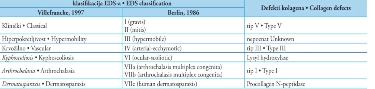
Potkraj 1960-ih istraživači su podijelili EDS u kategori-je – 1967. svrstali su ga u tri kategorikategori-je, 1968. u pet i 1972. u sedam. Sredinom 1980-ih bilo je dovoljno dokumentira-nih slučajeva toga poremećaja da se uspostavi formalna ne-sonimija. Istraživači su 1986. godine stvorili dijagram kori-štenjem rimskih brojeva i klasiicirali sindrom u deset tipova, ovisno o kliničkim pokazateljima i genskom nasljeđu. Kako je ta podjela zbunjivala, revidirana je 1997. godine u
Ville-Among the syndromes that present absence of lingual frenu-lum, there are: the Rapp-Hodgkin syndrome9, the Fraser
syndrome8 and the Ehlers-Danlos syndrome (3,10-17). he
Rapp-Hodgkin syndrome is associated with ectodermic dys-plasia, capillary alterations with tendency to alopecia and the presence of labial or palatine cleft (9). he Fraser syndrome presents as characteristics the cryptophthalmos (congenital partial or total absence of the eyelids), hand and foot syndac-tyly, and genital anomalies (8). he Ehlers-Danlos syndrome is more related with collagen alterations that may lead to tis-sue involvement, allowing hyperextensibility and hypermo-bility (3). Regarding the present familial study, EDS was the main genetic disorder considered because it might exhibit absence of the labial frenulum or, notoriously, absence of the LF, as potential pathognomonic indings.
EDS was previously described by the Danish dermatolo-gist Edvard Ehlers in 1901, who reported a patient with char-acteristics of loose joints, tendency to bleed and cutis laxa. In 1908, the French dermatologist Henri-Alexandre Danlos de-scribed a patient who presented thin and hyperelastic skin, in addition to molluscoid pseudotumors. From these reports, in the middle of the 1930s, the name Ehlers-Danlos syn-drome was designated to describe patients who presented the characteristics cited by Edvard Ehlers and Henri-Alexandre Danlos (10). At the end of the 1960s, the investigators di-vided the EDS into three categories in 1967, into ive types in 1968, and into seven types in 1972. In the middle of the Tablica 2. Morfologija LF-a ovisno o raznim genskim sindromima
Table 2 Morphology of LF according to different genetic syndromes.
Stanje • Condition
Frenumsko stanje • Frenum’s conditions
Hipoplastično •
Hypoplastic
Hiperplastično / Hipertroično • Hyperplastic /
Hipertroic
Odsutno • Absent Biid • Bifid Multipli •
Multiples
Ehlers-Danlosov sindrom •
Ehlers-Danlos syndrome lingvalni • lingual
-lingvalni; donji
labijalni • lingual;
labial inferior -
-Holoprosencephaly - - gornji labijalni • labial superior -
-Oral-facial-digital sindrom •
Oral-facial-digital syndrome - lingvalni • lingual - -
-Rapp-Hodgkinov sindrom •
Rapp-Hodgkin syndrome - - lingvalni • lingual -
-Fraserov sindrom • Fraser syndrome lingvalni • lingual - lingvalni • lingual -
-Pallister-Hallov sindrom • Pallister-hall
syndrome - - -
-gornji labijalni;
donji labijalni •
labial superior; labial inferior
Ellis-van Creveldov sindrom • Ellis -
Van Creveld syndrome
-gornji labijalni;
donji labijalni •
labial superior; labial inferior
- -
-Optizov trigonocefalijski sindrom •
Optiz trigonocephaly syndrome - - -
-gornji labijalni;
donji labijalni •
labial superior; labial inferior
W-sindrom • W syndrome - - - gornji labijalni • labial superior
-www.ascro.hr
1980s, there were enough documented types of EDS to es-tablish a formal disease. In the year 1986, the investigators organized a diagram using Roman numerals and classiied the syndrome in ten types considering clinical manifesta-tions and genetic inheritance. his classiication system was very confusing, and was reassessed in 1997, in Villefranche – France, which originated the current classiication system that divides the syndrome into six types according to minor and major criteria (10,11), (Table 3).
franchu u Francuskoj, što je rezultiralo trenutačnim susta-vom klasiikacije koja sindrom dijeli na šest tipova, ovisno o manjim i većim simptomima (10, 11), (tablica 3.).
EDS čini skup poremećaja od ukupno šest tipova s hete-rogenim kliničkim i genskim karakteristikama zbog defekta u sintezi i strukturi vlakana kolagena te, do nekog stupnja, općom hiperpokretljivošću zglobova. Ta karakteristika, u ve-ćoj ili manjoj mjeri, nalazi se kod svih tipova sindroma (10). Etiologija sindroma tipa IV (vaskularni tip) rezultat je
auto-klasiikacija EDS-a • EDS classiication
Defekti kolagena • Collagen defects Villefranche, 1997 Berlin, 1986
Klinički • Classical I (gravis)II (mitis) tip V • Type V
Hiperpokretljivost • Hypermobility III (hypermobile) nepoznat Unknown
Krvožilno • Vascular IV (arterial-ecchymotic) tip III • Type III
Kyphoscoliosis • Kyphoscoliosis VI (ocular-scoliotic) Lysyl hydroxylase
Arthrochalasia • Arthrochalasia VIIa (arthrochalasis multiplex congenita)VIIb (arthrochalasis multiplex congenita) tip I • Type I
Dermatosparaxis • Dermatosparaxis VIIc (human dermatosparaxis) Procollagen N-peptidase
Tablica 3. Kliničke varijante EDS-a, ovisno o klasiikaciji defekata kolagena – modiicirano prema Abelu i Carrascu (10).
Table 3 Clinical variants of EDS according to classiications and collagen defects. Adapted from: Abel and Carrasco10.
somno dominantne heterozigotne mutacije gena COL3A1 koja uzrokuje strukturne promjene u lancu kolagena Pro 1 (III) tip II. Pritom je karakteristično smanjenje termičke ot-pornosti, smanjena sekrecija i abnormalna proteolitička raz-gradnja (13). EDS tipa III (hiperpokretljivi tip) posljedica je nepoznate promjene gena, ali fenotipski se nasljeđuje auto-somno dominantno. Primjerice, roditelji imaju neke blage sustavne manifestacije, što je najčešći oblik EDS-a, bez ve-ćih promjena na koži, pa se takvi slučajevi često ne dijagno-sticiraju (10).
U prikazanom slučaju članovi obitelji bili su na testovima kako bi se isključio EDS tipa II, no ustanovljeno je da nitko od njih nema taj poremećaj.
Pacijenti s EDS-om klinički mogu imati sljedeća obiljež-ja: hiperrastezljivost kože, hiperpokretljivost zglobova, po-vremenu luksaciju zglobova, Metenierov znak (rastezljivost vjeđa), krhkost krvnih žila i mekih tkiva, produženo cijelje-nje tkiva, distroične ožiljke (ožiljci poput cigaretnog papi-ra), potkožne čvoriće pseudotumore i često spontano stvara-nje hematoma.
Glavne opisane oralne manifestacije su sljedeće: Gorli-nov znak (hiperpokretljivost jezika), gotsko nepce, odsutnost LF-a i donjega labijalnog frenuluma, rani parodontitis, na-kupine ibrina u parodontu, krvarenje gingive, ulceracije slu-znice, pokretljivost zuba, zubna hipoplazija, strukturalne ne-pravilnosti dentina, kratki nepravilni ili dilacerirani korijeni zuba, kalciikacije pulpe, nedostatak zuba, prekobrojni zubi i promjene u temporomandibularnom zglobu (hiperpokret-ljivost, trismus, krepitacije, preskakanje i zapinjanje) (3, 10, 16). Analizom članova obitelji u prikazanom slučaju, samo je jedan član imao hiperpokretljive zglobove i hiperelastič-nu kožu te nije imao LF. No kod njega ipak nije pronađena većina kliničkih obilježja EDS-a. Naime, pacijenti s promje-nama poput hiperpokretljivosti zglobova, hiperrastezljivosti
EDS constitutes a group of disorders, comprising a total of six types, with clinical and genetic heterogeneous charac-teristics, resulting in defects in the synthesis and structure of ibrillar collagen, with some degree of generalized joint hy-permobility. his characteristic is present in all types of the syndrome and expressed in greater or lesser degrees10. he
etiology of type IV syndrome (vascular type) is the result of autosomal dominant heterozygous mutations in COL3A1 gen, causing structural defects in the Pro1 (III) chain of type III collagen, which are characterized by decrease in thermal stability, secretion decrease, and abnormal proteolytic pro-cessing (13). Type III EDS (hypermobile type) presents un-known genetic alterations, but the phenotype has autosomal dominant inheritance and the patients may have some subtle systemic manifestations. In addition, it represents the most common form of EDS and lacks major skin involvement, which frequently contributes to the occurrence of undiag-nosed cases10. In the family described in the present case, to
rule out type III EDS, adequate medical evaluation was per-formed, conirming that none of the evaluated individuals presented the disease.
calci-www.ascro.hr
ications of the pulp, absence of teeth, supernumerary teeth, and alterations related to the temporomandibular joint (hy-permobility, trismus, crepitation, clicking and closed lock), (3,10,16). Analyzing the family member in the present study, only one presented joint hypermobility and hyperelas-ticity of the skin, in addition to absence of LF. However, the great majority of the other clinical indings of the EDS were not identiied in this patient. In this context, patients who present alterations as hypermobility of joints, tissue hyper-extension and/or other tissue alterations have this character-istic clinical aspect due to deiciency of collagen processing enzymes or dominant negative efect on the mutant collagen chains. he collagen is a ibrous protein secreted mainly by connective tissue cells, and is an important structural com-ponent of the human body (10). herefore, we believe that alterations in its production can result in structural altera-tions in tissues composed by this substance, as the lingual frenulum, for example.
It is believed that the diagnosis of EDS can be obtained early through the investigation of oral indings, such as the absence of the lingual frenulum, which can be easily ob-served during clinical examination performed by physicians or dentists (12,15). Making an early diagnosis of EDS is very important to the evolution of the clinical aspects of the pa-tients, and also to enable the implementation of an appropri-ate therapy for each case. In this context, according to Ong et al., the irst alterations are observed at around 20 years of age, approximately 90% of the patients present an alter-ation before 40 years of age and the average survival rate is 40-50 years of age (13). he medical therapy can include an-tirheumatic drugs (for the treatment of joint pain), antibi-otic prophylaxis (for the prevention of bacterial endocardi-tis) and DDAVP (desmopressin may be useful preoperatively for hemorrhage control). Other commonly used drugs are ascorbic acid and zinc therapies, in addition to a program of physical therapy, important for children with hypotonia, de-layed motor development, and also important to promote the development of muscles and coordination (10). Until now, no treatment has proven eicient to prevent clinical events. Among new therapeutic options proposed, the Ce-liprolol, a cardioselective β-blocker with vasodilator β2 ago-nist, has been used in patients with EDS in order to reduce the mechanical stress on the ibers of the vascular endotheli-um collagen. herefore, the Celiprolol can reduce these vas-cular complications (such as rupture or dissection), showing eicacy in preventing vascular complications in patients with EDS by providing more stable hemodynamic conditions and promoting a less fragile arterial wall (13).
It is believed that EDS can be diagnosed early considering speciic oral indings, such as the absence of LF (15), which can be easily observed during clinical exams by dentists and physicians (12). However the scientiic literature shows that there is no agreement about the association of EDS with the absence of LF (11,15,16,23). he association of EDS with absent lingual or labial frenulum was suspected clinically but was irst demonstrated by de Felice et al., who, after compar-ing 12 EDS patients and 154 non-syndromic patients, argued that the absence of the inferior labial frenulum and/or LF is tkiva i/ili drugim tkivnim promjenama imaju
karakteristič-nu kliničku sliku zbog greške u enzimima pri stvaranju ko-lagena ili dominantno negativnog učinka zbog mutacije u lancima kolagena. Kolagen je ibrozni protein koji izlučuju uglavnom stanice vezivnog tkiva i važna je strukturna kom-ponenta u ljudskom tijelu (10). Zato se pretpostavlja da mjene u njegovu nastanku mogu uzrokovati strukturne pro-mjene u tkivima koja su većinom sastavljena od tih molekula poput, primjerice, lingvalnog frenuluma.
Smatra se da se rana dijagnoza EDS-a može postaviti pre-gledom oralnih nalaza, poput nedostatka LF-a koji se mo-že uočiti tijekom kliničkog pregleda liječnika ili stomatolo-ga (12, 15). Postavljanje rane dijagnoze EDS-a vrlo je važno za određivanje kliničkog stanja pacijenta i omogućuje pravo-dobnu primjenu odgovarajućih terapijskih postupaka. U ve-zi s tim, kako ističu Ong i suradnici, prve promjene uočavaju se kod 20-godišnjaka, a u oko 90 posto slučajeva nastaju pri-je dobi od 40 godina, uz srednju dob preživljavanja od 40 do 50 godina (13). Medicinska terapija uključuje antireumati-ke (protiv bolova u zglobovima), antibiotsku proilaksu (pre-vencija bakterijskog endokarditisa) te DDAVP (dezmopresin je koristan predoperativno zbog kontrole krvarenja). Drugi lijekovi koji se propisuju jesu askorbinska kiselina i cinkove masti kao dodatak izikalnoj terapiji važnoj kod djece s hipo-doncijom i zakašnjelim motoričkim razvojem jer stimulira razvoj mišića i koordinaciju (10). Istaknimo da se u prevenci-ji kliničkih posljedica do danas ni jedna terapija nije pokaza-la učinkovitom. Među novim terapijskim mogućnostima su Celiprolol, kardioselektivni β-blokator s vazodilatorom β2 agonistom koji se ordinira pacijentima s EDS-om kako bi se smanjio mehanički stres na kolagena vlakna u endotelu krv-nih žila. Naime kod njih Celiprolol može smanjiti vaskularne komplikacije (poput ruptura ili disekcije) te sprječava vasku-larne komplikacije i osigurava stabilnije hemodinamske uvje-te stvarajući manje krhke stijenke aruvje-terija (13).
lin-www.ascro.hr
gvalnog frenuluma potvrđena je u mnogobrojnim prikazi-ma slučajeva Macheta i suradnika (12) – opisani su slučaje-vi 43 pacijenta s dijagnosticiranim EDS-om od ukupno 129 proučavanih. U tom istraživanju potvrđeno je da je odsut-nost lingvalnog frenuluma speciični klinički nalaz pacijena-ta s EDS-om (12).
Zaključak
U ovom istraživanju složili smo se s ostalim autorima da je odsutnost LF-a manji znak EDS-a, za razliku od odluču-jućeg čimbenika za postavljanje dijagnoze toga poremećaja. Premda u obrađenim slučajevima klinički nalazi nisu doveli do dijagnoze EDS-a, liječnici i stomatolozi moraju biti opre-zni tijekom kliničkog pregleda kako bi uočili rane znakove EDS-a i uklonili sumnju na to genetsko stanje.
associated with EDS. hey concluded that the absence of the inferior labial frenulum corresponded to 100% of sensibility and 99.4% of speciicity. As for the absence of LF, it corre-sponded to 71.4% of sensibility and 100% of speciicity (15). Shankar et al., after evaluating the study performed by Böhm etal., concluded that the study of De Felice did not consid-er a signiicant study sample, and that the sample was not in agreement with the statistical data reported. he authors sug-gested, therefore, that more studies with larger samples are necessary to establish an association between the absence of LF and EDS (16). Perrinaud et al., (11) after analyzing the diferences between the studies of De Felice (15), and Shan-kar (16), and after observing two clinical cases, reported that systematic oral exam is important when there is a suspicion of EDS. In this context, the association of EDS with the ab-sence of the lingual frenulum was conirmed by a broad case-control study performed by Machet et al., (12). comprising 43 patients diagnosed with EDS from a total of 129 studied patients, in which the absence of the lingual frenulum was a speciic clinical inding of patients with EDS (12).
Conclusion
Considering the present study, we agree with other au-thors that the absence of LF represents a minor sign of EDS, as opposed to a determinant factor for the diagnosis of the disease. Although clinical indings did not lead to EDS di-agnosis in the present case, physicians and dentists must be alert during clinical examinations, with an aim to recognize early signs that may lead to a suspicion and diagnosis of this genetic condition.
Received:December 18, 2012
Accepted: June 11, 2013
Address for correspondence
Francisco Samuel Rodrigues Carvalho
Rua Belo Horizonte, 2830, apto. 122, bloco B, Jóquei Clube; CEP 60440-195, Fortaleza, Ceará, Brazil.
Phone: +55(85) 96224146. Fax : +55(88) 36132603 [email protected] Abstract
One of the most interesting and not yet understood anatomical structures of the oral cavity is the frenum or frenulum. The lingual frenum (LF) is an important structure that anatomically relates to the tongue and mandible, and, therefore, might be directly related to dental malocclusions. Oral frenula altered in morphology or in number have been directly associated with a number of ge-netic conditions, specially the Ehlers-Danlos syndrome. Some authors describe that the absence
of the LF is a pathognomonic inding for this disorder. The aim of this study was to report a case
of familial hypoplastic lingual frenum diagnosed during routine dental exam, and also discuss the association of non-syndromic conditions and/or genetic syndromes with the occurrence of
abnor-mal LF. Dentists and physicians must be alert during clinical exam, with the objective to recognize
early the signs that might indicate the diagnosis of the Ehlers-Danlos syndrome, since this condi-tion interferes directly with the orthodontic treatment.
Key words
Lingual Frenum; Ehlers-Danlos Syndrome
References
1. Brito SFd, Marchesan IQ, Educação CSe, Bosco CMd, Educação
CSe, et al. Lingual frenulum: classiication and conduct according
to speech language pathologist, odontologist and otorhinolaryn-gologist perspective. Rev CEFAC. 2008;10(3):343-51.
2. Ross RO, Brown FH, Houston GD. Histologic survey of the frena of the oral cavity. Quintessence Int. 1990 Mar;21(3):233-7.
3. Mintz SM, Siegel MA, Seider PJ. An overview of oral frena and
their association with multiple syndromic and nonsyndromic conditions. Oral Surg Oral Med Oral Pathol Oral Radiol Endod. 2005 Mar;99(3):321-4.
4. Jang SJ, Cha BK, Ngan P, Choi DS, Lee SK, Jang I. Relationship be-tween the lingual frenulum and craniofacial morphology in adults. Am J Orthod Dentofacial Orthop. 2011 Apr;139(4 Suppl):e361-7. 5. Whitman CL, Rankow RM. Diagnosis and management of ankylo-glossia. American Journal of Orthodontics. 1961;47(6):423-8.
6. Fletcher SG, Meldrum JR. Lingual function and relative length of the lingual frenulum. J Speech Hear Res. 1968 Jun;11(2):382-90. 7. Messner AH, Lalakea ML. Ankyloglossia: controversies in man-agement. Int J Pediatr Otorhinolaryngol. 2000 Aug 31;54(2-3):123-31.
8. Diniz MB, Lima LM, Sacono NT, de Paula AB, dos Santos-Pinto L. Clinical manifestations and oral indings in Fraser syndrome. J
Dent Child (Chic). 2007 Sep-Dec;74(3):231-5.
9. Kantaputra PN, Pruksachatkunakorn C, Vanittanakom P. Rapp-Hodgkin syndrome with palmoplantar keratoderma, glossy tongue, congenital absence of lingual frenum and of sublingual
caruncles: newly recognized indings. Am J Med Genet. 1998 Oct
12;79(5):343-6.
10. Abel MD, Carrasco LR. Ehlers-Danlos syndrome: classiications,
www.ascro.hr
Med Oral Pathol Oral Radiol Endod. 2006 Nov;102(5):582-90. 11. Perrinaud A, Matos M, Maruani A, Mondon K, Machet L. [Absence
of inferior labial or lingual frenula in Ehlers-Danlos syndrome: a new diagnostic criterion?]. Ann Dermatol Venereol. 2007 Nov;134(11):859-62.
12. Machet L, Hüttenberger B, Georgesco G, Doré C, Jamet F, Bon-nin-Goga B, et al. Absence of inferior labial and lingual frenula in Ehlers-Danlos syndrome: a minor diagnostic criterion in French patients. Am J Clin Dermatol. 2010;11(4):269-73.
13. Ong KT, Perdu J, De Backer J, Bozec E, Collignon P, Emmerich J, et al.
Effect of celiprolol on prevention of cardiovascular events in vas-cular Ehlers-Danlos syndrome: a prospective randomised, open, blinded-endpoints trial. Lancet. 2010 Oct 30;376(9751):1476-84.
14. Celletti C, Castori M, La Torre G, Grammatico P, Morico G, Cam-erota F. Reassessment of oral frenula in Ehlers-Danlos syndrome: a study of 32 patients with the hypermobility type. Am J Med Gen-et A. 2011 Dec;155A(12):3157-9.
15. De Felice C, Toti P, Di Maggio G, Parrini S, Bagnoli F. Absence of the inferior labial and lingual frenula in Ehlers-Danlos syndrome. Lancet. 2001 May 12;357(9267):1500-2.
16. Shankar S, Shirley E, Burrows NP. Absence of inferior labial or lingual frenula is not a useful clinical marker for Ehlers-Dan-los syndrome in the UK. J Eur Acad Dermatol Venereol. 2006 Nov;20(10):1383-4.
17. Arun T, Nalbantgil D, Sayinsu K. Orthodontic treatment pro-tocol of Ehlers-Danlos syndrome type VI. Angle Orthod. 2006 Jan;76(1):177-83.
18. Klockars T. Familial ankyloglossia (tongue-tie). Int J Pediatr Oto-rhinolaryngol. 2007 Aug;71(8):1321-4.
19. Beighton P, Solomon L, Soskolne CL. Articular mobility in an Afri-can population. Ann Rheum Dis. 1973 Sep;32(5):413-8. 20. Castori M. Ehlers-danlos syndrome, hypermobility type: an
un-derdiagnosed hereditary connective tissue disorder with muco-cutaneous, articular, and systemic manifestations. ISRN Derma-tol. 2012;2012:751768.
21. Queiroz Marchesan I. Lingual frenulum: classiication and speech
interference. Int J Orofacial Myology. 2004 Nov;30:31-8. 22. Horton CE, Crawford HH, Adamson JE, Ashbell TS. Tongue-tie.
Cleft Palate J. 1969 Jan;6:8-23.