True Thymic Hyperplasia / Gerçek Timik Hiperplazi
Diagnosis and Management of True Thymic Hyperplasia;
Description with Cases in Two Sisters
Gerçek Timik Hiperplazinin Tanı ve Yönetimi;
İki Kız Kardeşteki Olgularla Tanımlama
DOI: 10.4328/JCAM.2392 Received: 07.03.2014 Accepted: 30.06.2014 Printed: 01.10.2013 J Clin Anal Med 2013;4(suppl 5): 503-5
Corresponding Author: Koray Aydoğdu, Atatürk Chest Disease and Chest Surgery, Training and Education Hospital, Department of Thoracic Surgery, Ankara, Turkey. T.: +90 3125677247 F.: +90 312355135 E-Mail: [email protected]
Özet
Timik hiperplazi anterior mediastene lokalize olan timusun son derece nadir bir
neoplazmıdır. Genellikle farklı hastalıklara sekonder olarak gelişmektedir. Bazıla
-rı idiopatik olarak gelişir. Bugüne kadar 50’ den fazla olgu bildirilmiş olup bunla-rın çoğunluğu 1 ve 17 yaş arası çocuklardan oluşmaktadır. Bazı olgular ise yetişkin
-lerde bildirilmiştir. Ama bugüne kadar aynı erkek kardeş veya kız kardeşte bildiri
-len hiçbir vaka yoktur. Bu nedenle biz burada timik hiperplazinin kalıtsal olup ola
-mayacağı konusuna işaret etmek istedik.
Anahtar Kelimeler
Timus; Mediastinal Tümörler; Genetik
Abstract
Thymic Hyperplasia is an extremely rare neoplasm of thymus which is localized in the anterior mediastinum. Generally it occurs secondary to the diferent diseases. Some of them occurs idiopathicly. Until today there are more than 50 cases re
-ported and most of them are children between the ages of 1 and 17. A few cases are adults. But until today there is no reported case with the same brothers and sisters. Therefore we want to point on whether thymic hyperplasia can be he
-reditary or not.
Keywords
Thymus; Mediastinal Neoplasms; Genetics
Koray Aydoğdu1, Göktürk Fındık1, Funda İncekara1, Furkan Şahin1, Funda Demirağ2, Sadi Kaya1 1Department of Thoracıc Surgery, 2Department of Pathology,
Atatürk Chest Disease and Chest Surgery Training and Education Hospıtal, Ankara, Turkey
| Journal of Clinical and Analytical Medicine
Introduction
Thymic hyperplasia is an extremely rare neoplasm which is lo-calized in the anterior mediastinum [1,2]. Etiology and prog-nosis of thymic hyperplasia is not well deined. Fewer than 50 cases have been reported untill today. Most of the patients are infants and children between the ages of 1 and 17 [2-5]. True thymic hyperplasia is rarely observed in adults. It occurs twice as oten in boys than girls [6,7]. In our report our patients are two sisters and both of them submitted with the same sym-tomps such as cough, respiratory distress, shortness of breath and oten pulmonery infections. And both of their computed to-mography (CT) indings were huge masses that were illing the medistinum in large areas. Ater a surgery for total excision of masses, the histopathological examinations were adjusted with idiopthic true massive thymic hyperplasia.
Case Presentation Case 1
A 4 years old girl had submitted to a pediatrics clinic with the complaints of cough, respiratory distress, shortness of breath and 39 degree persistant fever. All the laborotory examinations were normal except lymphcytosis in the blood analysis. Accord-ing to the posteroanterior chest X-ray of her irst submit, nearly whole of the let lung was consolided, so she was diagnosed with pnomonia (Figure 1:A). Medical treatment had started but despite treatment clinical and radiological improvement had not provided. Despite all the researches her disease could not be diagnosed and due to increasing respiratory distress she was referred to our thoracic surgery clinic for the purpose of both diagnosis and treatment.
In the physical examination in our clinic, let lung sound was charecterized with rhoncus and rales. So we planned a thorax CT for diagnosis. CT showed a huge mass was illing the an-terior mediastinum and huge part of let hemithorax in a solid form (Figure 1:B). For a further analysis abdominal ultrasonog-raphy (USG) and cranial CT were seen and both of them were normal. Fine needle aspiration biopsy was suggested for diag-nosis but her parents were not agreed because patients respi-ratory distress had increased and would lose time waiting for pathology results. Surgery was performed for both diagnosis and treatment as advised, especially to relief the mediastinal compression. Let posterolateral thoracotomy was preferred because the mass was generally in the let side. The approach to the mass for total excision with let thorocotomy was easier than median sternotomy. During the operation, we saw that the mass had illed nearly whole of let hemithorax and adhered to the arround tissues, and the let lung was consolided because of compression. The mass was in the dimension of 16x14x9cm. Mediastinal mass composed entirely normal thymic tissue. The mass was excised totaly.
The histopathological examinations showed that the mass con-tained well developed cortical areas, thymic medulla and Has-sal’s corpuscules. Thymic cortex was divided in to the lobules by septas. The septa extended to the corticomedullary junction (Figure 2:A). Hassal’s corpuscules expressed high molecular weight cytokeratin cortex contained CD1 a positive T cells. CD 20 positive B cell component was largely coninded to the thy-mic medulla (Figure 2:B). B cells were surrounded with Hassals
corpuscules.
There was no problem on her postoperative control posteroan-terior chest X-ray (Figure 1:C)
Case 2
Our irst patient’s sister submitted to a diferent hospital with cough, respiratory distress, 39,5 degree fear and shortness of breath, almost the same complaints with her elder sister when she was 6 months old in 2007. Her posteroanterior chest X-ray showed consolidation on bilateral upper lobes, right lobe and lingula (Figure 3:A). Bilateral lung sound was corelated with rales and rhoncus. First diagnose was pnomonia. So, medical treatment had started but despite treatment clinical and ra-diological improvement had not provided. So she had referred to our hospital. We thought that all of her complaints could be secondary to a mediastinal mass such as her elder sister. And also we took care whether that mass was hereditary or not. So we immediately planned a thorax CT (Figure 3:B). CT showed a huge mass that was illing the both lung parancymal distances begining from anterosuperior compartment of mediastinum and surrounding the hearth. Abdominal USG and peripherical blood analysis were normal. We immediately planned surgery, our choise was median sternotomy that time. Because the mass was illing both hemithorax and it was easier to aproach to the both side masses. Surgery was performed and the mass
Figure 3. Preoperative posteroanterior chest X-ray showed consoladation on bi
-lateral upper lobes, right lobe and lingula(A). Preoperative CT scan showed a huge mass that is illing the both lung parancymal distances begining from anterosu
-perior compartment of mediastinum and surrounding the hearth(B). Postperative
posteroanterior chest X-ray(C).
Figure 1. Preoperative posteroanterior chest X-ray showed nearly whole of the let lung was consolided(A). Preoperative thorax CT scan showed a huge mass that illing the anterior mediastinum and a great part of let hemithorax in a solid form(B). Postperative posteroanterior chest X-ray(C).
Figure 2. Histhopathological studies: Normal thymic tissue which containes well developed cortical areas, thymic medulla and Hassal’s corpuscules.Thymic cortex was divided in to the lobules by septas.The septa extends to the corticomedul
-lary junction(A). Hassal’s corpuscules expressed high molecular weight cytokeratin cortex contained CD1 a positive T cells. CD 20 positive B cell component was largely coninded to the thymic medulla(B).
A
A
B
B
C
C
A
B
| Journal of Clinical and Analytical Medicine
504
| Journal of Clinical and Analytical Medicine
excised totaly. There was no problem on her postoperative con-trol posteroanterior chest X-ray graph (Figure 3:C)
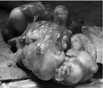
The excised mass was in the dimension of 14x7x5 cm and in the weight of 160 grams. Its
surface was grey-white colored and lobulated (Figure 4). All the hysthopathological study indings were adjusted with Idiopathic True Massive Thymic Hyperplasia, and all of lymph nodes were reactive. During postoperative controls, there were no compli-cations. All these results directed us to another baby of this family whom is a boy and was born from another mother .We looked for such a mediastinal mass. But he was safe. Our pa-tients were safe in their postoperative controls and all of their preoperative complaints were cured.
Discussion
Thymic hyperplasia is a rare cause of anterior mediastinal mass in children [1-3]. Etiology and prognosis of thymic hyperplasia is not well deined [2-5]. It is seperated in two categories such as lymphoid or foliculer hyperplasia that charecterised with ac-tivated germinal centers and lymph follicules. The other is True thymic hyperplasia which has a normal thymic architecture and normal germinal centers and lymph follicules expected for age and occures in two forms as rebound thymic hyperplasia and idiopathic true massive thymic hyperplasia [6,7]. Rebound hy-perplasia occurs ater especially graves disease, Cushing Syn-drome, burns, ater steroid therapy, association with endocrine abnormalities, testiculer tumors, sarcoidosis, lymphoma, and Beckwith Wiedeman Syndrome [8]. Idiopathic true thymic hyper-plasia has a well deined clinicopathological proile prevalence in children or young patients, absance of associated autoim-mune disease and oten presence of cough, respiratory distress, shortness of breath, peripheral blood lymphocytosis. All of these patients have normal immun systems. CT generally showes a mass located in anterior mediastinum. There have been cases reported about rebound massive thymic hyperplasia generally secondary to the non hodgkin lymphoma, hodgkin lymphoma and ater chemotherapy. And also there are a few reported cases of idiopthic true massive thymic hyperplasia especially in children and young patients. [8]. Treatment is surgical removal
of the thymic mass. Linegar and coworkers suggested median sternotomy, clamshell incision or single-sided posterolateral thoracotomy as surgical approach.
When we examine the literature mentioned with multiple cases, we did not detect familial intimacy or brotherhood in any of them. Howewer, that is strange that our cases are two sisters and they have the same complaints, both of them were oper-ated and both of their histhopathological study results were idi-opthic true massive thymic hyperplasia form. We want to point on whether it can be hereditary or not.
Competing interests
The authors declare that they have no competing interests.
References
1. Linegar AG, Odell JA, Fennell WM, et al. Massive thymic hyperplasia. Ann Thorac Surg 1993;55(5):1197-201.
2. Rice HE, Flake AW, Hori T, et al. Massive thymic hyperplasia: characterization of a rare mediastinal mass. J Pediatr Surg 1994;29(12):1561-4.
3. Lee Y, Moallem S, Clauss RH. Massive hyperplastic thymus in a 22-month-old infant. Ann Thorac Surg 1979;27(4):356-8.
4. Lamesch AJ. Massive thymic hyperplasia in infants. Z Kinderchir
1983;38(1):16-8.
5. Lack EE. Thymic hyperplasia with massive enlargement: report of two cases with review of diagnostic criteria. J Thorac Cardiovasc Surg 1981 ;81(5):741-6. 6. Tani T, Okumura F, Nakamura A, et al. Case of thymic hyperplasia showing slow gowth as revealed by chest X ray. Nihon Kyobu Shikkan Gakkai Zasshi
1994;32(2).194-8.
7. Yoshitake T, Itoyama S, Masunaga A, et al. Focal thymic hyperplasia in an adult: report of a case. Surg Today 1994;24(1):72-4.
8. Scharifker D. True Thymic Hyperplasia associated with a unilocular thymic cyst: an unusual combination not previously reported. Ann of Diagnostic pathology
2006;10(1):32-5.
How to cite this article:
Aydoğdu K, Fındık G, İncekara F, Şahin F, Demirağ F, Kaya S. Diagnosis and Man
-agement of True Thymic Hyperplasia; Description with Cases in Two Sisters. J Clin
Anal Med 2013;4(suppl 5): 503-5.
Figure 4. The excised mass is in the dimension of 14x7x5 cm and in the weight of 160 grams.It’s surface is grey -white colored and lobulated.