was made possible by the use of a transgenic line of mice expressing eGFP under the tyrosine hydroxylase promoter. The effects of temperature and different protocols on the Ihkinetics showed that, at 37uC and minimizing the disturbance of the intracellular milieu with perforated patch, this current actually activates at potentials more positive than what is generally indicated, with a half-activation potential of277.05 mV and with a significant level of opening already at rest, thereby substantially contributing to the control of membrane potential, and ultimately playing a relevant function in the regulation of the cell excitability. The implications of the known influence of intracellular cAMP levels on Ihamplitude and kinetics were examined. The direct application of neurotransmitters (DA, 5-HT and noradrenaline) physiologically released onto SNc neurons and known to act on metabotropic receptors coupled to the cAMP pathway modify the Ihamplitude. Here, we show that direct activation of dopaminergic and of 5-HT receptors results in Ih inhibition of SNc DA cells, whereas noradrenaline has the opposite effect. Together, these data suggest that the modulation of Ihby endogenously released neurotransmitters acting on metabotropic receptors –mainly but not exclusively linked to the cAMP pathway- could contribute significantly to the control of SNc neuron excitability.
Citation:Gambardella C, Pignatelli A, Belluzzi O (2012) The h-Current in the Substantia Nigra pars Compacta Neurons: A Re-examination. PLoS ONE 7(12): e52329. doi:10.1371/journal.pone.0052329
Editor:Kevin Currie, Vanderbilt University Medical Center, United States of America
ReceivedAugust 10, 2012;AcceptedNovember 12, 2012;PublishedDecember 21, 2012
Copyright:ß2012 Gambardella et al. This is an open-access article distributed under the terms of the Creative Commons Attribution License, which permits unrestricted use, distribution, and reproduction in any medium, provided the original author and source are credited.
Funding:The research was supported by a grant from the Ministero della Universita` e della Ricerca Scientifica (MIUR) - PRIN 2009. The funders had no role in study design, data collection and analysis, decision to publish, or preparation of the manuscript.
Competing Interests:The authors have declared that no competing interests exist.
* E-mail: [email protected]
Introduction
The presence of the h-current is an hallmark of midbrain dopaminergic (DA) neurons, including those of the substantia nigra pars compacta (SNc), up to the point that its occurrence is considered by many authors the main discriminating criterion to decide if a given neuron in this area is dopaminergic or not [1]. Many studies have confirmed the close relationship between the DAergic phenotype and Ih expression [2–6]. Apart from the
abundant electrophysiological evidence ([7–9], to cite a few), the presence of h-channels in SNc neurons is also supported by qualitative RT-PCR experiments on single cells, which revealed that SNc neurons co-express three of the four types of HCN subunits: HCN2, HCN3, and HCN4 [10].
As expected, the presence of a current typically associated with the pacemaking process (see [11] for a review) suggests that it could play its archetypal role also in SNc neurons, cells characterized by autorhythmicity. However, several studies reported that Ih has neither a significant role in spontaneous
pacemaker activity nor does it contribute substantially to the setting of the resting potential [9,12–15].
Overall, the present knowledge of the h-current in SNc neurons is not entirely satisfactory, and this is all the more surprising for a population of neurons which is object of so many studies. The inconsistencies in the description of Ih are probably due to the
strong dependence of the kinetics of this current on experimental conditions (e.g., temperature, patch configuration, ionic
compo-sition of solutions, modulation by cytoplasmic cyclic nucleotides, protocols used, etc.). This circumstance may explain why, even for a single cell type, different kinetics were found by different laboratories, and consequently different roles were proposed. In addition, there might be a problem in the cell identification: as a rule, cells in the midbrain are identified as dopaminergic on the basis of a series of electrophysiological characteristics, confirminga posteriori the identification in few randomly chosen cells with immunohistochemistry to ascertain the presence of TH. However, some of the more commonly used identification criteria are not really discriminative. For example, the presence of Ih-considered a
benchmark- can be misleading, as if the absence of this current in a midbrain neuron is a trustworthy predictor that the cell is not DAergic, its presence does not reliably predict TH co-labeling [16,17]. A novelty of this study is in the use of a transgenic line of animals that expresses a reporter protein (eGFP) under the TH promoter, allowing the exact identification of each studied neuron as DAergic.
modulate the h-current, thereby affecting the overall excitability profile of these cells.
Results
The results of this study are based on observations made in 357 voltage-clamped SNc neurons, all showing inward rectification at voltages negative to the resting membrane potential.
In order to isolate the h-current, except where otherwise stated, all the other main ionic currents were blocked; in particular, the sodium current with 0.6mM tetrodotoxin (TTX), the delayed rectifier potassium current with 20 mM tetraethylammonium (TEA), the A-type potassium current with 3 mM 4-aminopyridine (4AP), the calcium and calcium-dependent currents with 100mM cadmium and the KIRcurrent with 0.5 mM barium.
Biophysical Properties
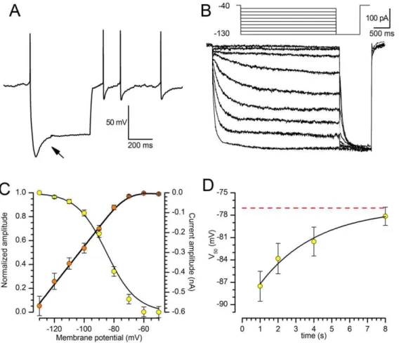
In this first section, all the recordings were carried out in slices at 37uC in whole-cell configuration. The input resistance was 413.64630.17 MV (n = 188; 5–95 percentile range 136– 651 MV), and the access resistance was 12.960.3 MV(n = 199; 5–95 percentile range 7.9–18.0 MV). In normal recording medium containing 2.5 mM-K+the resting membrane potential of SNc neurons was –60.5460.92 mV (n = 91; 5–95 percentile range250.2–71.0 mV), calculated as the potential corresponding to the zero injected current in voltage-clamp conditions. About 95% of the neurons fired spontaneously with a frequency of 4.960.67 Hz (n = 57, frequency measured in cell-attached mode). All of the examined SNc neurons showed a time-dependent sag in response to the injection of outward currents (Figure 1A, arrow), becoming evident at potentials more negative than 260/ 270 mV; the termination of the current pulses was usually followed by a depolarizing overshoot; the depolarizing sag became progressively bigger as the membrane was stepped to more negative potential.
Activation. Under voltage clamp, hyperpolarizing commands from a holding potential of 240 mV evoked slow inward relaxations over the same membrane potential range as the ones producing sags and depolarizing overshoots in hyperpolarizing electrotonic potentials (Figure 1B). The h- current activated slowly and increased the magnitude and rate of activation as the cells were progressively hyperpolarized, with no sign of inactivation. Two current components were measured during the hyperpolar-izing voltage steps: (i) an instantaneous current (Iinst), obtained at
the beginning of the step; (ii) a steady-state current (Iss), obtained at
the end of the step. The instantaneous current was almost linear along the explored voltage, while the steady-state current increased its magnitude as the membrane potential was made more negative; the h-current amplitude, measured as Iss-Iinst(see
methods) is plotted against voltage in Figure 1C (orange symbols). The steady-state activation curve (Figure 1C, yellow symbols) was obtained by interpolating the relative amplitudes of the tail currents with the Boltzmann function (Eqn. 2, explanation in Methods), finding - for 2 s hyperpolarizing step - values for half-activation (V50) of 284.1761.31 mV (n = 13) and for k of
7.7460.40 mV (n = 13).
The point of half activation of the h-current critically depends on the hyperpolarizing pulse length [18]: conditioning pulses of short duration do not allow the gating process to reach the steady-state condition, therefore the probability of opening can be seriously underestimated, leading to evaluations of V50 more
negative of their actual value. These measurement errors are more pronounced for slow HCN channels than for the fast ones, and are highly dependent on temperature [19,20]. Therefore, we have
analyzed the dependence of the midpoint from the duration of the conditioning command. In nine cells studied with the double pulse protocol described above, the first command had durations of 1, 2, 4 and 8 s; we also tried the next point in the log scale, 16 s, but the membrane did not withstand the prolonged hyperpolarizations at the more negative potentials. Increasing the duration of the conditioning pulse induces a significant shift of the steady-state activation curves in the depolarizing direction: the values of V50is
changed from287.5262 mV for 1 s stimuli to278.1561.22 mV for 8 s (Figure 1D); we did not observe any change in the corresponding slopes. The V50values as a function of the duration
of the first step can be described by the exponential function
V50ð Þt~AzV50e
{t=t
ð1Þ
where V50(t) is the value assumed by the midpoint of the
steady-state activation curve for conditioning potentials lastingtseconds; V50 is the range of variation of V50 as a function of the
conditioning periodt, and has a value of 13.9061.58 mV;tis the time constant of the process, 3.2161.42 s. Finally, A is the asymptote (i.e. the value to whichV50tends fortR‘; dashed line
in Figure 1D). The significant value obtained from this analysis is equal to277.05 mV 61.54; this means that, once the channel reaches its steady-state conditions, the point of half-activation is about 10 mV more positive than what is generally believed.
De-activation. The de-activation time constant was mea-sured using the envelope test [21] shown in Figure 2: from a holding potential of 240 mV, two hyperpolarizing pulses to 2130 mV lasting 4 s were imposed, separated by a repolarization to240 mV of variable length (Figure 2A). In Fig. 2B, the Ih
de-activation at240 mV and the envelope of re-activation records at 2130 mV shown in panel A are displayed together, in order to evidence the likeness of their exponential time course. The values of the amplitudes of the tail currents recorded upon re-activation at2130 mV were normalized, plotted as a function of depolar-izing step duration (Figure 2C), and the de-activation time constant was calculated by interpolating the experimental points with the exponential function.
Ið Þt~1zIe
{t= t
ð2Þ
whereIð Þtis the normalized current amplitude at timet,Ithe range
of change of the normalized current,tthe time constant of de-activation at the potentials indicated. In a group of 5 cells,twas 0.4560.07 s (Fig. 2C).
Reversal potential. The h-current is carried by cation channels permeable to Na+ and K+ ions [22–24]; in fact, increasing the extracellular concentration of potassium from 2.5 to 32.5 mM produced a reversible increase in the amplitude of Ih.
The mean amplitude in Ihduring exposure to 32.5 mM K+was
476%640 of control (n = 5, Figure 3A).
As shown in Figure 3A (arrow), we failed to observe any increase in the instantaneous current in high K+
[9]. The explanation for this discrepancy is complicated by the fact that the nature of Iinstis
not well defined yet [25], contrary to the slower component, which is certainly sustained by cations passing through the well-characterized pore of HCN channels. In addition, Iinst usually
has a small amplitude, and is not observed in any measurement of Ih. Speculations on the nature of Iinstrange from models where this
current represents a leak conductance or an experimental artifact, to models in which Iinstis caused by a second pore within the same
HCN or a second channel population associated with HCN channels [26–28]. Midbrain DAergic neurons also have another hyperpolarization-activated current, KIRtype, that could
contrib-ute to the Iinstamplitude, and which is enhanced by an increase of
the external K+
concentration. It cannot be excluded, therefore, that the difference in the results might be consequence of different degrees of blockage of the KIR current, in addition to possible
differences in the animal species used (mouse and rat).
The classical procedure to calculate the reversal potential of a voltage sensitive conductance is from the tail currents reversal, but in SNc neurons this method was rather problematic due to the activation of several outward rectifiers in the membrane potential range over which reversal was expected.
For a more precise calculation of the Ih reversal potential (Eh)
we used the method of the instantaneous (chord) and ‘steady-state’ current-voltage relationships [29]. At the membrane potential of 295 mV gh is strongly activated (Figure 3C) and does not show
time-dependent inactivation; the reversal potential of Ih is then
obtained from the intersection of the instantaneous (chord) current-voltage relationships recorded at holding potentials of 260 mV and 295 mV (Figure 3D), i.e. in the absence and presence of Ih.
In 8 neurons the mean value obtained for the reversal potential at 37uC was244.0363.10 mV (range229.4 to256.9 mV).
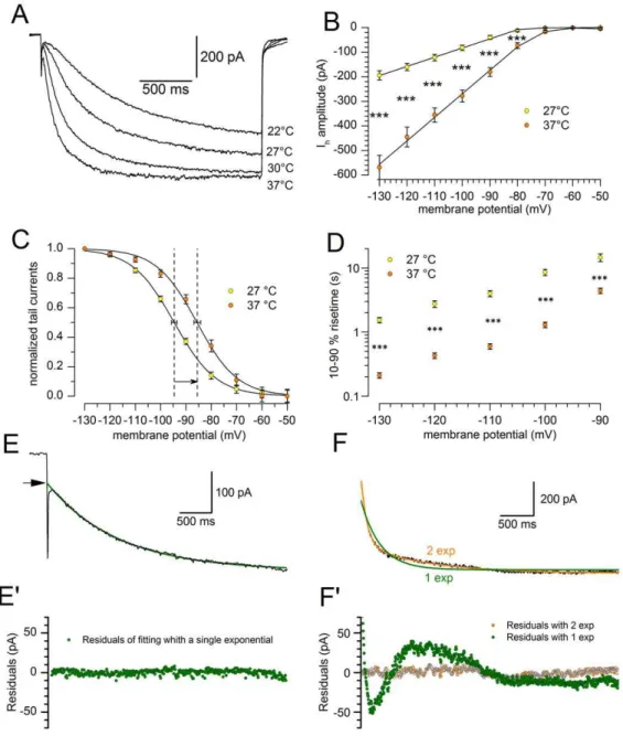
Effect of temperature. It has been shown in various types of preparation that the kinetics of Ih is particularly sensitive to
thermic conditions [20,30,31]. The temperature at which electro-physiological recordings are made, affecting both the amplitude and the kinetics of Ih(Figure 4A), is one of the limiting factors in
comparing the results; therefore, in this study most of the reported recordings were realized in precisely controlled temperature conditions.
Figure 4B shows the effect of a 10uC temperature increment on the Ihamplitude at different potentials. I/V graphs show the mean
current amplitudes recorded at 27uC (
N
) and 37uC (#) as afunction of membrane potential. At2130 mV, a 10uC increase causes a rise in amplitude from2194.2618.5 pA at 27uC (n = 19) to2569.1648.9 pA at 37uC (n = 23). The average value of Q10
for Ihamplitude between280 and2130 mV is 3.7660.53. The
resulting maximal conductance gh at 27 and 37uC is 2.26 and
6.62 nS, respectively.
We next explored whether the increase of Ihat2130 mV could
be explained by a shift in the voltage dependency. As seen from the graph (Figure 4C), the transition from 27uC (
N
) to 37uC (#) causes a shift of the steady-state activation curve by about+10 mV: the V50, calculated fitting the Boltzmann equation to the
experimental points (4 s conditioning pulses), is294.9161.72 mV at 27uC (n = 13) and 284.2361.28 mV at 37uC (n = 18), Figure 1. Basic properties of the h-current at 376C.A: Response of a SNc DA neuron under current-clamp condition to the injection of a 300 pA hyperpolarizing current pulse. Note the appearance of the archetypal sag (arrow) due to the activation of Ih; resting potential255 mV, bath solution for this recording was standard ACSF. B: Family of responses of a SN DA neuron under voltage-clamp conditions to the application of the double-pulse protocol shown in the inset above; explanation in the text. C: Current-voltage relationship of the h-current (%, right y-axis) and voltage-dependence of the activation curve (#, left y-axis) obtained from tail analysis of double-pulse experiments as shown in panel B; mean value6S.E. (n = 20). D: Dependence of the midpoint (V50) on the duration of the hyperpolarizing pulse; the dashed line highlights the asymptotical behavior of the midpoint for a conditioning pulse of infinite duration - see text for explanation.
(P,0.0001, two-tailed Student t test for unpaired data). No significant changes were observed in the steepness of the relationship: the slope is 8.0260.37 mV at 27uC and 7.7460.37 mV at 37uC.
The temperature does not affect only the total conductance of the h-current (Figure 4A) but -and to a much higher degree- also its activation kinetics, which is modified under two aspects. First, the tracings at 27uC can be satisfactorily fitted by a single exponential (Figure 4E, E9), but two exponentials are always needed for an adequate fit at 37uC (Figure 4F, F9). Second, the current development rate is strongly affected by the temperature. Since at 27uC there is only an exponential, and at 37uC two, a comparison of the time courses was possible only comparing the 10–90% rise time. Since not always the steady state was reached, due to the instability of the membrane at the more negative potentials, we used the following equations, obtained by solving equation 3 (see methods) for y = 10 and y = 100 after normaliza-tion of the amplitude to 100:
– for a single exponentialt90=tln(100/10) andt10=tln(100/90),
where t10 and t90 are the times at which the current is
developed for the corresponding percentage, andtis the time constant;
– for a double exponential the solution was less straightforward: first the amplitudes of the two exponentials (A1and A2) were
normalized so that their sum was 100; then, Eqn. 1 was solved
numerically fort[32,33] settingf(t)= 90 and = 10 (the Matlab code used can be found in the Supplementary material as Code S1), obtainingt10andt90, respectively.
The comparison of the t10–t90 times at 27 and 37uC is
represented graphically in Fig. 4D, and the corresponding Q10,
in the range 290 2130 mV, is 6.4, as calculated with Eqn. 3 settingrateas (t10–t90).
The de-activation time constant is also affected by temperature: the double-pulse protocol described above was applied in five cells at 27uC. The normalized amplitudes of the currents to the second pulse as a function of the delay between the two pulses can be described by the exponential function indicated in equation 2, with a time constant of 1.4660.1 s, which would give a Q10 of 3.2
when compared with the value at 37uC indicated in a section above.
Basic Pharmacology
The h-current is sensitive to low concentrations of Cs+ (1– 2 mM) [34] and to a certain number of organic compounds blocking selectively the h-channels, like ZD7288 [35] and S-16257 (ivabradine) [36,37]. Cs+1 mM effectively blocks the h-current (Figure 5A). However, as already observed in calf Purkinje cells [38], the action of Cs+is clearly voltage-dependent: in the negative region of the I-V curve Cs+induces a channel blockade, whereas at more positive potentials it is ineffective, and sometimes it can Figure 2. De-activation kinetics.A: Envelope test during deactivation at240 mV. After current activation at2130 mV, pulses to240 mV of variable duration were followed by re-activating steps to2130 mV (see protocol in the top panel). B: The tail at240 mV was also re-plotted after appropriate scaling (grey trace) to better compare its time course with that of the re-activation records envelope shown in panel A. C: Analysis of the deactivation time constant for a group of five cells; the average time dependence (dashed line) was fitted by the equation I(t) =1–0.69*exp(-t/0.45) (dashed line; continuous line delimitate the 95% confidence interval).
doi:10.1371/journal.pone.0052329.g002
even produce the opposite effect, i.e. a current increase. More selective, and completely voltage-independent blockages, can be obtained with ivabradine 10mM (Figure 5B) and ZD7288 (30mM, Figure 5C).
Role of Ihin Autorhythmicity
One of the hallmarks of SN neurons is the autorhytmicity: these neurons fire spontaneously action potentials characterized by an unusually long duration (.2.5 ms), a rather depolarized threshold (. 240 mV) and a marked afterhyperpolarization [1,39]. The role of the h-current in spontaneous activity has been thoroughly analyzed by several authors, and the conclusion has been that it is neither a significant factor underlying the spontaneous pacemaker activity nor does it contribute substantially to the setting of the normal resting potential level of the membrane [9,12–15].
Our data only partially confirm this viewpoint: recording at 37uC and in perforated patches, the block of the h-current by focal application of ivabradine 10mM does stop the spontaneous activity; the effect is rapid and reversible, and is paralleled by an important hyperpolarization (11.8362.07 mV, n = 7; Figure 6A). The blockage of spontaneous activity following a membrane hyperpolarization of 10 mV is not surprising, as the cell firing is based on a delicate interplay of conductances [8,14,40–42] that can be easily disrupted by the injection of outward currents ([43], and personal observation). We then tested whether this blockade represented the evidence for an essential role played by the h-current in the pacemaking mechanism, or if it was only the consequence of the hyperpolarization following the suppression of the h-current. In the presence of ivabradine, the injection of a depolarizing current sufficient to restore the membrane potential to the value antecedent the Ih block (arrowhead in Figure 6),
resumes immediately the spontaneous activity (Figure 6A, right).
This proves the absence of any direct role of the h-current in autorhythmicity, but also demonstrates that this current has a relevant role in determining the membrane potential.
Since the influence of the h-current on membrane potential is somewhat controversial, and since ivabradine is a relatively new drug, for which side effects have been described on currents other than Ih[44,45], we repeated the same experiment using the more
classical blocker ZD7288 (30mM). The results were substantially similar to those obtained with ivabradine (Fig. 6B), although the hyperpolarizing effect was less pronounced (6.8760.78 mV, n = 15) and in 7 out of 15 cases we were unable to obtain a substantial recovery after 209 washout. Interestingly, the hyper-polarizing effect of the h-current blockage was correlated with the resting membrane potential (Fig. 6C; p value,0.02, ANOVA), as expected for a conductance whose effect is increasingly influential at more negative potentials.
Modulation of h-current
Ih modulation by intracellular cAMP. The h current is
dually regulated by hyperpolarization and by cyclic AMP, directly binding to a sequence (cyclic nucleotide binding domain, CNBD) located in the C-terminal segment [46,47]. We have therefore analyzed the modulatory effect of cAMP on the h-current using a recording configuration (perforated patch with amphotericin B) minimizing the perturbations of the intracellular medium.
The first experiments were conducted in current clamp conditions to determine the effects of increased intracellular cAMP on the resting membrane potential. The addition to the extracellular solution of 10mM forskolin, a classical activator of adenylyl cyclase [48], caused a depolarization of 3.561.3 mV (n = 5) after 4 min of application. Then the effect declined, vanishing completely in the next 5 min with a return to the resting Figure 3. Analysis of Ih reversal potential.A: Response to hyperpolarizing steps from240 to2130 mV using the indicated concentration of K+
ions in the external saline; T = 27uC. B,C: Recordings of the slow current relaxations recorded at holding potentials of260 and295 mV, respectively, in response to the indicated protocols represented in the insets of Figure D; T = 37uC. D: Instantaneous (chord) and ‘steady-state’ current-voltage relationships of a SNc DA neuron voltage-clamped at holding potentials of260 mV (chord conductance 2.60 nS) and290 mV (chord conductance 8.89 nS). Note that the chord conductance plots are approximately linear at both the holding potentials, despite the presence of strong inward rectification in the ‘steady-state’ current-voltage relationship measured at the end of 2 s hyperpolarizing voltage jumps from a holding potential of 260 mV. Inward rectification in the ‘steady-state’ current-voltage relationship is entirely accounted by these slow relaxations.
membrane potential, even maintaining the forskolin supply (Figure 7A, yellow dots). However, when the bathing solution was further enriched with 0.1 mM IBMX, a phosphodiesterase inhibitor [49], the depolarization induced by forskolin was more prominent (7.961.8 mV, n = 5), and persisted as long as the application of the drug was maintained (Figure 7A, orange dots). Under voltage-clamp conditions, the bath application of 10mM forskolin and 0.1 mM IBMX, induce a significant increase of Ih
amplitude (Figure 7B): at 2130 mV the current amplitude is 2178.5623.5 pA in control conditions (n = 8), and 2227.0634.2 pA (n = 8) in the presence of increased levels of
cAMP; at any tested potentials the increase in current amplitude was statistically significant (P,0.005 level, t-test for paired data).
The effect of forskolin on the h-current is twofold. First, it promotes a depolarizing shifts of the steady-state activation curve V50 in the depolarizing direction of 6.3360.78 mV (n = 8; a
variation significant at 0.0005 level; Figure 7C). In addition, following the increase of intracellular levels of cAMP, the Ih
activation time course becomes significantly faster: from 17636340 ms in control to 14356298 ms for the 10–90% rise time (n = 8; p,0.025) at2100 mV (Figure 7D).
Figure 4. Effect of temperature on h-current amplitude and kinetics.A: Family of current tracings recorded in a single cell in response to hyperpolarizing pulses from240 to2130 mV, repeated at the temperatures indicated. B: Comparison of the I/V curves recorded at 27 (yellow dots) and 37uC (orange dots); n = 20; the difference, tested with two-way ANOVA and post-hoc Bonferroni test, is significant at 0.001 level for the potentials more negative than270 mV. C: Shift of the steady-state activation curves for a change from 27uC (yellow) to 37uC (orange), average values from 13 cells6S.E for the V50. D: Effect of the temperature on the h-current 10–90% risetime at 27uC (yellow) and 37uC (orange); see explanation in the text. E, E9: Sample fit with a single exponential of an h-current tracing obtained at 27uC in response to a voltage step to2130 mV; below the analysis of the residuals (experimental data minus the corresponding values of the fitting curve). F, F9: Sample fit with both single (green) and double (orange) exponential of a h-current tracing obtained at 37uC in response to a voltage step to2130 mV; below the analysis of the residuals in the same grey scale code. All the recordings shown in this figure were obtained in perforated patch conditions.
doi:10.1371/journal.pone.0052329.g004
Ih modulation by endogenously released neurotransmitters. We next sought for possible modulation of the h-current in SNc neurons by endogenously released neurotransmitters acting on metabotropic receptors. Although modulation of Ih by endogenously released neurotransmitters
other than dopamine has not been demonstrated yet, these effects could contribute significantly to the regulation of neuronal excitability, given the role of the h-current in controlling the resting membrane potential and ultimately the excitability profile of these cells. The effect of the principal neurotransmitters on the Ihwas analyzed in perforated patch recordings at 37uC using high
potassium concentration in the external saline in order to increase the amplitude of the current.
Of the dopaminergic receptors cloned so far, the D2is the most
abundantly expressed in the substantia nigra pars compacta neurons [50–52], where it is densely packed in the pericarya [53]. Since the activation of D2-like family receptors is coupled to a Gai
protein, which reduces the intracellular levels of cAMP by inhibiting the activity of the adenylate cyclase [54], we investigated whether the activation of D2receptors had a modulatory effect on
the h-current.
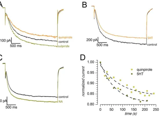
In the presence of the D2-like agonist quinpirole (30mM), the
current amplitude was reduced (Figure 8A): at 2130 mV, the measured current was 2653.36221.5 pA in control conditions (n = 6) and 2557.06189.2 pA in the presence of the agonist (n = 6), a difference significant at the 0.01 level (one-tailedt-test for paired data). On average, the D2-like agonist applied focally
induces a 15% reduction of the h-current amplitude in about three minutes, with a time constant of about 93 s (Figure 8D).
The application of the D2-like antagonist sulpiride (20mM)
suppressed the effect of quinpirole, and caused a 16.562.34% increase in Ih amplitude above the control point (Figure 8A),
increase significant at the 0.005 level (two tailed t-test for paired data n = 4); in other words, sulpiride not only quenched the effect of the antagonist, but resulted in an increase of the h current in the absence of exogenous D2 agonists. Since sulpiride is not an
activator of adenylate cyclase, the effect can be explained by the presence, reported in this brain area, of a spontaneous release of dopamine at rest [55], whose action on autoreceptors would result in a tonic inhibition of the adenylate cyclase.
Serotonin (5-HT) is a critical neurotransmitter in the generation and regulation of emotional behavior and plays a prominent role in the inhibition of impulses. Substantia nigra receives serotonin-ergic input via fibers from the dorsal raphe nucleus [56–58], and 5-HT containing terminals make direct contacts over DA-containing neurons [59].
In midbrain DA neurons, serotonin has been reported to inhibit Ihin the VTA dopaminergic neurons in concentration-dependent
manner [60], and to enhance the current in the substantia nigra pars compacta [61]. Therefore, we examined whether the 5-HT (100mM) had any effect on the h-current performing the experiments in perforated patch-clamp configuration at 37uC. Using a single-pulse protocol from 240 mV to 2130 mV, following application of serotonin and in the presence of Ba2+to avoid collateral effects on the KIR current (see discussion), we
observed a reduction of the h-current amplitude in 5 out of 8 cells (Figure 8B), whereas in the remaining cells we detected no significant changes (3 cells). In the first group the reduction in Ih
amplitude was about 20%, and was significant at the 0.05 level (t -test, single queued). The progression of the effect in the time domain is shown in Figure 8D - the time constant measured was of 67 s.
A diffuse network of noradrenaline (NA)-containing nerve endings in the neuropil of SNc has been demonstrated. The noradrenergic input is from the locus cœruleus [62–64] and -to a lesser extent- from other NA-containing neurons in the brainstem [65,66]. NA modulates the h-current in several type of neurons, among which thalamic relay neurons [67], neurons of the medial nucleus of the trapezoid body (MNTB) [68], dorsal root ganglia neurons [69] and VTA neurons [70,71]. In the thalamus, NA, acting onb-adrenergic receptors, increases the intracellular levels of cAMP and shifts the Ih steady-state activation curve to more
depolarized potentials, as it does in MNTB neurons. On the contrary, activation ofa2adrenergic receptors in the DRG and
VTA neurons causes a significant reduction in amplitude of Ih.
We therefore tested whether the NA (100mM) could determine any change in the h-current of DA neurons of SNc. We performed experiments in perforated patch configuration at 37uC, applying a single hyperpolarizing pulse from 240 mV to2130 mV. Upon application of NA, in 8 out of 10 cells we observed an increase in the amplitude of the h-current by about 11.861% (Figure 8C), whereas no change was detected in the remainder of SNc cells. The increase in amplitude was statistically significant, with a P-value,0.025 using single-tailed t-test for paired data.
Discussion
Two classes of hyperpolarization-activated inwardly rectifying currents have been reported in SNc neurons. One type has fast kinetics, is permeable primarily to K+
, is blocked by extracellular Ba2+ and Cs+, and has a voltage-dependence that is itself dependent on extracellular K+
([K+
]o) concentration. Ih (or Ifin
cardiac tissue), the second type of inward rectifier, is a mixed Figure 5. Basic pharmacology of h-current.A: Blockage by Cs+(1 mM): current-clamp responses to a repeated hyperpolarizing current step of 300 pA from a holding potential of260 mV. Note the progressive suppression of the sag after the indicated times of Cs+application; T 37uC. B: Blockage by ivabradine (10mM): voltage clamp responses to a repeated hyperpolarizing step to2130 mV from a holding potential of240 mV at the
indicated times of drug application; T 37uC. C: Blockage by ZD7288 (30mM); holding potential240 mV, test potentials ranging from270 to
2130 mV; T 27uC, Ba2+0.5 mM present throughout.
cation current, with a reversal potential substantially positive to EK
[72]. Ih has a relatively slow activation kinetics, is insensitive to
Ba2+
, and does not show a voltage sensitivity dependent on [K+ ]o
[25]. The sensitivity to drugs very selective for Ihlike ivabradine or
ZD7288, the Ba2+
insensitivity, the slow kinetics of activation and the reversal potential suggest that the current described here belongs to the latter class.
From the methodological point of view, a first novelty of this study is in the use of a transgenic line of animals that expresses a reporter protein (eGFP) under the TH promoter, allowing the precise recognition of DA neurons inin vitroslices. This is of some importance as, in these preparations, neurons are often identified as DAergic according to the expression of Ih, a current which is
not -or much less- expressed in putative non-DA neurons
[1,9,73,74]. Notably, while the absence of Ih in a DA neuron is
a reliable predictor that the cell is not DA-containing, the presence of Ih does not reliably predict TH co-labeling [16,17]. In other
words, since a significant number of neurons expressing the h-current is not DAergic, this means that one of the principal markers assumed to identify midbrain DAergic neurons is not associated exclusively, or even significantly, with confirmed DAergic neurons. The identification of the neurons by the expression of eGFP under the TH promoter, marks a first important difference of this study with respect to other functional studies, where the expression of TH (when done) was carried out only in few sample cells.
A second methodological hallmark of this work is the choice of working systematically at 37uC and in perforated patch configu-Figure 6. Role of Ih in autorhythmicity.A: Current-clamp recording (perforated patch, 37uC) showing the hyperpolarizing effect of a selective, non voltage-dependent blocker of the h-current (ivabradine 10mM, applied focally at the moment indicated by the arrow) on spontaneous activity.
The two insets below illustrate the method used for the determination of the ‘‘resting’’ membrane potential in spontaneously active cells: the value was actually measured as the prevailing potential, measured by fitting a Gaussian to the membrane potential values in the time intervals marked by the bars of the same color. At the time marked by the large arrow to the right, the membrane was manually depolarized to the value antecedent the ivabradine application, restoring the spontaneous activity and thereby showing that the h-current is not essential for the pacemaking mechanism. B: Same experiment using ZD7288 30mM. C: Relationship of the hyperpolarizing effects obtained with ZD7288 with the resting membrane potential,
showing a significant correlation (p,0.02; ANOVA). doi:10.1371/journal.pone.0052329.g006
ration, especially when the modulation of the h-current was studied. These working conditions are more respectful of the intracellular milieu and, in our view, lead to significant differences in the observed results.
In this preparation, Ivabradine was used for the first time as a blocker of Ih. This drug, originally developed ad a bradicardic
agent [44], proved to be an excellent blocker of all types of HCN subunits [37,75]. The reason of the preference accorded to this drug resides in the rapidity and complete reversibility of its action, whereas the more classical Ihblocker ZD7288 is slower and often
irreversible [76,77].
Biophysical Properties
Steady-state activation. At 37uC, and a 4 s hyperpolarizing pulses, we found a midpoint of activation at282.7361.17 mV with a slope of 7.2960.28 mV (n = 12); however, as first shown by ref. [18], the point of half-activation depends critically on the used voltage protocol. In particular, short voltage steps do not allow channel activation to come to completion and then the steady-state activation curve derived from tail current amplitudes is seriously underestimated. Using different pulse duration, and extrapolating to the asymptote the trend of the midpoint shift in the depolarizing direction as a function of the hyperpolarizing
pulse duration (Figure 1D), we calculated a V50 of277.05 mV,
i.e. from 10 to 28 mV more depolarized than the values reported in SNc neurons at 37uC [9,78].
The functional implication of these values becomes obvious if one considers that assuming a slope of 7.43 (the slope is 7.41, 7.36, 7.29 and 7.65 at 1, 2, 4 and 8 s, respectively), at265 mV about 16.5% of the h-channels are open, which corresponds to a conductance of 1.023 nS. Since we found an average input resistance of these cells of 414 MV, at265 mV the h-current gives a contribution of+8.9 mV to the resting membrane potential, a value matching very well the hyperpolarizations found upon blocking the h-current with ivabradine in the present experiments (211.8362.07 mV). Such an hyperpolarization would be impos-sible with the kinetic coordinates reported in most of the previous papers and the value reported in literature. Since the hyperpo-larizing effect of the Ihblockage is controversial, we repeated the
experiments using the more classical blocker ZD7288 (30mM), obtaining results substantially similar to those obtained with ivabradine, although the hyperpolarizing effect was less pro-nounced (26.8760.78 mV, n = 15).
There could be several explanations to justify the discrepancy of our data with others reported in literature, which do not show any effect of the h-current on the resting membrane potential [9,12– 15,79].
Figure 7. Effect of an increase of intracellular cAMP stimulated by forskolin on h-current.A: Effect of forskolin (10mM) alone (yellow) and
in association with IBMX (0.1 mM, orange) on resting membrane potential; explanation in the text. B: I/V relationship of the h-current in control conditions (green) and in the presence of forskolin+IBMX (blue); two-way ANOVA and Bonferroni post-hoc test: * indicates P,0.01, **P,0.001. C: Effect of forskolin+IBMX on steady-state activation curve. The shift of the midpoint in the depolarizing direction (6.3360.68 mV, n = 8) is significant at the 0.0005 level. D: effect of forskolin+IBMX on the 10–90% activation rise time of the h-current; two-way repeated measures ANOVA and Bonferroni
First, it cannot be excluded that ivabradine could also act on conductances different from the h-current. In fact, a inhibitory effect of high concentrations of ivabradine on L- and T-type calcium currents [44,45] has been reported; however, at the concentrations used in the present experiments (10mM), to the best of our knowledge, no side effects have ever been described, although these cannot be certainly excluded, considering the still scarce knowledge of this drug in nerve cells. However, we see a hyperpolarizing effect also of ZD7288; furthermore, notice that our result (a hyperpolarization of 6.9 mV) is almost coincident with that reported in a recent paper (7 mV; [80]).
Second, as shown by previous studies, Ih amplitude correlates
with calbindin (CB) expression in nigral dopamine cells, with larger currents in CB-negative cells [13,81], and dissimilar proportions of CB-positive and CB-negative cells could have been analyzed in different studies - in this regard, direct analysis by molecular methods would help to characterize calbindin expres-sion when recording from SN neurons.
Temperature dependence. For what concerns current amplitude, the Q10values reported by various authors lie in the
range between 1.35 [20,82] and 6 [83], with an average around 3. We found a value of 3.7660.53, which is well aligned with most of the reports. This increase is generally ascribed to depolarizing shifts in the steady-state activation curve for the h-current which, for a 10uC temperature increase, range from 2–3 mV [82] to 12– 13 mV [31,84] without change in the slope. In SNc neurons, rising the temperature from 27 to 37uC we observed a depolarizing shift in the midpoint of the steady-state activation curve (V50) of 10 mV
in the depolarizing direction, without any change in the slope. The main difference in temperature sensitivity with respect to the current literature was observed in the activation rise time, for which we found a Q10of 6.4, while values found by other authors
are around 3 [30,34,82] - a diversity that can be explained by differences in the preparation and consequently in the subunit composition of the channels.
The high temperature-dependence of the Ih in SNc neurons
underscores the importance of controlling this often neglected parameter in the electrophysiological recordings of this current. This dependence can be at the basis of many of the discrepancies found in literature.
The high sensitivity of the h-current in substantia nigra neurons might at least in part explain the previously observed warming-induced increase in firing frequency, decrease in input resistance, and an inward current reversing its polarity between 25 and 217 mV, which is dependent on extracellular Na+
[85]. In this context, it might also be useful to recall that changes of up to several degrees centigrade in the brain temperature are observed not only during fever, but also during different behavioral states [86]. Furthermore, neurons in different brain regions, including the substantia nigra, were reported to show high temperature sensitivity [87], a behavior whose underlying mechanism is unknown. Our data indicate that the h-current might have a role in the process.
Pharmacological Properties
The fundamental involvement of Ih in the control of resting
membrane potential makes the h-channel a privileged target for neurotransmitter systems aiming at the regulation of SNc neurons activity. In general, the excitability profile of a cell expressing an h-current can be controlled either by moving the membrane potential in and out of the range of Ih activation, or by moving
the Ih activation within the range of membrane potentials of
physiological interest by directly modulating the h-channel itself. Figure 8. Sample tracings showing the effect of different neurotransmitters on the h-current; responses to hyperpolarizations to 2130 mV from a holding potential of240 mV; perforated patches, 376C.The external saline included TTX, 4AP, TEA and Ba2+. A: Effects of the D2 agonist quinpirole (30mM) and of the D2 antagonist sulpiride (20mM); note that in the presence of the antagonist the response is greater than
controls. B: Effect of 5-HT (100mM). C: Effect of NA (100mM). D: Time constant of the development of the effects of quinpirole and 5-HT.
doi:10.1371/journal.pone.0052329.g008
other, as those mediated by cyclic nucleotide pathways, have been less extensively investigated. In this work we have attempted a preliminary exploration of the possible modulation of the h-current by some neurotransmitter known to act on SNc neurons. One known possible source of error is that the modulation (inhibition) of the h-current occurs secondary to GIRK channel activation [9,78,92], since between GIRK and Ihchannels there is
a close link [93,94]. We can exclude this possibility since, in experiments where the modulation of the h-current was studied, we were always in the presence of Ba2+
at concentrations that can entirely block the inward rectifier K+-currents, GIRK included.
Ih modulation by cAMP. The h-channels are directly
activated by cyclic nucleotides [46], but when tested in SNc neurons, the adenylate cyclase activator forskolin gave contradic-tory results on the amplitude of the h-current, going from a complete absence of effect [78] to a 25% increase, due to a
+5.33 mV shift of the steady-state activation curve towards more positive potentials [95]. Our results are almost exactly superim-posable with those of the latter authors, with a 27.5% increase in current amplitude and a+6.33 mV shift of V50. It should be noted
that initially we were unable to see any influence because we tested the forskolin effect 10 minutes after application, and without IBMX. In these conditions, the modification of the resting membrane potential is transient and vanishes after 7–8 min (Figure 7A), but in the presence of the phosphodiesterase inhibitor the effect is larger and stable in time, and this might explain some discrepancy in the literature about this point.
D2 receptors. Since its discovery by Aghajanian in 1977 [96,97], the presence of D2 receptors in midbrain DA neuron, is considered one of the hallmarks of these cells, and as such it has received much attention. DA D2-like receptors are present as auto-receptors on the DA neurons in SN and VTA and play an important role in the regulation of DA neuronal firing activity by means of auto-inhibition (for a review see [98]. These G protein-coupled receptors are activated by dendritically released DA [99,100] through a still controversial mechanism [101], and the hyperpolarization following their activation is classically described as mediated through an activation of potassium channels, GIRK type [43,78,102], and A-type [103,104]. In the present work, we show that, in addition, also the h-current is modulated by the activation of D2-like type DA receptors. This is not surprising, as D2 receptors are coupled to a G-protein Gai which directly
inhibits the formation of cAMP by inhibiting the enzyme adenylate cyclase [54]. Our observation confirms a relatively recent study, conducted in brain slices at 34uC showing that DA, released endogenously following a single action potential, hyper-polarizes neighboring DA neurons by inhibiting h-channels [105]. Thus, it is possible that DA autoreceptors are linked to different effector systems. Receptors that are coupled to h-channels may be preferentially located in the vicinity of DA release sites, whereas those coupled to G-protein-gated K+ channels may be extra-synaptic.
Serotoninergic receptors. The bulk of available neuroana-tomical data clearly indicate that the midbrain DA- neurons
the effect is direct or indirect.
Noradrenergic receptors. Controversial data were reported concerning the effects of NA in SNc. Several studies indicate that electrical stimulation of the locus cœruleus evokes an initial excitatory response in SNc neurons, frequently followed by a period of inhibition of firing [64,113]. In vivo studies report an hyperpolarization in rat SNc neurons by NA [43], which slows the frequency of spontaneous action potentials [114]. In SNc neurons other authors have found that NA apparently induced an inhibition of Ih, but then they report that in the presence of
300mM external barium or internal cesium, NA did not affect Ih,
suggesting that the effect on Ihis secondary to the activation of KIR
channels [92]. Contrary to what was found by these authors, we can exclude an indirect effect mediated by KIR channels as we
were systematically recording in barium 500mM. We did not look for better specification of the pathway involved in the NA stimulation of the h-current, which Cathala and Pupardin-Tritsch suggest being PKC, nor of the receptor involved, for which both a1 anda2 adrenergic types seem to be excluded [92].
Nevertheless, the observation of Cathala and Pupardin-Tritsch that NA inhibits the KIRcurrent, in addition to enhance the Ih, is
rather interesting, as there is an increasing evidence that also in other systems amine-activated pathways can modulate both Ihand
KIRacting in opposite direction. A synchronous and symmetrical
control of the balance between Ihand IKIRwas also described for
example in rat spinal motoneurons, where 5-HT increases the cell excitability inhibiting an IKIR and enhancing an Ih [115]; in
salamander motoneurons, where muscarinic modulation inhibits the Ih and enhances the IKIR[116]; in DAergic neurons of the
olfactory bulb, where cAMP increases the Ihand inhibits the IKIR
(personal unpublished observation).
In SNc neurons, where Ihis enhanced and IKIRis inhibited by
NA, this synchronous and symmetrical modulation might underlay a mechanism that could shorten hyperpolarizing events. Since the activation and deactivation kinetics of IKIRare much faster than
that of Ih, the net result would be a sharper response of the neuron
in response to hyperpolarizing events. The enhancement of Ihby
NA could then accelerate the recovery from hyperpolarizations, ultimately shortening their duration and increasing firing frequen-cy, a combined action that might be shared, not necessarily with the same sign, also in other systems.
Functional Implications and Conclusions
excitabil-ity through D2 autoreceptors. We show that the Ih, with its role in
the control of membrane potential, seems to be an important target of the afferent inputs to SNc neurons. We propose that this current may be one of the main actors responsible for the rich signaling repertoire displayed by these cells which, through their effects on forebrain dopamine levels, influences much of the functioning of the basal ganglia as a whole.
Materials and Methods
Animals and Surgical Procedures
Experimental procedures were carried out to minimize animal suffering and the number of mice used. The procedures employed were in accordance with the Directive 86/609/EEC on the protection of animals used for experimental and other scientific purposes, and were approved by the Campus Veterinarian of the Ferrara University. A total of 192 mice have been used, most of them in the 14–20 postnatal day range, and 20 over 3 months old. All experiments were performed using the transgenic mice TH-GFP/21–31 line carrying the eGFP gene under the control of the TH promoter [118,119]. Transgenic mice were identified either by PCR on the genomic DNA extracted from tail biopsies, or -at postnatal day 3 or 4- looking at the fluorescence of the olfactory bulbs transilluminated with a UV source (FBL/Basic-B & N-01; BLS, Hungary; FHS/F-01) and observed with an emission filter (FHS/EF-2G2; BLS, Budapest, Hungary). Transgenic lines were maintained as heterozygous by breeding with C57BL/6J inbred mice.
Preparation of Midbrain Slices
Mice were anaesthetized (intraperitoneal injection of 60 mg/kg of sodium pentobarbital) and decapitated. The brain was removed from the skull in less than 1.5 min and put into ice-cold (2–4uC) dissection solution of the following composition (in mM): 3.0 KCl, 1.25 NaH2PO4, 2 MgCl2, 1.6 CaCl2, 10.0 glucose,
21.0 NaHCO3, 215 sucrose; saturated with 95% O2and 5% CO2.
A brain section containing the substantia nigra pars compacta was obtained as follows. Two coronal cuts were performed, the caudal to remove the cerebellum and the rostral half of the cerebral hemispheres. The resulting block was glued on rostral surface with n-butyl cyanoacrylate adhesive (3 MTMVetbondTM, Segrate, Italy) to the support of vibratome (Campden HA 752, Loughborough, England) and then submerged by ice-cold dissection solution. The coronal slices (thickness, 150mm) were cut starting from the caudal surface. Slices containing the substantia nigra pars compacta were identified by illuminating the specimen with a desk UV light source (BLS), stored in an incubation chamber containing artificial cerebrospinal fluid (ACSF) continuously bubbled with carboxygen (95% O2, 5%
CO2), and then kept at room temperature for about 10 hours.
Slices were placed in the recording chamber and superfused with ACSF at a rate of 2 ml/min.
Current and Voltage Recordings
The temperature of the 1-ml recording chamber was controlled using a couple of 39.7 W Peltier devices (RS Components, Milan, Italy) and measured with a high-precision, low mass thermocouple (RS Components, Milan, Italy).
Current and voltage recordings were acquired with an Axopatch 200B amplifier (Molecular Devices, Sunnyvale, CA), and a 12 bit A/D–D/A converter (Digidata 1440A; Molecular Devices); the holding potentials were corrected for the junction potential, calculated using the related function of the acquisition software (pClamp 10, Molecular Devices). Borosilicate glass
pipettes (1.5 o.d., 0.87 i.d., with filament; Hilgenberg, Malsfeld, Germany) were pulled with a Zeitz-DMZ puller (Martinsried, Germany) and had a resistance of 4–5 MV when filled with standard intracellular (IC) solution; the seal formation was realized with the help of an air pressure controller (MPI, Lorenz Messgera¨tebau, Katlenburg-Lindau, Germany); the seal resistance was always greater than 3 GV. A 70–80% compensation of the series resistance and correction for junction potential was routinely used.
Solutions
The solutions used had the following composition (mM):
standard ACSF extracellular (EC) saline: 125 NaCl, 2.5 KCl, 26 NaHCO3, 1.25 NaH2PO4, 2 CaCl2, 1 MgCl2, and 15 glucose;
high K EC solution: 115 NaCl, 12 KCl, 26 NaHCO3, 1.25 NaH2PO4, 2 CaCl2, 1 MgCl2, and 15 glucose.
high sucrose EC dissection solution: 3.0 KCl, 1.25 NaH2PO4,
2 MgCl2, 1.6 CaCl2, 10.0 glucose, 21.0 NaHCO3, 215 sucrose.
All EC solutions were continuously bubbled with 95% O2and
5% CO2; the osmolarity was adjusted at 305 mOsm with glucose.
standard pipette-filling intracellular (IC) solution: 120 KCl, 10 NaCl, 2 MgCl2, 0.5 CaCl2, 5 EGTA, 10 HEPES, 2 Na-ATP, 10
glucose. The free calcium concentration with this internal solution was calculated to be 16 nM (http://www.stanford.edu/,cpatton/ downloads.htm).
For perforated patches, amphotericin B was included in the recording electrode filling solution as perforating agent (200mg/ ml plus 300mg pluronic F-127). In order to make sure of the integrity of the perforated patch, EGTA was omitted from this solution and the concentration of CaCl2was raised to 3 mM. Data
were collected after the series resistance fell to,50 MV. In all IC solutions the osmolarity was adjusted to 295 mOsm with glucose, and the pH to 7.2 with KOH.
Ivabradine was a generous gift from Servier (Suresnes, France).
Analysis of Current Recordings
Offline analysis was performed using version 10.2 of pClamp (Molecular Devices) and version 8 of Origin (OriginLab Corpo-ration, Northampton, MA).
The Ihamplitude was measured as the difference between the
steady-state current at the end of test voltage pulses (Iss) and the
instantaneous current and the beginning (Iinst); the latters were
measured extrapolating the exponential fitting the h-current (single or double, see below) to the time of the onset of the hyperpolarizing pulse, as indicated by the arrow in Figure 4E.
Rates of Ih activation were determined using the following
function (Clampfit 10.2, Molecular Devices):
f tð Þ~XAie {t=
tizC ð3Þ
where i =1 or 2 (a single or double exponential fit), A is the amplitude of the fitting component(s),tis the time constant, andC is the shift of the fitted trace from zero.
The activation curve of Ih was constructed using a two-step
protocol [120]: the Ihwas first activated to a variable degree by a
conditioning step, and then fully activated by a second pulse to 2130 mV (Figure 1B). The resulting tail current amplitudes were then normalized and fitted by the equation:
Itail
Itailmax
~f1zexp½ðVm{V50Þ=kg
{1 ð4Þ
Q10~
rate Tð 1Þ
ð5Þ
thus, for every 10uC of change in temperature there is aQ10-fold
change of the rate analyzed.
Unless otherwise stated, data are presented as means6s.e.m. Statistical significance of the results was assessed with one-way or two-way analysis of variance (ANOVA), or Student’s t test for paired samples, as indicated; the software used was Prism 5
The authors wish to thank Prof. Dario DiFrancesco for the useful discussion and Prof. Gaetano Zanghirati for his help with the numerical methods.
Author Contributions
Conceived and designed the experiments: OB. Performed the experiments: CG AP. Analyzed the data: OB CG. Contributed reagents/materials/ analysis tools: OB. Wrote the paper: OB.
References
1. Grace AA, Onn SP (1989) Morphology and electrophysiological properties of immunocytochemically identified rat dopamine neurons recorded in vitro. J Neurosci 9: 3463–3481.
2. Ford CP, Mark GP, Williams JT (2006) Properties and opioid inhibition of mesolimbic dopamine neurons vary according to target location. J Neurosci 26: 2788–2797.
3. Klink R, de Kerchove dA, Zoli M, Changeux JP (2001) Molecular and physiological diversity of nicotinic acetylcholine receptors in the midbrain dopaminergic nuclei. J Neurosci 21: 1452–1463.
4. Korotkova TM, Ponomarenko AA, Brown RE, Haas HL (2004) Functional diversity of ventral midbrain dopamine and GABAergic neurons. Mol Neurobiol 29: 243–259.
5. Labouebe G, Lomazzi M, Cruz HG, Creton C, Lujan R et al. (2007) RGS2 modulates coupling between GABAB receptors and GIRK channels in dopamine neurons of the ventral tegmental area. Nat Neurosci 10: 1559–1568. 6. Lammel S, Hetzel A, Hackel O, Jones I, Liss B et al. (2008) Unique properties of mesoprefrontal neurons within a dual mesocorticolimbic dopamine system. Neuron 57: 760–773.
7. Silva NL, Pechura CM, Barker JL (1990) Postnatal rat nigrostriatal dopaminergic neurons exhibit five types of potassium conductances. J Neurophysiol 64: 262–272.
8. Kang Y, Kitai ST (1993) A whole cell patch-clamp study on the pacemaker potential in dopaminergic neurons of rat substantia nigra compacta. Neurosci Res 18: 209–221.
9. Mercuri NB, Bonci A, Calabresi P, Stefani A, Bernardi G (1995) Properties of the hyperpolarization-activated cation current Ih in rat midbrain dopaminergic neurons. Eur J Neurosci 7: 462–469.
10. Franz O, Liss B, Neu A, Roeper J (2000) Single-cell mRNA expression of HCN1 correlates with a fast gating phenotype of hyperpolarization-activated cyclic nucleotide-gated ion channels (Ih) in central neurons. Eur J Neurosci 12: 2685–2693.
11. Wahl-Schott C, Biel M (2009) HCN channels: structure, cellular regulation and physiological function. Cell Mol Life Sci 66: 470–494.
12. Seutin V, Massotte L, Renette MF, Dresse A (2001) Evidence for a modulatory role of Ih on the firing of a subgroup of midbrain dopamine neurons. Neuroreport 12: 255–258.
13. Neuhoff H, Neu A, Liss B, Roeper J (2002) I(h) channels contribute to the different functional properties of identified dopaminergic subpopulations in the midbrain. J Neurosci 22: 1290–1302.
14. Puopolo M, Raviola E, Bean BP (2007) Roles of subthreshold calcium current and sodium current in spontaneous firing of mouse midbrain dopamine neurons. J Neurosci 27: 645–656.
15. Chan CS, Guzman JN, Ilijic E, Mercer JN, Rick C et al. (2007) ‘Rejuvenation’ protects neurons in mouse models of Parkinson’s disease. Nature 447: 1081– 1086.
16. Margolis EB, Lock H, Chefer VI, Shippenberg TS, Hjelmstad GO et al. (2006) Kappa opioids selectively control dopaminergic neurons projecting to the prefrontal cortex. Proc Natl Acad Sci U S A 103: 2938–2942.
17. Margolis EB, Lock H, Hjelmstad GO, Fields HL (2006) The ventral tegmental area revisited: is there an electrophysiological marker for dopaminergic neurons? The Journal of Physiology 577: 907–924.
18. Seifert R, Scholten A, Gauss R, Mincheva A, Lichter P et al. (1999) Molecular characterization of a slowly gating human hyperpolarization-activated channel predominantly expressed in thalamus, heart, and testis. Proc Natl Acad Sci U S A 96: 9391–9396.
19. Cuevas J, Harper AA, Trequattrini C, Adams DJ (1997) Passive and active membrane properties of isolated rat intracardiac neurons: regulation by H- and M-currents. J Neurophysiol 78: 1890–1902.
20. Pena F, Amuzescu B, Neaga E, Flonta ML (2006) Thermodynamic properties of hyperpolarization-activated current (Ih) in a subgroup of primary sensory neurons. Exp Brain Res 173: 282–290.
21. DiFrancesco D, Ferroni A, Mazzanti M, Tromba C (1986) Properties of the hyperpolarizing-activated current (if) in cells isolated from the rabbit sino-atrial node. J Physiol 377: 61–88.
22. DiFrancesco D (1986) Characterization of single pacemaker channels in cardiac sino-atrial node cells. Nature 324: 470–473.
23. DiFrancesco D (1981) A study of the ionic nature of the pace-maker current in calf Purkinje fibres. J Physiol 314: 377–93: 377–393.
24. Wollmuth LP, Hille B (1992) Ionic selectivity of Ih channels of rod photoreceptors in tiger salamanders. J Gen Physiol 100: 749–765. 25. Biel M, Wahl-Schott C, Michalakis S, Zong X (2009)
Hyperpolarization-activated cation channels: from genes to function. Physiol Rev 89: 847–885. 26. Macri V, Accili EA (2004) Structural elements of instantaneous and slow gating
in hyperpolarization-activated cyclic nucleotide-gated channels. J Biol Chem 279: 16832–16846.
27. Macri V, Proenza C, Agranovich E, Angoli D, Accili EA (2002) Separable gating mechanisms in a Mammalian pacemaker channel. J Biol Chem 277: 35939–35946.
28. Proenza C, Angoli D, Agranovich E, Macri V, Accili EA (2002) Pacemaker channels produce an instantaneous current. J Biol Chem 277: 5101–5109. 29. Mayer ML, Westbrook GL (1983) A voltage-clamp analysis of inward
(anomalous) rectification in mouse spinal sensory ganglion neurones. J Physiol 340: 19–45.
30. Hart G (1983) The kinetics and temperature dependence of the pace-maker current if in sheep Purkinje fibres. J Physiol 337: 401–416.
31. Yanagida H, Inoue R, Tanaka M, Ito Y (2000) Temperature-sensitive gating of cation current in guinea pig ileal muscle activated by hyperpolarization. Am J Physiol Cell Physiol 278: C40–C48.
32. Brent R (1973) Algorithms for Minimization Without Derivatives. Prentice-Hall.
33. Forsythe GE, Malcom MA, Moler CB (1976) Computer Methods for Mathematical Computations. Englewood Cliffs, NJ: Prentice-Hall. 259 p. 34. DiFrancesco D, Ojeda C (1980) Properties of the current if in the sino-atrial
node of the rabbit compared with those of the current iK, in Purkinje fibres. J Physiol 308: 353–367.
35. BoSmith RE, Briggs I, Sturgess NC (1993) Inhibitory actions of ZENECA ZD7288 on whole-cell hyperpolarization activated inward current (If) in guinea-pig dissociated sinoatrial node cells. Br J Pharmacol 110: 343–349. 36. Bucchi A, Baruscotti M, DiFrancesco D (2002) Current-dependent block of
rabbit sino-atrial node I(f) channels by ivabradine. J Gen Physiol 120: 1–13. 37. Bucchi A, Tognati A, Milanesi R, Baruscotti M, DiFrancesco D (2006)
Properties of ivabradine-induced block of HCN1 and HCN4 pacemaker channels. J Physiol 572: 335–346.
38. DiFrancesco D (1982) Block and activation of the pace-maker channel in calf purkinje fibres: effects of potassium, caesium and rubidium. J Physiol 329: 485– 507.
39. Lacey MG, Mercuri NB, North RA (1989) Two cell types in rat substantia nigra zona compacta distinguished by membrane properties and the actions of dopamine and opioids. J Neurosci 9: 1233–1241.
41. Nedergaard S, Greenfield SA (1992) Sub-populations of pars compacta neurons in the substantia nigra: the significance of qualitatively and quantitatively distinct conductances. Neuroscience 48: 423–437.
42. Yung WH, Hausser MA, Jack JJ (1991) Electrophysiology of dopaminergic and non-dopaminergic neurones of the guinea-pig substantia nigra pars compacta in vitro. J Physiol 436: 643–667.
43. Lacey MG, Mercuri NB, North RA (1987) Dopamine acts on D2 receptors to increase potassium conductance in neurones of the rat substantia nigra zona compacta. J Physiol 392: 397–416.
44. Bois P, Bescond J, Renaudon B, Lenfant J (1996) Mode of action of bradycardic agent, S 16257, on ionic currents of rabbit sinoatrial node cells. Br J Pharmacol 118: 1051–1057.
45. Suenari K, Cheng CC, Chen YC, Lin YK, Nakano Y et al. (2012) Effects of Ivabradine on the Pulmonary Vein Electrical Activity and Modulation of Pacemaker Currents and Calcium Homeostasis. J Cardiovasc Electrophysiol. 46. DiFrancesco D, Tortora P (1991) Direct activation of cardiac pacemaker
channels by intracellular cyclic AMP. Nature 351: 145–147.
47. Wainger BJ, DeGennaro M, Santoro B, Siegelbaum SA, Tibbs GR (2001) Molecular mechanism of cAMP modulation of HCN pacemaker channels. Nature 411: 805–810.
48. Seamon KB, Daly JW (1981) Forskolin: a unique diterpene activator of cyclic AMP-generating systems. J Cyclic Nucleotide Res 7: 201–224.
49. Beavo JA, Rogers NL, Crofford OB, Hardman JG, Sutherland EW et al. (1970) Effects of xanthine derivatives on lipolysis and on adenosine 39,59 -monophos-phate phosphodiesterase activity. Mol Pharmacol 6: 597–603.
50. Meador-Woodruff JH, Mansour A, Bunzow JR, Van Tol HH, Watson SJ Jr. et al. (1989) Distribution of D2 dopamine receptor mRNA in rat brain. Proc Natl Acad Sci U S A 86: 7625–7628.
51. Mengod G, Martinez-Mir MI, Vilaro MT, Palacios JM (1989) Localization of the mRNA for the dopamine D2 receptor in the rat brain by in situ hybridization histochemistry. Proc Natl Acad Sci U S A 86: 8560–8564. 52. Mansour A, Meador-Woodruff JH, Bunzow JR, Civelli O, Akil H et al. (1990)
Localization of dopamine D2 receptor mRNA and D1 and D2 receptor binding in the rat brain and pituitary: an in situ hybridization-receptor autoradiographic analysis. J Neurosci 10: 2587–2600.
53. Levey AI, Hersch SM, Rye DB, Sunahara RK, Niznik HB et al. (1993) Localization of D1 and D2 dopamine receptors in brain with subtype-specific antibodies. Proc Natl Acad Sci U S A 90: 8861–8865.
54. Neves SR, Ram PT, Iyengar R (2002) G protein pathways. Science 296: 1636– 1639.
55. Geffen LB, Jessell TM, Cuello AC, Iversen LL (1976) Release of dopamine from dendrites in rat substantia nigra. Nature 260: 258–260.
56. Imai H, Steindler DA, Kitai ST (1986) The organization of divergent axonal projections from the midbrain raphe nuclei in the rat. J Comp Neurol 243: 363–380.
57. Mori S, Matsuura T, Takino T, Sano Y (1987) Light and electron microscopic immunohistochemical studies of serotonin nerve fibers in the substantia nigra of the rat, cat and monkey. Anat Embryol (Berl) 176: 13–18.
58. Parent M, Wallman MJ, Gagnon D, Parent A (2011) Serotonin innervation of basal ganglia in monkeys and humans. J Chem Neuroanat 41: 256–265. 59. Nedergaard S, Bolam JP, Greenfield SA (1988) Facilitation of a dendritic
calcium conductance by 5-hydroxytryptamine in the substantia nigra. Nature 333: 174–177.
60. Liu Z, Bunney EB, Appel SB, Brodie MS (2003) Serotonin reduces the hyperpolarization-activated current (Ih) in ventral tegmental area dopamine neurons: involvement of 5-HT2 receptors and protein kinase C. J Neurophysiol 90: 3201–3212.
61. Nedergaard S, Flatman JA, Engberg I (1991) Excitation of substantia nigra pars compacta neurones by 5-hydroxy-tryptamine in-vitro. Neuroreport 2: 329– 332.
62. Gulley RL, Smithberg M (1971) Synapses in the rat substantia nigra. Tissue Cell 3: 691–700.
63. Jones BE, Moore RY (1977) Ascending projections of the locus coeruleus in the rat. II. Autoradiographic study. Brain Res 127: 25–53.
64. Collingridge GL, James TA, MacLeod NK (1979) Neurochemical and electrophysiological evidence for a projection from the locus coeruleus to the substantia nigra [proceedings]. J Physiol 290: 44P.
65. Lindvall O, Bjo¨rklund A (1983) Dopamine- and Norepinephrine-containing neuron systems: their anatomy in the rat brain. In: Emson PC, editors. Chemical Neuroanatomy. New York: Raven Press. 229–255.
66. Moore RY, Card JP (1984) Noradrenaline-containing neuron systems. In: Bjo¨rklund A, Ho¨kfelt T, editors. Handbook of Chemical Anatomy, Vol. 2 -Classical transmitters in the CNS - Part I. New York: Elsevier. 123–156. 67. Mccormick DA, Pape HC (1990) Noradrenergic and serotonergic modulation
of a hyperpolarization-activated cation current in thalamic relay neurones. J Physiol 431: 319–342.
68. Banks MI, Pearce RA, Smith PH (1993) Hyperpolarization-activated cation current (Ih) in neurons of the medial nucleus of the trapezoid body: voltage-clamp analysis and enhancement by norepinephrine and cAMP suggest a modulatory mechanism in the auditory brain stem. J Neurophysiol 70: 1420– 1432.
69. Yagi J, Sumino R (1998) Inhibition of a hyperpolarization-activated current by clonidine in rat dorsal root ganglion neurons. J Neurophysiol 80: 1094–1104.
70. Arencibia-Albite F, Paladini C, Williams JT, Jime´nez-Rivera CA (2007) Noradrenergic modulation of the hyperpolarization-activated cation current (Ih) in dopamine neurons of the ventral tegmental area. Neuroscience 149: 303–314.
71. Inyushin MU, Arencibia-Albite F, Va´zquez-Torres R, Ve´lez-Herna´ndez ME, Jime´nez-Rivera CA (2010) Alpha-2 noradrenergic receptor activation inhibits the hyperpolarization-activated cation current (Ih) in neurons of the ventral tegmental area. Neuroscience 167: 287–297.
72. Hibino H, Inanobe A, Furutani K, Murakami S, Findlay I et al. (2010) Inwardly rectifying potassium channels: their structure, function, and physiological roles. Physiol Rev 90: 291–366.
73. Seutin V, Engel D (2010) Differences in Na+conductance density and Na+
channel functional properties between dopamine and GABA neurons of the rat substantia nigra. J Neurophysiol 103: 3099–3114.
74. Yanovsky Y, Zhang W, Misgeld U (2005) Two pathways for the activation of small-conductance potassium channels in neurons of substantia nigra pars reticulata. Neuroscience 136: 1027–1036.
75. Mistrik P, Mader R, Michalakis S, Weidinger M, Pfeifer A et al. (2005) The murine HCN3 gene encodes a hyperpolarization-activated cation channel with slow kinetics and unique response to cyclic nucleotides. J Biol Chem 280: 27056–27061.
76. Berger F, Borchard U, Gelhaar R, Hafner D, Weis T (1994) Effects of the bradycardic agent ZD 7288 on membrane voltage and pacemaker current in sheep cardiac Purkinje fibres. Naunyn Schmiedebergs Arch Pharmacol 350: 677–684.
77. Ghamari-Langroudi M, Bourque CW (2000) Excitatory role of the hyperpo-larization-activated inward current in phasic and tonic firing of rat supraoptic neurons. J Neurosci 20: 4855–4863.
78. Watts AE, Williams JT, Henderson G (1996) Baclofen inhibition of the hyperpolarization-activated cation current, Ih, in rat substantia nigra zona compacta neurons may be secondary to potassium current activation. J Neurophysiol 76: 2262–2270.
79. Harris NC, Constanti A (1995) Mechanism of block by ZD 7288 of the hyperpolarization-activated inward rectifying current in guinea pig substantia nigra neurons in vitro. J Neurophysiol 74: 2366–2378.
80. Mrejeru A, Wei A, Ramirez JM (2011) Calcium-activated non-selective cation currents are involved in generation of tonic and bursting activity in dopamine neurons of the substantia nigra pars compacta. The Journal of Physiology 589: 2497–2514.
81. Brown MTC, Henny P, Bolam JP, Magill PJ (2009) Activity of neurochemically heterogeneous dopaminergic neurons in the substantia nigra during spontane-ous and driven changes in brain state. J Neurosci 29: 2915–2925.
82. Orio P, Madrid R, de la Pen˜a E, Parra A, Meseguer V et al. (2009) Characteristics and physiological role of hyperpolarization activated currents in mouse cold thermoreceptors. The Journal of Physiology 587: 1961–1976. 83. Robinson RB, Siegelbaum SA (2003) Hyperpolarization-activated cation
currents: from molecules to physiological function. Annu Rev Physiol 65: 453–480.
84. Vargas G, Lucero MT (1999) Dopamine modulates inwardly rectifying hyperpolarization-activated current (Ih) in cultured rat olfactory receptor neurons. J Neurophysiol 81: 149–158.
85. Guatteo E, Chung KK, Bowala TK, Bernardi G, Mercuri NB et al. (2005) Temperature sensitivity of dopaminergic neurons of the substantia nigra pars compacta: involvement of transient receptor potential channels. J Neurophysiol 94: 3069–3080.
86. Kiyatkin EA (2011) Brain temperature homeostasis: physiological fluctuations and pathological shifts. Front Biosci 15: 73–92.
87. Brown SJ, Gisolfi CV, Mora F (1982) Temperature regulation and dopaminergic systems in the brain: does the substantia nigra play a role? Brain Res 234: 275–286.
88. Zolles G, Klo¨cker N, Wenzel D, Weisser-Thomas J, Fleischmann BK et al. (2006) Pacemaking by HCN channels requires interaction with phosphoino-sitides. Neuron 52: 1027–1036.
89. Fogle KJ, Lyashchenko AK, Turbendian HK, Tibbs GR (2007) HCN pacemaker channel activation is controlled by acidic lipids downstream of diacylglycerol kinase and phospholipase A2. J Neurosci 27: 2802–2814. 90. Zong X, Eckert C, Yuan H, Wahl-Schott C, Abicht H et al. (2005) A novel
mechanism of modulation of hyperpolarization-activated cyclic nucleotide-gated channels by Src kinase. J Biol Chem 280: 34224–34232.
91. Lee CR, Tepper JM (2009) Basal ganglia control of substantia nigra dopaminergic neurons. J Neural Transm Suppl 71–90.
92. Cathala L, Paupardin-Tritsch D (1999) Effect of catecholamines on the hyperpolarization-activated cationic Ih and the inwardly rectifying potassium I(Kir) currents in the rat substantia nigra pars compacta. Eur J Neurosci 11: 398–406.
93. Svoboda KR, Lupica CR (1998) Opioid inhibition of hippocampal interneu-rons via modulation of potassium and hyperpolarization-activated cation (Ih) currents. J Neurosci 18: 7084–7098.
94. Takigawa T, Alzheimer C (1999) G protein-activated inwardly rectifying K+
(GIRK) currents in dendrites of rat neocortical pyramidal cells. J Physiol 517 (Pt 2): 385–390.