An Efficient and Versatile System for
Visualization and Genetic Modification of
Dopaminergic Neurons in Transgenic Mice
Karsten Tillack☯, Helia Aboutalebi☯, Edgar R. Kramer
*
Development and Maintenance of the Nervous System, Center for Molecular Neurobiology, University Medical Center Hamburg-Eppendorf, Hamburg, Germany
☯These authors contributed equally to this work.
Abstract
Background & Aims
The brain dopaminergic (DA) system is involved in fine tuning many behaviors and several human diseases are associated with pathological alterations of the DA system such as Par-kinson’s disease (PD) and drug addiction. Because of its complex network integration, detailed analyses of physiological and pathophysiological conditions are only possible in a whole organism with a sophisticated tool box for visualization and functional modification.
Methods & Results
Here, we have generated transgenic mice expressing the tetracycline-regulated transacti-vator (tTA) or the reverse tetracycline-regulated transactitransacti-vator (rtTA) under control of the tyrosine hydroxylase (TH) promoter, TH-tTA (tet-OFF) and TH-rtTA (tet-ON) mice, to visual-ize and genetically modify DA neurons. We show their tight regulation and efficient use to overexpress proteins under the control of tet-responsive elements or to delete genes of interest with tet-responsive Cre. In combination with mice encoding tet-responsive lucifer-ase, we visualized the DA system in living mice progressively over time.
Conclusion
These experiments establish TH-tTA and TH-rtTA mice as a powerful tool to generate and monitor mouse models for DA system diseases.
Introduction
The DA system in the brain is essential for mental and physical health as it controls many basic processes including movement, memory, motivation and emotion. Alterations in the DA system can lead to diseases such as Parkinson’s disease, schizophrenia, attention-deficit OPEN ACCESS
Citation:Tillack K, Aboutalebi H, Kramer ER (2015) An Efficient and Versatile System for Visualization and Genetic Modification of Dopaminergic Neurons in Transgenic Mice. PLoS ONE 10(8): e0136203. doi:10.1371/journal.pone.0136203
Editor:Xiaoxi Zhuang, University of Chicago, UNITED STATES
Received:February 16, 2015
Accepted:July 30, 2015
Published:August 20, 2015
Copyright:© 2015 Tillack et al. This is an open access article distributed under the terms of the
Creative Commons Attribution License, which permits unrestricted use, distribution, and reproduction in any medium, provided the original author and source are credited.
Data Availability Statement:All relevant data are within the paper and its Supporting Information files.
Funding:This study was supported by grants from the German Research Foundation (DFG KR 3529/4-1 to ERK) and the town of Hamburg (Lexi to ERK).
hyperactivity disorder and drug addiction [1]. The ability to temporarily regulate gene expres-sion in DA neurons would greatly advance the field since it would allow the separation of developmental and adult gene functions, the induction of genetically neurodegenerative, neu-roprotective or regenerative processes, and the labelling and monitoring of DA neurons over time during development and aging.
In the last decade several Cre expressing mice have been generated to delete genes of interest in DA neurons using promoters of different genes. They include genes such as TH [2–4], the first and rate-limiting enzyme for dopamine synthesis and highly expressed in DA neurons from early embryonic day 9–10 onwards throughout life [5], dopamine transporter (DAT) [6– 10], required for dopamine re-uptake into DA neurons and expressed from embryonic day 9, and Pitx3 [11], a transcription factor involved in DA neurons differentiation. Moreover, even mice with estrogen-regulated Cre under the control of the DAT [12], Pitx-3 [13], or TH pro-moter [14] were generated for temporarily controlled gene deletions. In these mice, Cre recom-binase is fused to a truncated estrogen receptor (CreER) and Cre activation can be regulated with the estrogen receptor antagonist, tamoxifen, which enables translocation of CreER into the nucleus where it can trigger recombination [15]. However, the use of the CreER system is frequently hampered by leakiness, female infertility during tamoxifen treatment, and unex-pected toxicity of tamoxifen in the presence of the CreER construct [15–18]. In addition, Cre-mediated recombination is an irreversible event, allowing one to switch genes of interest on or off only once, which prevents switching back and forth between the on and off state.
To overcome this limitation, we established transgenic mice using the tet-system [19,20] that expresses the dependent transactivator (tTA) or the reverse tetracycline-dependent transactivator (rtTA) under control of mouse TH gene promoter elements. In the tet-OFF system, tTA binds and activates the associated tetO promoter in the absence of tetracy-cline or its derivative doxycytetracy-cline (DOX). In the tet-ON mice, rtTA binds and activates the tetO promoter in the presence of DOX. Here, we show that in TH-tTA and TH-rtTA mice, tTA and rtTA are expressed in the midbrain DA system and their activity is tightly regulated by DOX. We used these mice to genetically delete DA neurons and to detect and continuously monitor changes of the DA system over time in living mice. These experiments establish TH-tTA and TH-rTH-tTA mice as an excellent tool for genetically modulating and visualizing the DA system in mice.
Materials and Methods
Transgenic mice
All procedures were performed in accordance with the German and European Animal Welfare Act and approved by the Hamburg State Authority for Health and Consumer Protection (BGV Hamburg, license 31/11; 28/12). All surgery was performed under anesthesia, and all efforts were made to minimize suffering. To obtain tissue for histology, mice were anesthetized by intraperitoneal injection of Ketamine (240mg/kg) and Xylazine (30mg/kg) and subsequently transcardially perfused with buffer and fixative. For stereotactic injection andin vivoimaging, isoflurane anesthesia was used in combination with buprenorphine (0,05mg/kg) and carprofen (5mg/kg) analgesia for the stereotactic injection. Surgical anesthesia was ensured by the absence of any nociceptive responses to tail and toe-pinching. Mice not needed for histology were sacrificed by cervical dislocation.
To generate the TH-tTA mice, the tTA2S encoding sequence from pUHT61-1 was cloned by PCR downstream of a 8.9 kilobase mouse TH promoter fragment in pBS-SK(-)-TH-Cre (W. Wurst, Helmholtz Center Munich, Germany) digested with SmaI and XbaI to linearize the vector and to release the Cre coding sequence (Fig 1,S1 Fig). To generate the TH-rtTA mice, rtTA3G was PCR cloned from pUC57 (GenScript USA Inc.) with primers encoding restriction sites for XmaI at the ends and cloned into XmaI digested pBS-TH-tTA2S to release tTA2S. TH-tTA2S and TH-rtTA3G expression cassettes were released from the pBS vector backbone by SalI digestion and purified by agarose DNA gel electrophoresis for pronucleus injection into C57BL/6JxCBA F1 hybrid zygotes. Positive founders were identified by PCR genotyping using the following primers: gTH2f- AGAACTCGGGACCACCAGCTTG, gTH2r- CACTTTAGCC CCGTCGCGATG and backcrossed to C57BL6 mice. Depending on the founder, the TH-tTA and TH-rtTA mice can be officially called C57BL/6-Tg(TH-tTA2S)1 to 4 and C57BL/6-Tg (TH-rtTA3G)1 to 5, respectively.
LC1 mice [21], Rosa26R reporter mice [22] and ROSA26-dt-a mice [23] were described pre-viously (Figs1–6).
Stereotactic injections of recombinant AAV vectors
The pAAV-tetO-Venus vector was generated by PCR amplification of the tetO sequence fused with the cytomegalovirus (CMV) minimal promoter from pUHD10-3 [24] with primers encoding restriction sites for XbaI 5’and NheI 3’. The PCR product was cloned into pAAV-hsyn1-Venus [25] substituting the synapsin promoter sequence with the tetO promoter after XbaI and NheI digestion (Fig 2). The AAV-LSL-mcherry DNA using the cytomegalovirus enhancer/chicken beta actin (CBA) promoter was published previously [26].
Before recombinant AAV production, inverted terminal repeat sequences of all rAAV vec-tors were checked by SmaI digestion. The production of rAAV2/5 vecvec-tors was conducted at the Vector Core Facility of the UKE using standard protocols. Typical titers used forin vivo trans-ductions in this study were 1013vg ml-1. For substantia nigra injections, glass pipettes were used attached to a 10μl syringe and an automatic pump at coordinates: bregma: -3.15 mm,
lat-eral: 1.2 mm, ventral: 4.2 mm. A volume of 1μl virus solution was injected at a rate of 0.2μl per
minute. Injection pipette was left in place for additional 5 minutes before retraction. Adult mice (2–12 months) were maintained after stereotactic injection with the AAV vectors in the ON state for the indicated time points (5 weeks inFig 2and 1 to 6 weeks inFig 4) before fluo-rescent protein expression was investigated. Some TH-tTA/LC1 mice were treated with DOX-containing food after stereotactic injection as indicated (seeFig 4).
Histological and immunohistochemical stainings
Mouse brain tissue processing and stainings were conducted as described before [27]. A mouse anti-TH antibody (1:1000; Acris #22941) was used for immunostaining of DA neurons. DAB stainings were mounted in aqueous mounting medium and fluorescent stainings or transgenic sections were mounted in anti-fading reagent (Fluoromount G).
To stain forβ-galactosidase activity, sections were first incubated in 0.5% EGTA (v/v), 20 mM MgCl2solution in PBS at room temperature for 15 min and subsequently washed 3 times
in washing buffer (2.5 mM MgCl2, 0.02% (v/v) Tween 20 in PBS) for 10 min each at room
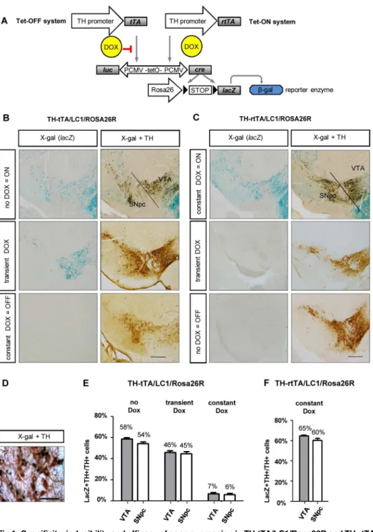
Fig 1. Specificity, inducibility and efficacy of gene expression in TH-tTA/LC1/Rosa26R and TH-rtTA/ LC1/Rosa26R mice. (A)Genetic scheme of DOX-regulated reporter gene expression in TH-tTA and TH-rtTA mice crossed with LC1 and Rosa26R reporter mice.(B and C)Coronal midbrain brain sections with
substantia nigra pars compacta (SNpc) and ventral tegmental area (VTA) DA neurons of TH-tTA/LC1/ Rosa26R (B) and TH-rtTA/LC1/Rosa26R (C) mice raised with or without doxycycline (DOX) or transiently with DOX to have the system switched off during development (TH-tTA mice treated with DOX during pre-and postnatal development till 6 weeks of age; TH-rtTA mice raised without DOX) pre-and on during analysis (TH-tTA mice from the age of 6 weeks without DOX; TH-r(TH-tTA mice from the age of 6 weeks with DOX). Sections were stained forβ-galactosidase activity with X-gal to visualize cells with activated tet-system. Adjacent sections were X-gal stained and co-stained with tyrosine hydroxylase (TH) antibodies and DAB substrate (brown) to mark DA neurons.(D)Zoom in picture of TH and lacZ double positive cells in the SNpc.(E and F)
(transient DOX) or without DOX (no DOX) in the tet-OFF (E) and tet-ON mice (F). mean + s.e.m.; n = 3. Scale bars: 500μm (B and C), 50μm (D).
doi:10.1371/journal.pone.0136203.g001
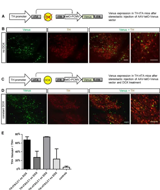
Fig 2. Cre independent adult gene expression in DA neurons of TH-tTA and TH-rtTA mice using a AAV-tetO-Venus vector. (A)Scheme of fluorescent DA neuron labelling in non-DOX treated TH-tTA mice stereotactically injected with AAV-tetO-Venus vector in the ventral midbrain.(B)Confocal fluorescent pictures of Venus expression (green) in DA neurons co-stained for tyrosine hydroxylase (TH; red) in the substantia nigra pars compacta (SNpc) of sagittal TH-tTA mouse brain sections.(C)Scheme of fluorescent DA neuron labeling in DOX-treated TH-rtTA mice stereotactically injected with AAV-tetO-Venus vector in the ventral midbrain.(C)Scheme of fluorescent DA neuron labelling in DOX-treated TH-rtTA mice
stereotactically injected with AAV-tetO-Venus vector in the ventral midbrain.(D)Confocal fluorescent pictures of Venus expression in DA neurons co-stained for TH in the substantia nigra pars compacta (SNpc) of sagittal TH-rtTA mouse brain sections.(E)Quantification reveals that in the ON-state 67% and 73% and in the OFF-state 27% and 23% of TH+ cells are also Venus-positive in TH-tTA and TH-rtTA mice, respectively. In non-transgenic mice (controls) only 5% of TH+ cells were also Venus-positive. n = 3–5. Scale bars: 250μm and in high magnification picture 50μm.
Fig 3. Loss of DA neurons in newborn and adult TH-tTA/LC1/Rosa26dt-a mice. (A)Scheme of diphteria toxin-a (dt-a) expression in TH-tTA/LC1/Rosa26dt-a mice.(B)Coronal midbrain sections of newborn TH-tTA/ LC1/Rosa26dt-a mice and control mice, both stained for tyrosine hydroxylase (TH) to visualize DA neurons of the substantia nigra pars compacta (SNpc) and ventral tegmental area (VTA). No DOX was applied. Scale bar = 50μm.(C)Stereological quantification of TH-positive cells in the SNpc and VTA of newborn TH-tTA/ LC1/Rosa26dt-a and control mice without DOX treatment.(D)TH stained coronal midbrain sections of adult TH-tTA/LC1/Rosa26dt-a mice and control mice raised with DOX until 6 weeks of age followed by 30 weeks without DOX. Scale bar = 500μm.(E)Stereological quantification of TH-positive cells in the SNpc and VTA of adult TH-tTA/LC1/Rosa26dt-a and control mice raised with DOX until 6 weeks of age followed by 6 or 14–30 weeks without DOX. mean + SEM,*p<0.05,**p<0.01,***p<0.001 Student´st-test, n = 3.
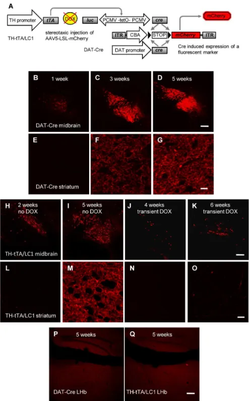
virus injection until analyzed (J, K, N, O). Scale bars = 200μm (H-K); 20μm (L-O).(P and Q)No fluorescent mCherry expression was detected in the lateral habenula (LHb) of AAV-LSL-mCherry injected DAT-Cre (P) and TH-tTA/LC1 (Q) mice after 5 weeks. Scale bars = 200μm.
doi:10.1371/journal.pone.0136203.g004
Fig 5.In vivobioluminescence imaging of DA neurons in TH-tTA/LC1 and TH-rtTA/LC1 mice. (A)Detection of luciferase activity in anesthetised TH-tTA/LC1 and LC1 control mice after fur removal and in brain tissue of dead mice after removal of the skull and dissection of brains using an IVIS200in vivo imaging device (Calipers Co.).(B)Comparison of thein vivobioluminescence signals in P3 TH-tTA/LC1 mouse pups expressing diphteria toxin A (dt-a) with TH-tTA/LC1 control mice and LC1 mice without luciferase expression. The radiance [p/sec/cm2/sr] RAW data luminescence signal intensity is shown in percentage compared to the maximal signal in the TH-tTA/LC1 control mice.(C)Temporal control of bioluminescence signal via DOX treatment in TH-tTA/ LC1 mice. Quantification of luciferase activity in anesthetised TH-tTA/LC1 and LC1 control mice. After initial measurement, mice were treated with DOX in the drinking water (2 mg/ml) for 8 days and afterwards kept without DOX for further 111 days. Bioluminescence was again measured at the indicated time points, n = 3.(D)Temporal control of bioluminescence signal via DOX treatment in TH-rtTA/LC1 mice. Quantification of luciferase activity in anesthetised TH-rtTA/ LC1 and LC1 control mice raised on DOX for 6 weeks and kept during adulthood without DOX. After initial measurement, mice were treated with DOX-containing food for 35 days and afterwards kept without DOX for further 14 days, n = 3–8.
Conventional microscopy
Transmitted light images were acquired using a SZX16 stereo microscope equipped with a DP72 camera and cellSens Entry 1.4.1 imaging software (Olympus). DAB stained sections and fluorescent cells were imaged with an epifluorescent upright microscope Axio Imager.M1 (Zeiss, Goettingen, Germany) equipped with an automated stage (Ludl MAC 6000 system), Hamamatsu camera C8484 and Axiovision software 4.8. Fluorescent sections were imaged with a Leica TCS SP2 confocal microscope system with 10x (0.3 NA) and 63x (oil 1.32 NA) objectives or Zeiss LSM 700 Confocal microscope system with 10x (0.25 NA) and 40x (oil 1.3 NA) objectives. Confocal images were acquired by using a 514-nm Argon line for Venus or a 561-nm photodiode laser for mCherry. Image stack were maximally projected and contrast and intensity levels were uniformly adjusted using ImageJ (NIH).
Quantifications
Stereological countings were performed on 30μm coronal serial sections analyzing every sixth
section for the SNpc and VTA. This was done using an oil immersion 63x objective, a counting frame of 50 x 50μm, and a grid size of 100 x 100μm, as described previously [27] using the
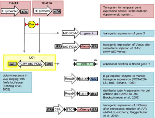
Fig 6. Schematic overview of utilized transgenic mice and recombinant AAV (rAAV) constructs. TH-tTA and TH-rTH-tTA mice express, under the tyrosine hydroxylase (TH) promoter, the tetracycline-regulated transactivator protein (tTA) or the reverse tetracycline-regulated transactivator protein (rtTA) in dopaminergic (DA) neurons. tTA binds to the tetracycline operon (tetO) in the absence of doxycycline (DOX) and rtTA binds in the presence of DOX, driving transient expression of gene X, for example the fluorescent protein Venus from a recombinant AAV vector (AAV-tetO-Venus), or luciferase and Cre from the LC1 construct. The LC1 encodes a luciferase enzyme, which can be used for bioluminescencein vivoimaging, and the Cre recombinase for deleting gene Y or removing floxed STOP codons (LSL). Cre is used here as a genetic switch to activate, from the ROSA locus, either expression of the reporter gene lacZ (encodingβ
-galactosidase) to visualize transgene expression or to activate expression of diphtheria toxin protein A (dt-a) to selectively induce cell death of DA neurons. Here we use Cre also to activate expression of the fluorescent protein mCherry from AAV-LSL-mCherry.
optical fractionator method of the StereoInvestigator software 8.0 (MicroBrightField). To quantify lacZ positive and TH positive cells (Fig 1) we counted at least 2 representative 30μm
serial coronal sections. Venus positive and TH positive cells (Fig 2) were quantified in at least 3 representative 50μm serial sagittal sections per mouse (every forth section) by applying a
binary mask and the“Analyze Particles”function in ImageJ (NIH).
In vivo
bioluminescence imaging
Bioluminescencein vivoimaging was performed on adult mice (2–12 months inFig 5A, 5C and 5D) and non DOX treated P3 mice (Fig 5B) by using the IVIS 200 system and Living Image 3.2 software (Perkin Elmer) according to manufacturer´s instructions. For biolumines-cence imaging, mice were i.p. injected with D-luciferin aqueous solution (15 mg ml-1; 4.5 mg per 30g of bodyweight).
Statistical analysis
Data are expressed as means + s.e.m. The statistical analyses were performed with GraphPad Prism 5.0 (GraphPad Software) using two-tailed, unpaired Student´s t-test or ANOVA, fol-lowed by Tukey‘s post hoc test to compare group means. In all analyses, p-values smaller than 0.05 were considered statistically significant.
Results
Generation and basic characterization of TH-tTA and TH-rtTA mice
To develop a tet-OFF and a tet-ON system for DA neurons of the mouse, we cloned the tTA2S or rtTA3G transactivator encoding sequences downstream of an 8.9 kilobase promoter frag-ment of the mouse TH gene and generated transgenic mice by pronuclear injections (Fig 1;S1 Fig). We generated 12 tTA founder mice of which 9 founders went germline and 9 TH-rtTA founders of which 8 went germline. The founders were functionally characterized by crossing them with LC1 mice [21] and with Rosa26R reporter mice [22] (Fig 1A).
In the triple-transgenic TH-tTA/LC1/Rosa26R mice, the tTA transactivator-induced Cre recombinase expression from the LC1 transgene triggered the deletion of a STOP cassette in front of the LacZ gene, encoding the reporter enzymeβ-galactosidase, in the Rosa26 locus. Using X-gal staining, we could detectβ-galactosidase activity in midbrain DA neurons in 4 of the 9 TH-tTA founder lines (founder 1 inFig 1B; founder 2–4 inS2 Fig). 5 founder lines did not show any lacZ expression. The four TH-tTA founder lines showed no difference in the expression pattern without DOX and no obvious physiological or behavioral abnormalities (data not shown).
To switch the tet-system permanently ON in TH-rtTA/LC1/Rosa26R mice, the parents were fed DOX-containing food before pregnancy and their offspring were kept on DOX until analysis. 5 founders showed X-gal staining under these conditions and were similar with regards to expression and physiology (founder 1 inFig 1C; founder 2–5 inS3 Fig).
on the other hand an advantage to enable investigation of targeted and non-targeted DA neu-rons in the same mice, which is preferred for example in electrophysiological studies.
Besides expression in DA neurons of the VTA and SNpc, DA neurons in the arcuate nuclei and retrorubral field and noradrenergic cells of the locus coeruleus (LC) were stained, too (S4 andS5Figs). TH expression has been described not only in catecholaminergic neurons, such as DA and noradrenergic neurons, but also transiently or permanent in many other cells inside and outside the nervous system. Transient TH protein expression during development in the absence of catecholamine synthesis has been reported in rodents for example in some interneu-rons of the cortex [28,29], mesospiny neurons in the striatum [30] and Purkinje cells of the cerebellum [31]. Also the accessory, deeper layer and periglomerular cells of the olfactory bulb express TH protein during development [1,32]. However, in adult mice TH protein expression is no longer detectable in these cell populations, although they continue to express TH mRNA [33]. As a consequence, X-gal staining was observed in the tTA/LC1/Rosa26R and TH-rtTA/LC1/Rosa26R mice with the tet-system constantly active in many distinct brain regions such as the cerebellum, pons, striatum, hippocampus, cortex, thalamus, and lateral midbrain (Fig 1B,S4andS5Figs). Also, the extent of expression in the different tissues was variable, ranging from activity in all Purkinje cells of the cerebellum and all medium spiny neurons in the striatum to only mosaic, sporadic activation in pyramidal cells in the hippocampus and cortex (S4andS5Figs).
In the SNpc and VTA region of TH-tTA/LC1/Rosa26R and TH-rtTA/LC1/Rosa26R mice raised with constitutive active tet-system, more than 60% of X-gal positive cells were also stained with antibodies against TH. Similarly, in transiently DOX treated TH-tTA/LC1/ Rosa26R mice, more than 90% of X-gal positive cells were stained (S2BandS3BFigs). There-fore, these cells were considered to be DA neurons. The X-gal-positive cells negative for TH protein expression might be due to cells that only express TH mRNA and no TH protein [34– 36] (as described earlier) or cells expressing TH only during development and not adulthood (see next two chapters) (S2B and S2C,S3B and S3CFigs). The endogenous TH enzyme starts to be expressed in the mouse ventral midbrain at embryonic day E9-10 [37,38] soon after the identification of the first midbrain DA neurons by the expression of the retinoic acid–synthe-sizing enzyme Aldh1a1 and before DA neurons are functionally connected to their postsynap-tic partners [5]. Thus, our TH-tet mice permit targeting of DA neurons very early on during development and requires tight regulation of the tet-system if they are used to alter expression during a later developmental phase, e.g. during adulthood.
Regulation of gene expression in TH-tTA and TH-rtTA mice
To test the regulation of the tet-system in the TH-tTA and TH-rtTA mice, we first tried to switch off the tet-system completely by constantly feeding TH-tTA/LC1/Rosa26R mice with DOX-containing food or feeding TH-rtTA/LC1/Rosa26R mice with non DOX-containing food. This resulted in very few X-gal positive cells in the ventral midbrain DA system (Fig 1B and 1C). This suggests that there is a tight regulation of the transcriptional activity in both TH-tet mice under these conditions with little leakage.
at least 6 additional weeks. However, no X-gal stained cells were observed (Fig 1C). The reduced number of responding DA neurons after DOX treatment in TH-tTA/LC1/Rosa26R mice, as well as the lack of DOX response of adult TH-rtTA/LC1/Rosa26R mice, might be due to genetic silencing of the LC1 construct that prevents re-activation, as previously described [39,40].
LC1 independent targeting of adult DA neurons in TH-tTA and TH-rtTA
mice
In order to overcome possible genetic silencing of the LC1 construct in TH-rtTA/LC1 mice, we generated an AAV vector expressing the fluorescent protein Venus directly under the control of the tetO promoter (AAV-tetO-Venus). Before virus production, we tested the AAV-tetO--Venus DNA construct in HEK293-T cells co-transfected with a tTA expression vector and found a DOX-dependent expression of Venus (S6 Fig). Subsequently, the AAV-tetO-Venus virus vector was stereotactically injected in the SN of adult non-DOX treated TH-tTA mice and DOX-treated TH-rtTA (Fig 2). We investigated 5 weeks after injection the expression of Venus in the mouse midbrain DA neurons visualized by TH antibody staining. In TH-tTA/ AAV-tetO-Venus mice, 67% of DA neurons were also Venus-positive (Fig 2A, 2B and 2E). With DOX treatment 10 days before AAV-tetO-Venus injection, we observed only 27% of Venus-positive DA neurons in TH-tTA mice (Fig 2E,S7 Fig). In DOX-treated TH-rtTA/AAV-tetO-Venus mice, 73% of DA neurons were expressing Venus, while only 23% expressed Venus without DOX (Fig 2C–2E,S7 Fig). In non-transgenic control mice, AAV-tetO-Venus injection resulted in only 5% of Venus-positive DA neurons, which was comparable with back-ground expression seen in AAV-tetO-Venus transfected HEK293-T cells (Fig 2E,S6andS7 Figs). This result confirms that it is possible to induce the tet-system in TH-rtTA mice with a transient postnatal DOX treatment. Furthermore, it provides evidence that in both transgenic mouse lines, the TH-tTA and TH-rtTA mice, the tet-system can efficiently activate expression from virally provided and tetO regulated genes of interest during adulthood.
Genetic deletion of DA neurons in TH-tTA mice
We made use of our TH-tTA mice to generate a genetic mouse model with reduced number of DA neurons. To do this, we replaced the Rosa26R with the Rosa26dt-a transgene in the triple-transgenic mice that, in the absence of DOX, induced cell death by the expression of diphtheria toxin-A [23] (Fig 3A). TH-tTA/LC1/Rosa26dt-a mice without DOX died shortly after birth in the first postnatal week and lost around 40% of midbrain DA neurons in the SNpc and VTA (Fig 3B and 3C).
The mice survived until adulthood if transiently treated with DOX during embryogenesis and early postnatal development until 6 weeks of age. We stereologically quantified the number of DA neurons in mice 6 weeks and between 14 to 30 weeks after removal of DOX and found 18 to 32% less DA neurons in the SNpc and 28 to 33% less DA neurons in the VTA, respec-tively (Fig 3D and 3E). This supports the tight regulation of the tet-system in our TH-tTA mice and the possibility to generate, with this approach, a genetic adult-onset DA system neurode-generation mouse model.
Fluorescent labeling of adult DA neurons in TH-tTA mice
label the DA cell bodies, but only weakly label the DA innervation in the target fields. This includes the striatum, which is important to investigate in the DA system degeneration or regeneration experiments related to PD. This is most likely due to the low copy number of fluo-rescent protein encoding cDNAs in the transgenic mice and the very long and manifold brunched axons of DA neurons.
Since the stereotactic injection of the AAV-tetO-Venus vector in the TH-tTA and TH-rtTA mice did not efficiently label the complete axonal network of DA neurons, we tried to enhance the fluorescent signal by using the Cre-lox system. We stereotactically injected adult TH-tTA/ LC1 mice in the SNpc with an AAV-LSL-mCherry virus construct, that uses the strong cyto-megalovirus enhancer/chicken beta actin (CAG) promoter followed by a floxed STOP codon (LSL) and the mCherry expression cassette [26] (Fig 4A). As an alternative DA neuron-specific Cre mouse line, we also injected DAT-Cre mice with the same AAV-LSL-mCherry virus [7] (Fig 4A). In both mouse lines the STOP codon was efficiently removed in midbrain DA neu-rons and allowed to label them with mCherry (Fig 4B–4O). To determine the minimal time required to label DA axons in the striatum, we analyzed the mice 1, 2, 3, 4, 5 and 6 weeks after virus injection. We found that 1 week was sufficient to label a few cell bodies, but after 5 weeks there was a strong fluorescent staining of midbrain DA neurons and of the DA fibers in the striatum for both AAV-LSL-mCherry injected mouse lines, DAT-Cre (Fig 4B–4G) and TH-tTA/LC1 mice (Fig 4H, 4I, 4L and 4M). In both Cre lines injected with AAV-LSL-mCherry, DAT-Cre and TH-tTA/LC1 mice, we did not observe labeled fibers in the lateral habenula (LHb), which is still an open question in the field (Fig 4P and 4Q) [34–36,45,46]. Treating the TH-tTA/LC1 mice after viral injection for 4 or 6 weeks with DOX strongly reduced the amount of labeled cells in the midbrain and the fiber staining in the striatum (Fig 4J, 4K, 4N and 4O). Together with the previous experiments this shows the tight regulation of the tet-system in our TH-tTA mice during adulthood. These data suggests that the combination of TH-tTA/LC1 mice with the AAV-LSL-mCherry virus is an excellent approach to efficiently label TH express-ing neurons and their axons in their target fields such as the striatum.
In vivo bioluminescence imaging of the DA system in tTA and
TH-rtTA mice
adulthood, we could show reversible gene activation during adulthood with faster ON-kinetics compared to the TH-tTA/LC1 mice (Fig 5D). TH-rtTA/LC1 mice without DOX showed only a very weak luciferase signal that increased after 2 weeks on DOX to the maximal luciferase sig-nal and decreased again to the origisig-nal levels 2 weeks after withdrawal from DOX (Fig 5D).
Thus, the bioluminescence signal in TH-tTA/LC1 and TH-rtTA/LC1 mice allowsin vivo monitoring of the integrity and gene expression activity of the DA system, and might therefore be applied to follow degeneration and regeneration processes of the DA system over time in the same mouse.
Discussion
The TH-tTA and TH-rtTA mouse lines described enable targeting of midbrain DA neurons with high efficiency during all developmental stages, show a tight DOX-dependent regulation and therefore can be used for a multitude of applications (Fig 6). We have shown that the TH-tTA and TH-rTH-tTA mice are an excellent tool for regulating gene expression in midbrain DA neurons. They can be used to overexpress or delete genes of interest and they can be combined with the Cre-lox recombination system (Figs1–6). Therefore, our TH-tTA and TH-rtTA mouse lines permit studying, for example, the loss of function phenotype of genes in the adult DA system that lead to embryonic lethality or possible activation of compensatory mechanisms during development, which might mask their function during adulthood. In addition, we show that the TH-tet mice can be efficiently used to visualize DA neurons and their innervation fluo-rescently in the brain (Figs2and4). Furthermore, they can be used to monitor the DA system in vivoas an entity over the whole life time of a mouse and therefore allow following alterations progressively over time (Fig 5).
In 2004, the first ratTH-rtTA2S-M2 construct was generated and successfully used in HEK293 cells and in the dopaminergic cell line MN9D [47]. However, three groups have only recently generated transgenic mice in which the tet-system was targeted to midbrain DA neu-rons [48,49]. In the first mouse line, a tTA knock-in was created into the endogenous DAT locus, which generates a heterozygote DAT knockout mouse showing low DAT levels and hyperactivity [50]. In a second line, a TH-rtTA mouse line, tetracycline-dependent regulation was subsequently lost [49](data not shown). In the third mouse line, carrying a Pitx3-tTA con-struct, tetracycline-dependent regulation has not yet been shown [48]. To preserve our func-tional tet-system mouse lines for the research community, we immediately conserved them by sperm freezing.
The TH-tTA and TH-rtTA mice can be combined with many different tetO-constructs at the same time and used to activate or inhibit gene expression during embryogenesis, adulthood and in aging mice since DOX is rapidly taken up into cells, shows low toxicity and penetrates the placenta and the blood-brain barrier [51–53]. Silencing of tetO-constructs in the mouse can be overcome, as shown here, by administering it at specific time points, for example, by electro-poration of DNA in embryos or by viral injections in adult mice. If a gene should only be expressed for a short time period, the rapid inducible tet-ON system might be preferred. How-ever, if a gene should only be switched off for a short time period the use of the tet-OFF system might be an advantage and can save DOX-food costs. In order to decide between each system, it is worthwhile mentioning that the tet-OFF systems shows a slower ON-kinetics (several weeks) compared to the tet-ON system (several days), as shown also in ourin vivoluciferase experi-ment. Furthermore, long DOX-treatment times might lead to an accumulation of DOX in the bone that is only slowly cleared after removal of DOX from the food or water [51–53].
with the rat TH promoter [2,4]. This seems to be the case even in mice with the transgene knocked into the endogenous mouse TH locus behind an internal ribosomal entry sequence (IRES) [3]. Besides the TH promoter specific features this appears to be at least in part also due to the reporter construct used as viral vectors and transgenic mice do not express their encoded reporter homogeneously in DA neurons [3]. In addition, we did not find any fluorescently labeled DA neurons when we crossed the TH-tTA/LC1 mice with Ai9 tdTomato reporter mice [54] (data not shown). This is surprising, because Ai9 mice use like the Rosa26R and Rosa26dt-a mice the RosRosa26dt-a locus for tdTomRosa26dt-ato expression [22]. Yet, Ai9 mice lRosa26dt-abeled DA neurons nicely when crossed with DAT-Cre mice [54](data not shown). Despite this, more research is needed to better understand the underlying biology of this phenomenon. The mosaic targeting can also be an advantage for some experiments. In studies where DA cells with different genetic alterations need to be comparedin vivoin the same environment—for example, in electrophys-iological studies—the partial targeting of DA neurons in the TH-tTA mice will turn out to be a very helpful condition, much like the MARCM clones inDrosophila melanogasterthat turned out to be an excellent tool to studyin vivogene function in the wildtype background of flies [55]. The mosaic expression in the TH-tTA/LC1 mice allowed us to establish viable TH-tTA/ LC1/Rosa26dt-a mice showing a very reproducible and progressive DA system degeneration in young adult mice reaching in 3–7 month old mice 30% of DA neuron loss. The DA system degeneration in TH-tTA/LC1/Rosa26dt-a mice is less variable and cumbersome to generate compared to the commonly used injections of neurotoxins such as MPTP, 6-OHDA or rote-none to generate PD animal models [56]. Therefore, the inducible DA system degeneration phenotype in TH-tTA/LC1/Rosa26dt-a mice might be a favorable alternative to test cell and tissue replacement therapies for the treatment of PD.
The neonatal lethality in TH-tTA/LC1/Rosa26dt-a mice is most likely due to the loss of nor-adrenergic neurons in the peripheral nervous system innervating for example the heart. This view is supported by the findings that DAT-Cre/Rosa26dt-a mice also show a reduced number of midbrain DA neurons but are viable and show a normal motor behavior despite the DA cell loss of around 90% [57]. In addition, TH deficiency in mice has been shown to be embryonal lethal apparently due to cardiovascular failure [58] and the lethality can be rescued by knock-ing-in TH into the noradrenergic-specific dopamine-β-hydroxylase locus [59]. DOX treatment of TH-tTA/LC1/Rosa26dt-a mice during development prevents postnatal lethality and allows deleting dopaminergic neurons during adulthood.
To enable the unbiased fluorescent detection of DA neuron axonal branches and to allowin vivodetection of the fluorescently labeled DA system in our TH-tet mice, we made use of a Cre-mediated expression of mCherry from an AAV vector (AAV-LSL-mCherry). We show as a proof of principle that for this purpose the AAV5-LSL-mCherry virus can be used in combi-nation with both mouse lines expressing Cre recombinase in DA neurons, the DAT-Cre and the TH-tTA/LC1 mice. The advantage of the TH-tTA/LC1 mice becomes apparent if you want to label DA neurons in which genetic alterations should not only be induced by Cre recombi-nase events but also by the tet-O promoter enabling reversible gene activation.
from the midbrain with our AAV-LSL-mCherry virus in the same DAT-Cre mouse line used before and in our TH-tTA/LC1 mice. Taken together these results support the idea that not only the Cre expressing mouse line but also the transgenic or viral reporter construct dramati-cally influences the outcome of targeting and that the outcome of a specific combination of Cre and reporter is so far not predictable.
We can imagine that combining several AAV-LSL-XFP viruses encoding different fluores-cent proteins (XFP) might allow labeling and tracing single DA neurons in almost unique col-ors. This could facilitate the detailed analysis of the heterogeneous population of DA neurons [1,60] for which only 8 neurons have been traced in detail from the rat brain and none from the mouse brain [61]. Further experiments are needed to test this hypothesis in detail.
We provide here the proof of principle thatin vivoimaging of the DA system in our TH-tet mice is feasible using bioluminescence signals.In vivoimaging might allow detecting progres-sive degeneration and regeneration processes of the DA system over time in individual mice and therefore might be suitable for testing drugs for their beneficial or detrimental effect. This method should allow cutting down the number of animals in longitudinal studies and should avoid inter-subject variability problems. Since thein vivomeasurement takes only seconds to minutes, it might reduce the time for analysis dramatically and can replace or precede a detailed histological analysis. The special resolution and signal intensity of thein vivosignal might be further improved by replacing the skull with a glass plate, using more sensitive biolu-minescence substrates such as Cycluc1 [62], and by switching for biolubiolu-minescence imaging to a red-shifted luciferase variant [63].
In the future, TH-tet mice might also be used for altering and monitoring acute DA cell physiology by combining them with Ca2+ imaging and optogenetic tools, such as in AAV-LSL-channelrhodopsin or halorhodopsin vectors and the KENGE-tet system, using knockin-mediated enhanced gene expression by improved tetracycline-controlled gene induc-tion to express a highly light-sensitive channelrhodopsin-2 mutant at levels sufficient to drive the activities in different cells [64,65].
With the TH-tTA and TH-rtTA mice we generated a versatile tet-system available to geneti-cally modify the DA system of the mouse for studyingin vitroandin vivoDA neuron develop-ment and maintenance, physiology and pathophysiology, and biochemical or histological alterations. The establishment of the tet-OFF and tet-ON system in mouse DA neurons enlarged our tool kit for more sophisticated and fine-tuned manipulations of the DA system that should help to increase our knowledge about this complex and fascinating system and might shed more light on the etiology and possible treatments of human diseases such as PD, schizophrenia and drug addiction.
Supporting Information
S1 Fig. Schematic overview of transgenic TH-tTA mice. (A)Scheme of the TH-tTA DNA construct: transcription of the tetracycline transactivator protein tTA is controlled by a 8.9 kb mouse genomic fragment upstream of the AUG start codon of the tyrosine hydroxylase (TH) coding sequence.(B)Agarose gel analysis of the TH-tTA fragment isolated from the SalI cut plasmid used for pronucleus injection (PI) into C57Bl/6J single cell embryos to generate trans-genic mice. 1: GeneRuler 1kb DNA ladder; 2: TH-tTA DNA fragment of around 12 kb.(C) Principle of the Tet-OFF system: tTA protein is expressed via the TH-tTA construct and binds in the absence of doxycycline (DOX) to the tet-responsive promoter (tetO) and activates gene of interest expression.
S2 Fig. Specificity, inducibility and efficacy of gene expression in additional TH-tTA foun-ders after crossing them with LC1 and Rosa26R mice. (A)Coronal midbrain brain sections with SNpc and VTA DA neurons of TH-tTA/LC1/Rosa26R for founders 2, 3, and 4, respec-tively. Mice were raised without (no) DOX, with (constant) DOX or with DOX until the age of 6 weeks followed by 6 weeks without (transient) DOX. Sections were stained for beta-galactosi-dase activity with X-gal to visualize cells with activated tTA, LC1 and ROSA26 locus. Scale bars: 500μm.(B)Ratio of lacZ and TH double positive cells to all lacZ positive cells in the
SNpc and VTA in TH-tTA/LC1/Rosa26R without or transient DOX treatment as indicated. (C)lacZ positive cells in the VTA, SNpc and ventral midbrain without VTA and SNpc in non DOX treated TH-tTA/LC1/Rosa26R mice.
(TIF)
S3 Fig. Specificity, inducibility and efficacy of gene expression in additional TH-rtTA foun-ders after crossing them with LC1 and Rosa26R mice. (A)Coronal midbrain brain sections with SNpc and VTA DA neurons of TH-rtTA/LC1/Rosa26R for founders 2, 3, 4, and 5, respec-tively. Mice were raised with (constant) DOX, without (no) DOX, or transiently without DOX until the age of 6 weeks followed by 6 weeks with (transient) DOX. Sections were stained for beta-galactosidase activity with X-gal to visualize cells with activated rtTA, LC1 and ROSA26 locus. Scale bars: 500μm.(B)Ratio of lacZ and TH double positive cells to all lacZ positive
cells in the SNpc and VTA in TH-rtTA/LC1/Rosa26R with constant DOX treatment.(C)lacZ positive cells in the VTA, SNpc and ventral midbrain without VTA and SNpc in constantly DOX treated TH-rtTA/LC1/Rosa26R mice.
(TIF)
S4 Fig. Specificity, inducibility and efficacy of gene expression outside midbrain DA neu-rons in adult mice of TH-tTA founder 1 after crossing with LC1 and Rosa26R mice. Coro-nal mouse brain sections stained with X-gal to visualizeβ-galactosidase expression in TH-tTA/ LC1/Rosa26R mice without (A,C, E, G) or transient DOX (B, D, F, H). Broad lacZ expression was detected in mice without DOX treatment in the cerebellum and pons (A), in the striatum (C), the olfactory bulb (E), the hippocampus and different layers of the cortex (G). Mice tran-siently treated with Dox during development till 6 weeks after birth and evaluated 6 weeks later showed still X-gal staining in the cerebellum (B) but not anymore in the pons (B), the striatum (D), hippocampus and cortex (H). Scale bars: 200μm (A-H), blowup 500μm (A-D).(I)
Quan-tification of TH+ lacZ+ double positive cells in the locus coeruleus of TH-tTA/LC1/Rosa26R mice without DOX revealed 17% targeted noradrenergic neurons.
(TIF)
S5 Fig. Specificity and efficacy of gene expression outside midbrain DA neurons in adult mice of TH-rtTA founder 1 after crossing with LC1 and Rosa26R mice.Coronal mouse brain sections stained with X-gal to visualizeβ-galactosidase expression in TH-rtTA/LC1/ Rosa26R mice with DOX. Broad lacZ expression was detected in the cerebellum and pons (A), hippocampus (B), striatum (C), in different layers of the cortex (B and C) and the olfactory bulb (D). Scale bars: 200μm (A and C), blowup 500μm (A-D).
(TIF)
S6 Fig. Transfection of new pAAV-tetO-Venus DNA construct in HEK293-T cells.Cells were transfected with the following DNA constructs and analyzed 48 hours later for Venus expression:(A)pUHD61-1-tTA vector encoding tTA under a CMV promoter;(B) pAAV-tetO-Venus vector encoding Venus under the tetO promoter; and(C)co-transfection of both pUHD61-1-tTA and pAAV-tetO-Venus vector. Scale bar: 200μm.
S7 Fig. Cre-independent adult gene expression in DA neurons of TH-tTA and TH-rtTA mice using a AAV-tetO-Venus vector. (A-C)Confocal fluorescent pictures of Venus expres-sion (green) in DA neurons co-stained for TH (red) in the substantia nigra pars compacta (SNpc) of sagittal mouse brain sections 5 weeks after AAV-tetO-Venus virus injection. TH-tTA mice were treated with DOX for 10 days before streotactic injections until analysis to switch the system off (A). TH-rtTA mice were here not DOX treated to keep the system inac-tive (B). Non-transgenic control mice were analyzed also 5 weeks after AAV-tetO-Venus virus injection (C). Scale bars: 250μm.
(TIF)
Acknowledgments
This study was supported by grants from the German Research Foundation (DFG KR 3529/4-1 to E.R.K.) and the town of Hamburg (Lexi to E.R.K.). We thank all mouse donors H. Bujard, K Schönig, D. Riethmacher, P. Soriano; W. Wurst for providing the mouse TH promoter; H. Bujard and K Schönig for tet-system related plasmids; S. Guggenhuber and M. Klugmann for the AAV-LSL-mCherry vectors; J. Kleinschmidt for AAV packaging plasmids; K. Duncan, I. Hermans-Borgmeyer, M. Moles and the whole Kramer lab for helpful comments on the manu-script. I. Hermans-Borgmeyer, A. Leingärtner, M. Horn-Glander, D. Isbrandt, P. Putthoff, F. Yan, V. Nanos, S. Homann, F. Morellini, the UKE vector core facility, the UKEin vivooptical imaging facility, the transgenic animal service and the members of the mouse facility at the UKE for excellent technical assistance.
Author Contributions
Conceived and designed the experiments: ERK. Performed the experiments: KT HA ERK. Ana-lyzed the data: KT HA ERK. Contributed reagents/materials/analysis tools: KT HA ERK. Wrote the paper: KT HA ERK.
References
1. Bjorklund A, Dunnett SB. Dopamine neuron systems in the brain: an update. Trends Neurosci. 2007; 30(5):194–202. Epub 2007/04/06. doi: S0166-2236(07)00067-7 [pii] doi:10.1016/j.tins.2007.03.006 PMID:17408759.
2. Gelman DM, Noain D, Avale ME, Otero V, Low MJ, Rubinstein M. Transgenic mice engineered to target Cre/loxP-mediated DNA recombination into catecholaminergic neurons. Genesis. 2003; 36(4):196–
202. doi:10.1002/gene.10217PMID:12929090.
3. Lindeberg J, Usoskin D, Bengtsson H, Gustafsson A, Kylberg A, Soderstrom S, et al. Transgenic expression of Cre recombinase from the tyrosine hydroxylase locus. Genesis. 2004; 40(2):67–73. Epub 2004/09/29. doi:10.1002/gene.20065PMID:15452869.
4. Savitt JM, Jang SS, Mu W, Dawson VL, Dawson TM. Bcl-x is required for proper development of the mouse substantia nigra. J Neurosci. 2005; 25(29):6721–8. Epub 2005/07/22. doi: 25/29/6721 [pii] doi: 10.1523/JNEUROSCI.0760-05.2005PMID:16033881.
5. Wurst W, Prakash N. Genetic Control of Meso-diencephalic Dopaminergic Neuron Development in Rodents. In: Iversen LL, IS D., Dunnett SB, Bjorklund A, editors. Dopamine Handbook: Oxford Univer-sity Press; 2010. p. 141–59.
6. Backman CM, Malik N, Zhang Y, Shan L, Grinberg A, Hoffer BJ, et al. Characterization of a mouse strain expressing Cre recombinase from the 3' untranslated region of the dopamine transporter locus. Genesis. 2006; 44(8):383–90. Epub 2006/07/26. doi:10.1002/dvg.20228PMID:16865686.
7. Zhuang X, Masson J, Gingrich JA, Rayport S, Hen R. Targeted gene expression in dopamine and sero-tonin neurons of the mouse brain. J Neurosci Methods. 2005; 143(1):27–32. Epub 2005/03/15. doi: S0165-0270(04)00350-4 [pii] doi:10.1016/j.jneumeth.2004.09.020PMID:15763133.
8. Ekstrand MI, Terzioglu M, Galter D, Zhu S, Hofstetter C, Lindqvist E, et al. Progressive parkinsonism in mice with respiratory-chain-deficient dopamine neurons. Proc Natl Acad Sci U S A. 2007; 104(4):1325–
9. Turiault M, Parnaudeau S, Milet A, Parlato R, Rouzeau JD, Lazar M, et al. Analysis of dopamine trans-porter gene expression pattern—generation of DAT-iCre transgenic mice. FEBS J. 2007; 274 (14):3568–77. Epub 2007/06/15. doi: EJB5886 [pii] doi:10.1111/j.1742-4658.2007.05886.xPMID: 17565601.
10. Parlato R, Rieker C, Turiault M, Tronche F, Schutz G. Survival of DA neurons is independent of CREM upregulation in absence of CREB. Genesis. 2006; 44(10):454–64. Epub 2006/09/19. doi:10.1002/dvg. 20236PMID:16981198.
11. Smidt MP, von Oerthel L, Hoekstra EJ, Schellevis RD, Hoekman MF. Spatial and temporal lineage analysis of a Pitx3-driven Cre-recombinase knock-in mouse model. PLoS One. 2012; 7(8):e42641. doi: 10.1371/journal.pone.0042641PMID:22870339; PubMed Central PMCID: PMC3411649.
12. Engblom D, Bilbao A, Sanchis-Segura C, Dahan L, Perreau-Lenz S, Balland B, et al. Glutamate receptors on dopamine neurons control the persistence of cocaine seeking. Neuron. 2008; 59(3):497–508. Epub 2008/08/15. doi: S0896-6273(08)00582-5 [pii] doi:10.1016/j.neuron.2008.07.010PMID:18701074.
13. Qiu HY, Guo C, Cheng XW, Huang Y, Xiong ZQ, Ding YQ. Pitx3-CreER mice showing restricted Cre expression in developing ocular lens and skeletal muscles. Genesis. 2008; 46(6):324–8. Epub 2008/ 06/11. doi:10.1002/dvg.20399PMID:18543300.
14. Rotolo T, Smallwood PM, Williams J, Nathans J. Genetically-directed, cell type-specific sparse labeling for the analysis of neuronal morphology. PLoS One. 2008; 3(12):e4099. Epub 2009/01/01. doi:10. 1371/journal.pone.0004099PMID:19116659.
15. Hayashi S, McMahon AP. Efficient recombination in diverse tissues by a tamoxifen-inducible form of Cre: a tool for temporally regulated gene activation/inactivation in the mouse. Dev Biol. 2002; 244(2):305–18. Epub 2002/04/12. doi:10.1006/dbio.2002.0597S001216060290597X [pii]. PMID:11944939.
16. Takebayashi H, Usui N, Ono K, Ikenaka K. Tamoxifen modulates apoptosis in multiple modes of action in CreER mice. Genesis. 2008; 46(12):775–81. Epub 2008/12/24. doi:10.1002/dvg.20461PMID:19105217.
17. Schmidt-Supprian M, Rajewsky K. Vagaries of conditional gene targeting. Nat Immunol. 2007; 8 (7):665–8. Epub 2007/06/21. doi: ni0707-665 [pii] doi:10.1038/ni0707-665PMID:17579640.
18. Forni PE, Scuoppo C, Imayoshi I, Taulli R, Dastru W, Sala V, et al. High levels of Cre expression in neu-ronal progenitors cause defects in brain development leading to microencephaly and hydrocephaly. J Neurosci. 2006; 26(37):9593–602. Epub 2006/09/15. doi: 26/37/9593 [pii] doi:10.1523/JNEUROSCI. 2815-06.2006PMID:16971543.
19. Traykova-Brauch M, Schonig K, Greiner O, Miloud T, Jauch A, Bode M, et al. An efficient and versatile system for acute and chronic modulation of renal tubular function in transgenic mice. Nat Med. 2008; 14 (9):979–84. doi:10.1038/nm.1865PMID:18724376; PubMed Central PMCID: PMC3446847.
20. Schonig K, Freundlieb S, Gossen M. Tet-Transgenic Rodents: a comprehensive, up-to date database. Transgenic Res. 2013; 22(2):251–4. doi:10.1007/s11248-012-9660-9PMID:23180363; PubMed Cen-tral PMCID: PMC3591533.
21. Schonig K, Schwenk F, Rajewsky K, Bujard H. Stringent doxycycline dependent control of CRE recom-binase in vivo. Nucleic Acids Res. 2002; 30(23):e134. Epub 2002/12/06. PMID:12466566.
22. Soriano P. Generalized lacZ expression with the ROSA26 Cre reporter strain. Nat Genet. 1999; 21 (1):70–1. Epub 1999/01/23. doi:10.1038/5007PMID:9916792.
23. Brockschnieder D, Pechmann Y, Sonnenberg-Riethmacher E, Riethmacher D. An improved mouse line for Cre-induced cell ablation due to diphtheria toxin A, expressed from the Rosa26 locus. Genesis. 2006; 44(7):322–7. Epub 2006/06/23. doi:10.1002/dvg.20218PMID:16791847.
24. Gossen M, Freundlieb S, Bender G, Muller G, Hillen W, Bujard H. Transcriptional activation by tetracy-clines in mammalian cells. Science. 1995; 268(5218):1766–9. PMID:7792603.
25. Shevtsova Z, Malik JM, Michel U, Bahr M, Kugler S. Promoters and serotypes: targeting of adeno-asso-ciated virus vectors for gene transfer in the rat central nervous system in vitro and in vivo. Exp Physiol. 2005; 90(1):53–9. doi:10.1113/expphysiol.2004.028159PMID:15542619.
26. Guggenhuber S, Monory K, Lutz B, Klugmann M. AAV vector-mediated overexpression of CB1 cannabi-noid receptor in pyramidal neurons of the hippocampus protects against seizure-induced excitoxicity. PLoS One. 2010; 5(12):e15707. Epub 2011/01/05. doi:10.1371/journal.pone.0015707PMID:21203567.
27. Kramer ER, Aron L, Ramakers GM, Seitz S, Zhuang X, Beyer K, et al. Absence of Ret signaling in mice causes progressive and late degeneration of the nigrostriatal system. PLoS Biol. 2007; 5(3):e39. Epub 2007/02/15. doi: 1544-9173-5-3-e39 [pii] doi:10.1371/journal.pbio.0050039PMID:17298183; PubMed Central PMCID: PMC1808500.
29. Satoh J, Suzuki K. Tyrosine hydroxylase-immunoreactive neurons in the mouse cerebral cortex during the postnatal period. Brain Res Dev Brain Res. 1990; 53(1):1–5. PMID:1972039.
30. Masuda M, Miura M, Inoue R, Imanishi M, Saino-Saito S, Takada M, et al. Postnatal development of tyrosine hydroxylase mRNA-expressing neurons in mouse neostriatum. Eur J Neurosci. 2011; 34 (9):1355–67. doi:10.1111/j.1460-9568.2011.07873.xPMID:22004548.
31. Fujii T, Sakai M, Nagatsu I. Immunohistochemical demonstration of expression of tyrosine hydroxylase in cerebellar Purkinje cells of the human and mouse. Neurosci Lett. 1994; 165(1–2):161–3. Epub 1994/ 01/03. PMID:7912416.
32. Min N, Joh TH, Kim KS, Peng C, Son JH. 5' upstream DNA sequence of the rat tyrosine hydroxylase gene directs high-level and tissue-specific expression to catecholaminergic neurons in the central ner-vous system of transgenic mice. Brain Res Mol Brain Res. 1994; 27(2):281–9. Epub 1994/12/01. PMID:7898312.
33. Baker H, Kobayashi K, Okano H, Saino-Saito S. Cortical and striatal expression of tyrosine hydroxylase mRNA in neonatal and adult mice. Cellular and molecular neurobiology. 2003; 23(4–5):507–18. PMID: 14514011.
34. Stamatakis AM, Jennings JH, Ung RL, Blair GA, Weinberg RJ, Neve RL, et al. A unique population of ventral tegmental area neurons inhibits the lateral habenula to promote reward. Neuron. 2013; 80 (4):1039–53. doi:10.1016/j.neuron.2013.08.023PMID:24267654; PubMed Central PMCID: PMC3873746.
35. Lammel S, Steinberg EE, Foldy C, Wall NR, Beier K, Luo L, et al. Diversity of transgenic mouse models for selective targeting of midbrain dopamine neurons. Neuron. 2015; 85(2):429–38. doi:10.1016/j. neuron.2014.12.036PMID:25611513.
36. Stuber GD, Stamatakis AM, Kantak PA. Considerations when using cre-driver rodent lines for studying ventral tegmental area circuitry. Neuron. 2015; 85(2):439–45. doi:10.1016/j.neuron.2014.12.034 PMID:25611514; PubMed Central PMCID: PMC4303766.
37. Diporzio U, Zuddas A, Cosenzamurphy DB, Barker JL. Early Appearance of Tyrosine-Hydroxylase Immunoreactive Cells in the Mesencephalon of Mouse Embryos. Int J Dev Neurosci. 1990; 8(5):523–
32. WOS:A1990EC81300002. PMID:1980787
38. Kawano H, Ohyama K, Kawamura K, Nagatsu I. Migration of Dopaminergic-Neurons in the Embryonic Mesencephalon of Mice. Dev Brain Res. 1995; 86(1–2):101–13. doi:10.1016/0165-3806(95)00018-9 WOS:A1995RC41800011.
39. Zhu P, Aller MI, Baron U, Cambridge S, Bausen M, Herb J, et al. Silencing and un-silencing of tetracy-cline-controlled genes in neurons. PLoS One. 2007; 2(6):e533. Epub 2007/06/21. doi:10.1371/journal. pone.0000533PMID:17579707.
40. Oyer JA, Chu A, Brar S, Turker MS. Aberrant epigenetic silencing is triggered by a transient reduction in gene expression. PLoS One. 2009; 4(3):e4832. doi:10.1371/journal.pone.0004832PMID: 19279688; PubMed Central PMCID: PMC2654015.
41. Sawamoto K, Nakao N, Kobayashi K, Matsushita N, Takahashi H, Kakishita K, et al. Visualization, direct isolation, and transplantation of midbrain dopaminergic neurons. Proc Natl Acad Sci U S A. 2001; 98(11):6423–8. Epub 2001/05/17. doi:10.1073/pnas.111152398111152398 [pii]. PMID:11353855.
42. Donaldson AE, Marshall CE, Yang M, Suon S, Iacovitti L. Purified mouse dopamine neurons thrive and function after transplantation into brain but require novel glial factors for survival in culture. Mol Cell Neurosci. 2005; 30(1):108–17. doi:10.1016/j.mcn.2005.06.004PMID:16024255; PubMed Central PMCID: PMC1949425.
43. Zhao S, Maxwell S, Jimenez-Beristain A, Vives J, Kuehner E, Zhao J, et al. Generation of embryonic stem cells and transgenic mice expressing green fluorescence protein in midbrain dopaminergic neu-rons. Eur J Neurosci. 2004; 19(5):1133–40. Epub 2004/03/16. doi:10.1111/j.1460-9568.2004.03206.x EJN3206 [pii]. PMID:15016072.
44. Zhou W, Lee YM, Guy VC, Freed CR. Embryonic stem cells with GFP knocked into the dopamine trans-porter yield purified dopamine neurons in vitro and from knock-in mice. Stem cells. 2009; 27(12):2952–
61. doi:10.1002/stem.216PMID:19750538.
45. Root DH, Mejias-Aponte CA, Qi J, Morales M. Role of glutamatergic projections from ventral tegmental area to lateral habenula in aversive conditioning. J Neurosci. 2014; 34(42):13906–10. doi:10.1523/ JNEUROSCI.2029-14.2014PMID:25319687; PubMed Central PMCID: PMC4198536.
46. Root DH, Mejias-Aponte CA, Zhang S, Wang HL, Hoffman AF, Lupica CR, et al. Single rodent mesoha-benular axons release glutamate and GABA. Nat Neurosci. 2014; 17(11):1543–51. doi:10.1038/nn. 3823PMID:25242304.
48. Lin X, Parisiadou L, Sgobio C, Liu G, Yu J, Sun L, et al. Conditional expression of Parkinson's disease-related mutant alpha-synuclein in the midbrain dopaminergic neurons causes progressive neurodegen-eration and degradation of transcription factor nuclear receptor related 1. J Neurosci. 2012; 32 (27):9248–64. doi:10.1523/JNEUROSCI.1731-12.2012PMID:22764233; PubMed Central PMCID: PMC3417246.
49. Chinta SJ, Kumar MJ, Hsu M, Rajagopalan S, Kaur D, Rane A, et al. Inducible alterations of glutathione levels in adult dopaminergic midbrain neurons result in nigrostriatal degeneration. J Neurosci. 2007; 27 (51):13997–4006. doi:10.1523/JNEUROSCI.3885-07.2007PMID:18094238.
50. Cagniard B, Beeler JA, Britt JP, McGehee DS, Marinelli M, Zhuang X. Dopamine scales performance in the absence of new learning. Neuron. 2006; 51(5):541–7. doi:10.1016/j.neuron.2006.07.026PMID: 16950153.
51. Xie W, Chow LT, Paterson AJ, Chin E, Kudlow JE. Conditional expression of the ErbB2 oncogene elic-its reversible hyperplasia in stratified epithelia and up-regulation of TGFalpha expression in transgenic mice. Oncogene. 1999; 18(24):3593–607. doi:10.1038/sj.onc.1202673PMID:10380881.
52. Kistner A, Gossen M, Zimmermann F, Jerecic J, Ullmer C, Lubbert H, et al. Doxycycline-mediated quantitative and tissue-specific control of gene expression in transgenic mice. Proc Natl Acad Sci U S A. 1996; 93(20):10933–8. PMID:8855286; PubMed Central PMCID: PMC38261.
53. Gingrich JR, Roder J. Inducible gene expression in the nervous system of transgenic mice. Annu Rev Neurosci. 1998; 21:377–405. doi:10.1146/annurev.neuro.21.1.377PMID:9530501.
54. Madisen L, Zwingman TA, Sunkin SM, Oh SW, Zariwala HA, Gu H, et al. A robust and high-throughput Cre reporting and characterization system for the whole mouse brain. Nat Neurosci. 2009; 13(1):133–
40. Epub 2009/12/22. doi: nn.2467 [pii] 10.1038/nn.2467. PMID:20023653. doi:10.1038/nn.2467
55. Lee T, Luo L. Mosaic analysis with a repressible cell marker for studies of gene function in neuronal morphogenesis. Neuron. 1999; 22(3):451–61. PMID:10197526.
56. Duty S, Jenner P. Animal models of Parkinson's disease: a source of novel treatments and clues to the cause of the disease. British journal of pharmacology. 2011; 164(4):1357–91. doi: 10.1111/j.1476-5381.2011.01426.xPMID:21486284; PubMed Central PMCID: PMC3229766.
57. Golden JP, Demaro JA 3rd, Knoten A, Hoshi M, Pehek E, Johnson EM Jr., et al. Dopamine-dependent compensation maintains motor behavior in mice with developmental ablation of dopaminergic neurons. J Neurosci. 2013; 33(43):17095–107. doi:10.1523/JNEUROSCI.0890-13.2013PMID:24155314; PubMed Central PMCID: PMC3807031.
58. Zhou QY, Quaife CJ, Palmiter RD. Targeted disruption of the tyrosine hydroxylase gene reveals that catecholamines are required for mouse fetal development. Nature. 1995; 374(6523):640–3. doi:10. 1038/374640a0PMID:7715703.
59. Zhou QY, Palmiter RD. Dopamine-deficient mice are severely hypoactive, adipsic, and aphagic. Cell. 1995; 83(7):1197–209. PMID:8548806.
60. Lammel S, Hetzel A, Hackel O, Jones I, Liss B, Roeper J. Unique properties of mesoprefrontal neurons within a dual mesocorticolimbic dopamine system. Neuron. 2008; 57(5):760–73. Epub 2008/03/18. doi: S0896-6273(08)00107-4 [pii] doi:10.1016/j.neuron.2008.01.022PMID:18341995.
61. Matsuda W, Furuta T, Nakamura KC, Hioki H, Fujiyama F, Arai R, et al. Single nigrostriatal dopaminer-gic neurons form widely spread and highly dense axonal arborizations in the neostriatum. J Neurosci. 2009; 29(2):444–53. Epub 2009/01/16. doi: 29/2/444 [pii] doi:10.1523/JNEUROSCI.4029-08.2009 PMID:19144844.
62. Evans MS, Chaurette JP, Adams ST Jr., Reddy GR, Paley MA, Aronin N, et al. A synthetic luciferin improves bioluminescence imaging in live mice. Nat Methods. 2014; 11(4):393–5. doi:10.1038/nmeth. 2839PMID:24509630; PubMed Central PMCID: PMC4026177.
63. Loening AM, Wu AM, Gambhir SS. Red-shifted Renilla reniformis luciferase variants for imaging in liv-ing subjects. Nat Methods. 2007; 4(8):641–3. doi:10.1038/nmeth1070PMID:17618292.
64. Tanaka KF, Matsui K, Sasaki T, Sano H, Sugio S, Fan K, et al. Expanding the repertoire of optogeneti-cally targeted cells with an enhanced gene expression system. Cell reports. 2012; 2(2):397–406. doi: 10.1016/j.celrep.2012.06.011PMID:22854021.