MiR-10
Represses
HoxB1a
and
HoxB3a
in Zebrafish
Joost M. Woltering*, Antony J. DurstonInstitute of Biology, Leiden University, Leiden, The Netherlands
Background. TheHoxgenes are involved in patterning the anterior-posterior axis. In addition to the protein coding Hox genes, themiR-10,miR-196and miR-615families of microRNA genes are conserved within the vertebrateHoxclusters. The members of themiR-10family are located at positions associated withHox-4paralogues. No function is yet known for this microRNA family but the genomic positions of its members suggest a role in anterior-posterior patterning. Methodology/ Principal Findings.Using sensor constructs, overexpression and morpholino knockdown, we show in Zebrafish thatmiR-10 targetsHoxB1aandHoxB3aand synergizes withHoxB4in the repression of these target genes. Overexpression ofmiR-10also induces specific phenotypes related to the loss of function of these targets.HoxB1aandHoxB3ahave a dominant hindbrain expression domain anterior to that ofmiR-10 but overlap in a weaker expression domain in the spinal cord. In this latter domain,miR-10knockdown results in upregulation of the target genes. In the case of aHoxB3asplice variant that includes miR-10c within its primary transcript, we show that the microRNA acts in an autoregulatory fashion. Conclusions/ Significance.We find thatmiR-10acts to repressHoxB1aandHoxB3awithin the spinal cord and show that this repression works cooperatively with HoxB4. As with the previously described interactions between miR-196 and HoxA7 and Hox-8 paralogues, the target genes are located in close proximity to the microRNA. We present a model in which we postulate a link between the clustering of Hox genes and post-transcriptional gene regulation. We speculate that the high density of transcription units and enhancers within theHoxclusters places constraints on the precision of the transcriptional control that can be achieved within these clusters and requires the involvement of post-transcriptional gene silencing to define functional domains of genes appropriately.
Citation: Woltering JM, Durston AJ (2008) MiR-10 Represses HoxB1a and HoxB3a in Zebrafish. PLoS ONE 3(1): e1396. doi:10.1371/journal. pone.0001396
INTRODUCTION
Hox genes participate in regionalizing the anterior-posterior body axis in metazoan animals. In mammals, this gene family comprises 39 closely related genes for homeodomain transcription factors, organized in 4 homologous clusters (A, B, C, D) [1–4]. The genes are expressed along the body axis in a sequence that corresponds to their genomic sequence within theHoxclusters. The more 39a gene is located in aHoxcluster the more anterior is its expression domain. This feature is commonly referred to as ‘spatial colinearity’. TheHox
genes have sharply defined anterior expression boundaries but their posterior boundaries are typically less clear and overlap with the expression of more posteriorHoxgenes.
The 4 mammalian Hox clusters arose from an ancestral Hox
cluster via two genome duplications. InTeleost fish, an additional genome duplication generated 8Hoxclusters (namedAa,Ab, etc.), there being 7 clusters containing 49 genes in Zebrafish [5,6,7] due to loss of one cluster during evolution.
In addition to theHoxcoding genes, themiR-10,miR-196and
miR-615microRNA gene families have been identified within the vertebrate Hox clusters [8–11]. MicroRNAs are small (,22 nt) non-coding RNAs which are derived from stemloop forming precursor transcripts, through processing by the RNAse III enzymes Dicer [12] and Drosha [13]. MicroRNAs function in post-transcriptional gene silencing by binding to imperfect target sites in messengerRNAs. They thereby induce translational inhibition and RNA destabilization [14,15].
In theHoxclusters,miR-10genes are closely associated with the positions of Hox-4paralogue members, miR-196is located 59 of
Hox-9paralogues and the more recently clonedmiR-615is located in the HoxC5intron in mammals but appears to be absent from
TeleostsandXenopus tropicalis. The latter microRNA could therefore be restricted either to mammals or to amniotes. In the Zebrafish genome,miR-10is present in 5 paralogues representing 4 different isoforms (a, b, c and d), which differ from each other at 1 to 3 positions [16,17]. We previously showed that the genomic location
of themiR-10dmicroRNA corresponds to the degeneratedHoxDb
cluster [16].
Mouse knockouts and a Zebrafish germlineDicermutant have revealed important functions for microRNAs in the coordination of normal embryonic development [18,19], but the individual vertebrate microRNAs are in general still enigmatic genetic objects and only a few have been characterized at a functional level. In Zebrafish, miR-430 [20] has been shown to silence maternal RNAs,miR-214is involved in proper somite specification [21] and
miR-375is necessary for the maintenance of embryonic pancreas integrity [22].
With respect to theHoxrelated microRNAs,HoxA7andHox-8
paralogues have been identified as targets ofmiR-196[23,24,25]. In chicken the interaction withHoxB8has been implicated in the mechanism that abolishes the competence of posterior lateral plate mesoderm for limb induction by retinoic acid [25]. InDrosophila, a
Academic Editor: David Raible, University of Washington, United States of America
ReceivedSeptember 8, 2007;AcceptedDecember 10, 2007;PublishedJanuary 2, 2008
Copyright:ß2008 Woltering, Durston. This is an open-access article distributed under the terms of the Creative Commons Attribution License, which permits unrestricted use, distribution, and reproduction in any medium, provided the original author and source are credited.
Funding:JMW and AJD have been supported by NWO ALW grant 811.38.006 and by the EU network of excellence grant LSHM-CT-2003-504468: ‘Cells Into Organs’. Neither organization had influence on the design and conduct of the study, on the collection, analysis, and interpretation of the data, or on the preparation, review, or approval of the manuscript.
Competing Interests:The authors have declared that no competing interests exist.
conserved or possibly convergent interaction exists for themiR-196
homologueIAB-4which targets theUbx Hoxgene [26]. Until now, the function of the miR-10 microRNA family has remained unclear but, based on its evolutionary conservation within the anterior part of theHox clusters, an associated role in anterior-posterior patterning seems likely.
The anterior Hox genes are strongly expressed in the central nervous system and play an important role in patterning the hindbrain, spinal cord and branchial arches [27,28,29]. In the hindbrain, the expression of these Hoxgenes follows the rhombo-meric boundaries.HoxB1a andHoxB1bare expressed in rhombo-mere (r) 4,HoxB2adefies the rule of colinearity and is expressed more anteriorly, in r 3 and r 4,HoxB3aandHoxA3aare expressed most strongly in r 5 and 6 with a weaker domain extending more posteriorly in the spinal cord. TheHox-4paralogues are expressed from r 7 onwards and throughout the spinal cord (reviewed 30). The pattern of Hox expression in the hindbrain contributes to the formation of localized neuronal structures like the rhombomere 4 specific Mauthner neurons and the distinct patterns of cranial motor nerves in different regions of the hindbrain.
Here, we address the function ofmiR-10with relation to a possible role in anterior-posterior patterning. We show thatmiR-10represses the nearbyHoxB1aandHoxB3agenes and that its overexpression also induces the associated loss of function phenotypes for both genes.MiR-10morphant embryos show upregulation of these target genes within the normalmiR-10expression domain, indicating that active repression occurs in the embryo. In overexpression experi-ments,miR-10synergizes withHoxB4in the repression of these target genes. In the case of a long rangeHoxB3atranscript that includes
miR-10cwithin its primary transcript, we show that the microRNA acts in an essentially auto regulatory fashion. In addition, we present a model in which we explain the need for post-transcriptional regulatory interactions within theHoxclusters on basis of the high density of enhancers and transcription units within the clusters.
RESULTS
MiR-10 is expressed in a Hox-4 like pattern
TheMiR-10paralogues are associated with the 59genomic region of Hox-4 genes and microRNA specific Locked Nucleic Acid (LNA)in situhybridization [16,31] and transgenic sensor lines [24] have revealed similar patterns of expression as for the Hox-4
paralogues. RT-PCR with primers located 59 of miR-10cand in theHoxB4acoding sequence shows thatmiR-10candHoxB4aare located on the same primary transcript (figure 1A) and RT-PCR for the individual genes shows the same temporal expression pattern (figure 1B).
The exact anterior boundary of the neural expression of miR-10cwas determined in double in situhybridization together with the anterior neighboring geneHoxB3a(figure 1C). Consistent with the transcriptionally implied co-regulation,miR-10chas the same anterior boundary of expression as described for HoxB4aand is expressed in a mutually exclusive domain with the anterior strong r 5/6 expression domain ofHoxB3a(singlein situ, figure S1A).
Under some circumstances, LNA probes are known to exhibit single nucleotide resolution [16,32]. In situ hybridization with probes matching each of themiR-10isoforms (figure S2) excludes that othermiR-10isoforms, which are possibly not detected by the
miR-10cLNA probe, are expressed in domains overlapping with or anterior to the main expression domain ofHoxB3a. The expression patterns of themiR-10isoforms differ in that the probes for miR-10b and miR-10d show a more posterior rostral boundary, with highest intensity staining caudal to the hindbrain, whilemiR-10a
andmiR-10cprobes have an anterior boundary at r6/7.
MiR-10 target sites are present in HoxB1a and
HoxB3a
MicroRNAs bind through a complementary fuzzy match to target sites in messengerRNAs. The specificity of this interaction resides in the sequence of nucleotides 2-7 of the microRNA, called ‘the seed’, which does not differ between the different isoforms of a microRNA family. This sequence forms the minimal requirement for a target site and is usually flanked at position 1 by an adenosine or a perfect match [33]. Accordingly, sequence information permits the prediction of target genes.In silicotarget analysis in
Teleostshas predicted the presence ofmiR-10target sites within the
Hoxclusters [34]. Inspection of the Zebrafish HoxBacluster with themiR-10seed sequence (nucleotide1-7) indicates the presence of putative target sites primarily in the 39 part of the cluster. Candidate target sequences are associated with two Hox
transcripts: 2 sites are located in the 39UTR of HoxB1a, 2 sites in the 39UTR ofHoxB3aand 3 sites are present in the HoxB3a
open reading frame (figure 2A).
E-YFP sensor constructs containing the HoxB1a and HoxB3a
39UTR target sequences were tested for their sensitivity to silencing by miR-10 (figure 2B). Constructs containing point mutations in the seed sequence were used as negative controls. Sensor construct RNA was injected with or without miR-10
siRNA. Co-injection with E-CFP RNA was used as a loading control. Embryos were analyzed for fluorescence at blastula/early gastrula stages. Both HoxB1a and HoxB3a wildtype sensor constructs are strongly repressed by the microRNA (figure 2C), while the seed point mutant construct proves insensitive to
Figure 1. Spatial and temporal expression profile ofmiR-10c.A) RT-PCR with primers located 59ofmiR-10cand within the coding region of exon 1 ofHoxB4ashows inclusion ofmiR-10candHoxB4aon the same transcript. PCR 35 cycles, -RT: no reverse transcriptase added. B) RT-PCR shows similar temporal expression during development ofHoxB4a(28 cycles) and miR-10cpre-miRNA (35 cycles). C) Whole mount in situ hybridization on different stage Zebrafish embryos shows mutually exclusive expression of theHoxB3arhombomere 5/6 domain (red) withmiR-10c(purple). doi:10.1371/journal.pone.0001396.g001
repression by miR-10. The seed mutant construct forHoxB1ain which the two target sites are mutated is still partially silenced however after co-injection with miR-10(data not shown). Closer inspection revealed a 3rdpossible target sequence corresponding to nucleotide 2-7 flanked by a T at position 1. After introduction of a point mutation into this seed sequence the construct is no longer repressed by injection ofmiR-10 (figure 2C).
A phenotypic sensor assay was used to validate the target sites located in theHoxB3aORF. Overexpression of 40pgHoxB3aRNA induces a very strong phenotype with both anterior and posterior truncations of the embryo (figure 2D). These defects are completely rescued by co-injection of miR-10 siRNA, indicating absence of overexpressed HoxB3a protein. These experiments identify the predicted HoxB1a and HoxB3a 39UTR and ORF target sites as mediators ofmiR-10repression.
Response of endogenous Hox genes to miR-10 gain
and loss of function
To investigate the role ofmiR-10in the regulation of endogenousHox
genes, we performedmiR-10gain and loss of function experiments. For gain of function, the injection of a siRNA into the zygote is an effective way to overexpress microRNAs [35]. Loss of function can be achieved via the injection of antisense morpholinos. Processing and production of a mature miRNA are effectively blocked by a morpholino directed against a microRNA precursor [22]. As morpholinos allow mismatches with the target sequence, it is possible to target several miRNA isoforms using fewer morpholino sequences. ClustalW alignment of the 5miR-10paralogue precursor sequences reveals a region of extended conservation in the stem loop
59to the mature microRNA (figure 3A), which allows the design of two morpholino reagents with only minor overlap to control against off-target effects. Morpholino reagent 1 (MO1) consists of a mix of two morpholinos directed against themiR-10aandmiR-10bmature sequences. Morpholino reagent 2 (MO2) is a single morpholino directed against the upstream conserved sequence (figure 3A). Either morpholino has maximally one nucleotide mismatch with any of the 5miR-10 paralogues. Injection of either MO1 or MO2 leads to absence or very strong reduction of the signal for each of the 4 miR-10 isoforms in northern blots (figure 3B), showing that their processing is efficiently inhibited. For MO2 we observe a slight recovery of the signal at 48 and 72 hpf (hours post fertilization), but overall, injection results in a very strong decrease of maturemiR-10
levels.In situhybridization withmiR-10LNA probes also shows no signal in morpholino injected embryos (figure 3C).
Besides interfering with translational processes, targeting by microRNAs leads to reduced transcript stability and decreases the amounts of transcript present [20,36]. RNA levels can therefore function as read out of a transcript/microRNA interaction [20].
Embryos were injected with either miR-10 siRNA or miR-10
morpholino and then analyzed at 24 hpf for the expression of the target genes HoxB1a and HoxB3a, and ofHoxB2a, HoxB4a and
HoxB5a, genes that are predicted not to be targets.
Overexpression ofmiR-10leads to downregulation ofHoxB1aand
HoxB3ain their strong anterior hindbrain expression domains but does not influence the expression of the otherHoxgenes (figure 4A markedmiR-10siRNA).In situhybridization with the ‘sensor part’ of
HoxB3ashows that this region responds identically to overexpression of miR-10 (figure S1B). In the morpholino injected embryos, increasedHoxB1a expression is observed in the hindbrain/spinal
Figure 2.miR-10target sites within theHoxBacluster.A) Schematic representation of the zebrafishHoxBacluster withMiR-10seed sequences (nucleotide 1-7) within the sense strand indicated as orange bars. Known and EST database inferred matureHoxtranscripts are indicated in blue. The
miR-10cmicroRNA gene is indicated in green. B) Schematic representation of theHoxB1aandHoxB3a E-YFPsensor constructs and theHoxB3a
overexpression construct. Red boxes indicate the position of the seed sequences. The light red box in theHoxB1a39UTR is a target site flanked at position one by a T instead of an A. C) Validation of theHoxB1aandHoxB3a E-YFPsensor constructs by injection of wildtype (WT) and seed mutant (mut) constructs in presence and absence ofmiR-10siRNA.E-CFPwas co-injected as a loading control. D) Phenotypic sensor assay to validate the
HoxB3aORFmiR-10target sites. Overexpression of 40pgHoxB3aresults in severe anterior and posterior truncations that are rescued by co-injection withmiR-10siRNA
doi:10.1371/journal.pone.0001396.g002
cord transition (figure 4A). HoxB3a expression shows a similar posterior upregulation in morphant embryos, although to a lesser extent (figure 4A). The otherHoxgenes, which are also not affected by the overexpression of miR-10 siRNA, show no change in expression levels in morphant embryos. Relative quantitativity of the method was assessed by control doublein situhybridization using
HoxB1aandHoxB4a, which shows that it is possible to visualize the different responses of these genes (figure 4B).
AdditionalHox-1andHox-3paralogue members are located in the
HoxAa,HoxBb,HoxCaandHoxDaclusters. InHoxA3a, one putative
miR-10 target site is present in the HoxA3a 39 UTR (777 nt downstream of the ORF), in HoxB1b one putative target site is located in the 39 UTR (125 nt downstream of the ORF) and in
HoxA1a, a seed sequence is located 5472 nt downstream of its ORF. No seed sequences are associated with theHoxC1a,HoxC3aor
HoxD3a coding regions or 39 UTRs. We also determined the responses of HoxA3a, HoxA1aand HoxB1bto overexpression and knockdown ofmiR-10. Surprisingly, bothHoxA1aandHoxB1bare strongly upregulated in the spinal cord in miR-10 morphant embryos (figure 4C, E), suggesting either direct de-repression by
miR-10 or activation by the now de-repressed HoxB1a gene. In additionHoxA1a also responds strongly to overexpression of the
miR-10siRNA. We don’t observe any changes in the expression of
HoxA3a resulting from overexpression or knockdown of miR-10
(figure 4C).
HoxB1a upregulation by retinoic acid is elevated in
miR-10 morphants
Anterior Hox genes are regulated in early developing central nervous system by retinoic acid (RA) [27] and possess cis-acting retinoid response elements [37]. The co-expression and implied co-regulation of miR-10c with HoxB4a suggest that it could be regulated in the same way. In situ hybridization shows that treatment with 1026M RA leads to upregulation of miR-10c
(figure 5A), in a manner similar to that ofHoxB4a, showing that transcription ofmiR-10cis indeed activated by RA.
In mouse,HoxB1 possesses several retinoid response elements and retinoids play a role in the establishment of the endogenous neural expression pattern [38,39,40,41]. The 39 DR[2] type retinoid response element, which has been shown to regulate neural RA responsiveness of the mouse HoxB1 gene [38], is conserved in the Zebrafish HoxB1a gene (figure 5B). In RA stimulated Zebrafish embryos, HoxB1a expression is indeed no longer restricted to a single strong domain (figure 5C) and this gene is expressed much more extensively throughout the embryo,
Figure 3. Morpholino knockdown ofmiR-10.A) ClustalW alignment of the 5 ZebrafishmiR-10precursor sequences. Indicated are the positions of the mature microRNA, the hairloop and themiR-10*(antisense pairing sequence in the hairpin). The target sequences for both morpholino reagent 1 and 2 are indicated with yellow bars (MO1 and MO2). B) Northern blot for all 4 differentmiR-10isoforms in morpholino injected embryos at 24, 48 and 72 hpf. There is an absence or very strong downregulation of the mature microRNA in the morpholino injected samples. C) LNAin situhybridization formiR-10bandmiR-10cin 72 hpfmiR-10morphants. The endogenous expression ofmiR-10bandmiR-10cis no longer detected.
doi:10.1371/journal.pone.0001396.g003
suggesting activation. There is, however, a decrease in expression level, which is remarkable in the light of the presumed activating activity of RA. We investigated whether the co-activation of miR-10 plays a role in this. When miR-10 knockdown embryos are stimulated with 1026 RA, an increased upregulation ofHoxB1a
but not HoxB4a is observed (figure 5C) compared to wildtype treated embryos. Apparently RA simultaneously activates expres-sion of both HoxB1a and miR-10 and miR-10 modulates the downstream response to retinoid signaling.
Overexpression of miR-10 induces phenotypes
associated with loss of HoxB1a and HoxB3a but not
HoxB1b
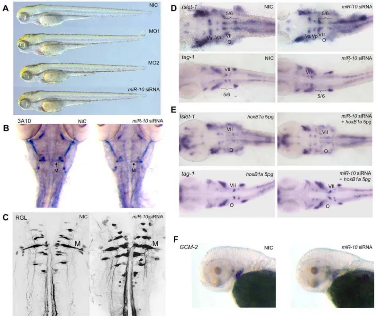
The phenotypes of morphant and overexpression embryos are remarkably normal during the first 5 days of development and they have no apparent defects (figure 6A, 72 hpf embryos shown). However, as miR-10appears to target HoxB1aand HoxB3a and possibly other1and3paralogue genes, the phenotype ofmiR-10 Figure 4. Effect ofmiR-10knockdown and overexpression on endogenousHoxtarget transcripts.A) Whole mountin situhybridization with probes for
hoxB1a,B2a,B3a,B4aandB5aon 24hpf embryos injected with morpholino reagent 1 or 2 (MO1 or MO2),miR-10siRNA or non injected controls (NIC). To allow quantitative detection, embryos hybridized with the same probe were stained equally long and staining was continuously monitored and stopped before reaching signal saturation.HoxB1aandHoxB3arespond to both gain and loss of function (arrows) ofmiR-10with a decrease and increase in expression levels respectively.HoxB2a,HoxB4aandHoxB5aare unresponsive tomiR-10overexpression or knockdown. B) Double whole mountin situ
hybridization on 24 hr embryos using probes forhoxB1aandhoxB4ashowing the different responses of the genes. Embryos were stained equally long till adequate staining was obtained for thehoxB4aprobe. C)In situhybridization withHoxA1a and HoxA3aon 24 hpf embryos injected with MO1, MO2 or
miR-10siRNA.HoxA1aresponds to both overexpression and knockdown. The transcript level ofHoxA3adoes not respond to either overexpression or knockdown. D)In situhybridization withHoxB1bon 24 hpf embryos morphant and overexpression embryos. There is strong upregulation ofHoxB1b
expression in the morphants but no downregulation is observed in themiR-10siRNA injected embryos. doi:10.1371/journal.pone.0001396.g004
overexpression is expected to combine at least the loss of function phenotypes for these genes.
In Zebrafish, morpholino studies have shown that HoxB1a is required for the correct patterning of rhombomere 4 together with
HoxB1b[42]. Single knockdown ofHoxB1aresults in a failure of the branchiomotor neurons of the VIIth cranial nerve to migrate out of rhombomere 4. In the double knockdown ofHoxB1aand
HoxB1b,there is additional absence of the rhombomere 4 primary Mauthner neurons.
Morpholino knockdown ofHoxB3aandHoxA3ahas been shown to result in downregulation of the gcm-2 gene in the branchial arches in Zebrafish [43].
Analysis of themiR-10 overexpression phenotype by immuno-labeling with the primary neuron specific 3A10 antibody shows that the Mauthner neurons are still present (figure 6B). Retrograde labeling in 5 days old embryos also reveals a normal pattern of reticulospinal neurons projecting from the hindbrain into the spinal cord (figure 6C).
Hindbrain branchiomotorneurons were visualized in in situ
hybridization withislet-1andtag-1. Islet-1stains the bodies of the Vth, VIIth, IXth and Xth nerves andtag-1is specifically expressed in the migrating VIIth nerve.
In situhybridization on 30 hpfmiR-10injected embryos shows that branchiomotor neurons of the VIIth nerve no longer migrate into rhombomere 5 and 6 but remain in rhombomere 4 (figure 6D). The pattern of the Vth, IXth and Xth branchiomotor nerves as visualized byislet-1 appears normal. To show that the VIIth nerve defect is directly due to targeting ofHoxB1abymiR-10, we rescued themiR-10
overexpression by co-injecting 5pgHoxB1aRNA from a construct that does not contain any of the target sites.
Injection of 5 pgHoxB1aalone does not induce any phenotype. Co-injection withmiR-10 siRNA restores migration of the VIIth nerve as show by bothislet-1andtag-1 in situhybridization (figure 6E). To show targeting ofHoxB3a, 72 hpf embryos were analyzed for the expression of gcm-2. In miR-10 overexpression embryos we observe downregulation ofgcm-2(figure 6F) in the branchial arch region as would be expected for embryos with impairedHoxA3a
and/orHoxB3aexpression [43].These analyses show thatmiR-10
is able to induce specific phenotypes associated with the loss of function ofHoxB1aandHoxB3a/HoxA3agenes but not ofHoxB1b. Analysis of the same genes inmiR-10morphant embryos shows patterns similar to wildtype embryos (figure S3).
MiR-10 acts synergistically with HoxB4
In Xenopus laevis, overexpression of HoxB4 has been reported to repress the expression ofHoxB1andHoxB3in neuralized animal caps [44]. Considering that these are the same genes that are targeted by miR-10 and that there exists a close association between the microRNA gene and theHoxB4open reading frame, this could indicate a synergistic action betweenmiR-10andHoxB4. To test this hypothesis we overexpressed HoxB4 and miR-10
individually and combined. Embryos were injected with 150pg
hoxB4 RNA, miR-10 siRNA or a combination of the two and analyzed forHoxB1aandHoxB3aexpression. Injection of 150 pg
Xenopus laevis HoxB4 RNA strongly represses the hindbrain rhombomere 4 expression of HoxB1a and rhombomere 5/6 expression ofHoxB3a(figure 7A).
However, forHoxB1athere is still a weak r4 domain detectable and for HoxB3a a discrete r5/6 stripe of expression is present. When coexpressed with miR-10, we observe a complete
disap-Figure 5. Retinoid induction ofmiR-10cand upregulation ofHoxB1ainmiR-10morphants.A) LNAin situhybridization formiR-10cin wildtype (WT) and 1026M retinoic acid (RA) treated embryos. RA treatement results inmiR-10cupregulation. B) Presence of a DR[2] type retinoic acid response element (RARE) 1kb 39of the ZebrafishHoxB1agene. This sequence is conserved in the mouse in which it has been shown to mediate the neural response ofHoxB1to RA [38] C) Different response ofHoxB1ato RA stimulation in wildtype ormiR-10morphant embryos.HoxB1ais strongly upregulated inmiR-10morphants. Injection with themiR-10siRNA has no effect.HoxB4aresponds similar to all conditions.
doi:10.1371/journal.pone.0001396.g005
pearance of the rhombomere 4 HoxB1a expression stripe (figure 7A). ForHoxB3a, there still is expression in the hindbrain but a discrete r5/6 domain is no longer discernable (figure 7A). In addition to this, we observe a stronger phenotype at 48 hpf, with more severe anterior and posterior truncations in embryos injected with the combination of HoxB4 RNA and miR-10 siRNA (figure 7B).In situhybridization for the endogenousHoxB4ashows that in theHoxB4+miR-10siRNA co-injected embryos, the parts anterior and posterior to the endogenous HoxB4a domain are reduced more strongly than in embryos injected withHoxB4aonly. These experiments indicate thatmiR-10synergizes withHoxB4in the repression ofHoxB1aand HoxB3aand also attains a greater posteriorizing activity in the presence ofmiR-10.
Evolutionary conservation of the target sites in the
HoxB cluster
Evolutionary conservation of sequence information is considered a good indicator of functionality and is used in microRNA target prediction programs [34] to assign confidence levels. We searched the anterior part of theHoxB(a)clusters in Medaka, Three spined stickleback, Tetraodon, Takifugu, Xenopus, Oppossum, Mouse, Rat, Cow and Human for the presence of putativemiR-10target sites (seed nucleotide 1-7). In figure 8A, the anterior parts of theHoxB
andHoxBa cluster homologues are shown with indication of the identified seed sequences. The conservation of themiR-10target sites in the 39UTR and coding regions ofHoxB3(a)genes is clear;
Figure 6. Overexpression ofmiR-10inducesHoxB1aandHoxB3aloss of function phenotypes.A) Wildtype (WT),miR-10morphant (MO1, MO2) andmiR-10siRNA overexpression embryos at 72 hpf show no apparent developmental differences. B) Mauthner neuron development as visualized by 3A10 neurofilament immunostaining in 72 hpf embryos shows no differences betweenmiR-10siRNA injected embryos and controls. C) Confocal images of reticulospinal hindbrain neurons in retrograde labeled, 5 day old embryos. Wildtype andmiR-10siRNA injected embryos are similar. D) Islet-1andtag-1 in situhybridization on 30 hpf wildtype andmiR-10siRNA injected embryos. Flatmounts of head regions are shown. In wildtype embryos the VIIth cranial nerve migrates into rhombomere 5/6 at the level of the otic vesicle. InmiR-10siRNA injected embryos the VIIth nerve does no longer migrate out of rhombomere 4. E) Co-injection of 5pgHoxB1aRNA rescues themiR-10siRNA induced migration defect of the VIIth cranial nerve as shown byislet-1andtag-1 in situhybridization. F)Gcm-2expression is downregulated inmiR-10siRNA injected embryos, which is consistent with repression ofHoxB3a.
doi:10.1371/journal.pone.0001396.g006
all species investigated have at least 2 target sites associated with the ORF or 39UTR region. The sites inHoxB1(a)show a weaker conservation profile and are most prominently present in Zebrafish. All of theTeleosts for which sequence information could be found (note that the available HoxBa Medaka contig stops 300 nt downstream ofHoxB1) posses a candidatemiR-10target site in the 39 UTR of theirHoxB1a gene. This further adds to the implied relevance of the repression ofHoxB1aandHoxB3abymiR-10.
A polycistronic transcript including both HoxB3a
and miR-10c is targeted by miR-10
A transcript,HoxB3asplv2 [45], has been described, which starts 39ofHoxB5a, has two exons originating 39ofHoxB4aand includes the main HoxB3a open reading frame (figure 8B). The primary unspliced form of this long transcript thus includes bothHoxB3a
and themiR-10cmicroRNA.
In situhybridization with a 59UTR probe shows that expression of this transcript obeys the rules of colinearity and that its rostral expression boundary thus corresponds to the position of its transcriptional start site (i.e. expression similar to that ofHoxB5a) (figure 8C) and [46]. This transcript is thus expressed more posteriorly than the main HoxB3a expression domain and completely within the domain of the miR-10c microRNA, a feature also expected from the presence ofmiR-10con theHoxB3a
splv2 primary transcript. This transcript includes the fullHoxB3a
open reading frame together with the 3 miR-10 target sites. Morpholino knockdown of miR-10 leads to upregulation of this transcript in bothin situhybridization and RT-PCR (figure 8D, E), confirming that it is indeed targeted bymiR-10in vivo. In this case themiR-10cmicroRNA apparently acts on parts of its own primary transcript and is therefore autoregulatory.
DISCUSSION
MiR-10is expressed in the hindbrain and spinal cord posterior to the rhombomere 6/7 boundary and occupies an axial domain similar to those ofHox-4paralogue genes. We reveal an interaction between miR-10 and the anterior HoxB1a and HoxB3a genes. TheseHox genes have strong anterior expression domains in the hindbrain and are expressed at a low level in the spinal cord where their expression overlaps withmiR-10expression (figure 9A). The upregulation of the target genes in the morphant embryos shows that the target genes are indeed repressed bymiR-10within this posterior domain.
Overexpression of miR-10 indeed induces the phenotypes associated with the loss ofHoxB1aandHoxB3a. The very specific phenotype induced in the overexpression experiments is striking. The embryos are virtually indistinguishable from wildtypes, apart from the VIIth nerve defect and alteredgcm-2expression. In the target prediction section of miRBase, where the output of the Miranda algorithm (http://microrna.sanger.ac.uk/) is listed, there are however 1969 predicted target genes for Zebrafish miR-10. This high number of predicted target genes seems incompatible with the very specific phenotypic defects observed in themiR-10
overexpression embryos and strongly suggest that, at least for miR-10, there is a high component of false positives in the outcome of target prediction algorithms.
We observe that the HoxB4 overexpression phenotype is significantly enhanced bymiR-10. The close genomic association of these two genes and their synergistic activity could indicate that these genes are part of the same genetic unit. The system ofHox
regulation is characterized by the phenomenon of posterior prevalence, meaning that there is a hierarchy in the functioning of theHoxgenes, such that posterior genes are always dominant in the
Figure 7.MiR-10acts in synergy withHoxB4.A) Embryos injected withHoxB4,miR-10siRNA andHoxB4+miR-10siRNA analyzed for the expression of
HoxB1aandHoxB3aat 24 hpf. Injection of 150pgHoxB4leads to downregulation ofHoxB1aand of downregulation of the hindbrain domain ofHoxB3a. The rhombomere 4 expression domain ofHoxB1aand the rhombomere 5/6 expression domain ofHoxB3aare still discernable though. When 150pgHoxB4
is expressed together withmiR-10siRNA the expression domain ofHoxB1adisappears and no clear rhombomere 5/6 stripe ofHoxB3aexpression can be detected. B) Embryos injected withHoxB4,miR-10siRNA andHoxB4+miR-10siRNA analyzed for the expression of endogenousHoxB4aat 48 hpf. The combination ofHoxB4together with themiR-10siRNA induces a stronger phenotype with more severe anterior and posterior truncations than injection withHoxB4alone. On the right groups of embryos injected withHoxB4or the combination ofHoxB4andmiR-10siRNA are shown.
doi:10.1371/journal.pone.0001396.g007
determination of a regional phenotype over coexpressed anterior genes [2]. In the overexpression experiments it looks as if miR-10
facilitates a full posteriorizing activity of HoxB4. This synergistic action betweenmiR-10andHoxB4suggests that post-transcriptional gene regulation by microRNAs plays a role in posterior prevalence. We find thatHoxB1aandHoxB3aare targeted bymiR-10. Other
Hox-1 and Hox-3 paralogues genes are present in different Zebrafish Hoxclusters but are not targeted. On the basis of our experiments and the presence of putative target sites, onlyHoxA1a,
HoxB1a,HoxA3aandHoxB3agenes are candidatemiR-10targets. Whether they are true targets remains to be seen;HoxA1a, despite not being near to a clear target site, responds strongly to the loss and gain ofmiR-10.HoxB1bpossesses a candidate target site and is upregulated in morphant embryos. What however argues strongly against the targeting ofHoxB1bis that the overexpression of miR-10 does not induce the same phenotypic changes as observed in the doubleHoxB1a/HoxB1bknockdown [42]. As there is extensive crossregulation betweenHoxparalogues it is also possible that the effects observed are a direct result of the derepression ofHoxB1a.
HoxA3a also possesses one candidate target site but seems unaffected by gain and loss ofmiR-10. MicroRNAs affect target genes both by inhibition of translation and by degradation of messengerRNA [20,36]. To what extent these processes are coupled and whether translational inhibition is always accompa-nied by an increase in messenger RNA decay is not yet known. It is thus theoretically possible that the effect ofHoxA3arepression will only be noticeable at the protein level.
There could be several reasons whymiR-10 targets particular
Hox-1andHox-3genes and not others. One explanation would be that there is a high degree of subfunctionalization within these paralogue groups, as was nicely illustrated for the ZebrafishHox-1
genes [42]. This could create needs for post-transcriptional silencing that differ from one paralogue member to another.
The inhibition ofmiR-10leads to posterior upregulation of the targeted genes. Since microRNAs cause downregulation of their target messenger RNAs, it has been a frequently debated issue whether low or absent levels of target gene expression within the microRNA domain reflect different domains of transcription or whether they are a direct consequence of the downregulation by the microRNA [e.g. 47]. Inhibition ofmiR-10leads to posterior target gene upregulation in case ofHoxB1a,HoxA1aand HoxB3a
but certainly not to the same high level found in their dominant anterior expression domains. It thus appears that restriction to the dominant expression domain occurs primarily at the transcrip-tional level and that it is within a posterior domain with an already low level of transcription that silencing by the microRNA occurs. This observation is consistent with the identification of rhombo-mere specific transcriptionalHoxenhancers in mouse [e.g. 48, 49]. In case of the microRNA target interactions described in this study, it seems that both different transcriptional domains and a direct repression by the microRNA shape the mRNA expression domains in the embryo. The effects at the transcript level likely reflect an on/off situation at the protein level where all translation is silenced although there is still a significant amount of messengerRNA detectable.
Why are there post-transcriptional gene regulatory
interactions within the Hox clusters?
In general it is poorly understood why functional domains of genes are sometimes restricted post-transcriptionally instead of by transcriptional silencing. The emerging view for the microRNAs
Figure 8. Evolutionary conservation ofmiR-10targetsites and autoregulation ofmiR-10c.A) PutativemiR-10target sites are indicated by seed sequences in the sense strand of the anterior vertebrateHoxB(a)clusters. Seed sequences are shown in green, open reading frames are indicated in light blue. Note conserved association of target sites with theHoxB3(a)ORF and conserved presence of a putative target site inTeleost HoxB1a. B) The
HoxB3asplv2 polycistronic transcript includes one exon betweenHoxB4aandHoxB5a, two exons betweenHoxB4aandHoxB3aand the mainHoxB3a
coding sequence. The primary transcript for this isoforms includesmiR-10c. The 59UTR sequence is shown in orange, this sequence corresponds to the probe used in C and D to specifically detect this splice isoforms. C) Comparison of theHoxB3aexon1 expression (red) and the expression of
HoxB3asplv2 (purple).HoxB3asplv2 is expressed posterior to the main rhombomere 5/6 expression domain ofHoxB3aas reported previously [46]. The staining reaction forHoxB3asplv2 was developed for much longer than the reaction for theHoxB3aexon1 probe and theHoxB3asplv2 is presumably expressed at a much lower level. D)In situhybridization withHoxB3asplv2. Expression is upregulated inmiR-10morphant embryos (arrows). E) Semi quantitative RT-PCR for theHoxB3asplv2 59UTR,ß-actinis used as loading control.HoxB3ais upregulated inmiR-10morphant embryos.HoxB3asplv2: 31 cycles,ß-actin: 22 cycles.
doi:10.1371/journal.pone.0001396.g008
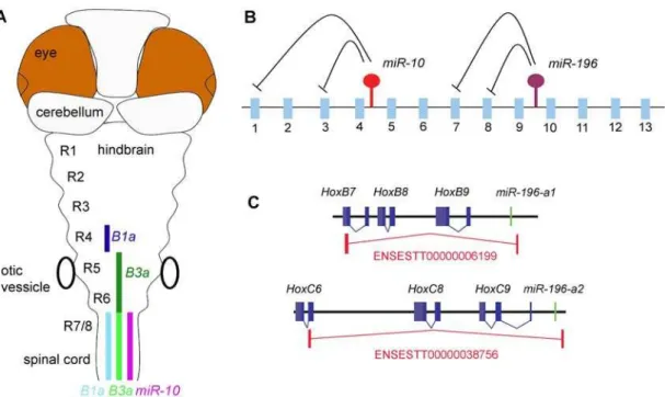
in theHoxclusters is that they target codingHoxgenes (figure 9B) that can even be located within the same clusters. These microRNAs thus seem to be involved in post-transcriptional gene regulatory interactions with genes that are located in their very close vicinity. The short genomic distances betweenmiR-196and
miR-10and their targets are remarkable;miR-10c is,25 kb from the target sites inHoxB3aand,48 kb from those inHoxB1aand (in mammals) a miR-196 paralogue is located at ,18 kb from HoxB8andHoxC8and at,14 kb fromHoxA7.
In this light, the presence ofmiR-10cand its targetHoxB3aon a single primary transcript is also interesting. ThemiR-10c/HoxB3a
polycistronic transcript includes both the microRNA and a target gene. In this case, the microRNA thus acts in an autoregulatory fashion on parts of its original precursor. The HoxB3a splv2 transcript itself appears to be expressed exclusively within the expression domain of the microRNA and is thus never expected to be translated into a functionalHoxB3aprotein. Similar transcripts are present in the EST database formiR-196a-1/HoxB8and miR-196a-2/HoxC8 (figure 9C). The inclusion of microRNAs and target genes on the same transcription unit is counterintuitive in the sense that one wonders why the target genes are not simply omitted from the transcript as they are silenced by the accompanying microRNA anyway.
A possible explanation for the presence of target gene/microRNA combinations within single transcription units and for the targeting of nearbyHoxgenes by bothmiR-10andmiR-196, may lie in the complexity of theHoxregulatory mechanisms which involve multiple global and local transcriptional elements. The high selective pressure to maintain the clustered genomic organization of vertebrate Hox
genes probably results from the presence of global enhancers located
outside of the clusters and from dependence on sharing of local enhancers [50]. As a result, theHoxclusters consist of closely spaced transcription units and enhancer regions. The high density of transcription units could easily cause them to interfere with one another and make the system prone to inappropriate enhancer sharing, resulting in ectopic expression. The extensive amount of ‘strange’ polycistronic and antisense transcripts being produced from theHoxclusters [51] could result from this. These transcripts do not necessarily have any function but could represent inherent transcriptional ‘noise’. It is interesting that it is specifically the nearest genes that are silenced post- transcriptionally as these are the ones most likely to be influenced by the same enhancers as the microRNA genes. The posterior expression domains of these anteriorHox genes could well be a consequence imposed on the transcriptional process by the clustered nature of the genes and they do not necessarily serve any function. We suggest that an inability to separate the transcriptional controls of several Hox genes is the selective force driving the post-transcriptional gene silencing relationships within theHoxclusters.
In vertebrates, Hox genes have stayed clustered throughout evolution. However, in the sister group of tunicates (Ciona and
Oikopleura), the Hox clusters have broken up and are present in separate regions of the genome [52,53]. It is interesting to note that the miR-10 microRNA has been lost from the tunicates [17,54]. This observation provides a possible phylogenetic link between post-transcriptional gene silencing and gene clustering.
It would be interesting to see whether it is possible to extrapolate these observations to other microRNA/(predicted) target pairs and see if similar constraints can be identified that possibly account for the involvement of post-transcriptional gene regulation.
Figure 9. Post-transcriptional regulatory interactions within the hox clusters.A) Schematic representation ofmiR-10and target gene expression in the Zebrafish hindbrain.MiR-10is expressed posterior from the rhombomere 6/7 boundary. The target genesHoxB1aandHoxB3aare expressed in a strong domain (dark colour) anterior in the anterior hindbrain and in a weaker domain (light colour) in the area where they overlap withmiR-10.
HoxB1a shows a gap in expression in r5 and 6, possibly due to stronger transcriptional repression. B) Schematic representation of the post-transcriptional relations within the hox clusters.MiR-196is known to repressesHoxB8,HoxC8,HoxD8andHoxA7and we have identifiedHoxB1aand
HoxB3aas targets formiR-10. The emerging view is that the microRNAs in the hox clusters target more anterior genes in their close proximity. C) Polycistronic transcripts identified from the EST database show inclusion ofmiR-196paralogues andHoxB8andHoxC8target genes on the same primary transcripts.
doi:10.1371/journal.pone.0001396.g009
MATERIALS AND METHODS
Zebrafish husbandry and embryo culturing
An AB6TL strain of Zebrafish was used for all experiments;
housing and embryo collection was according to standard procedures; embryos were cultured at 28uC.
RT-PCR
Whole embryo RNA was isolated using Tri-pure (Roche #1667165) and reverse transcribed with MuMlv Reverse transcriptase (Promega) using oligo-dT N = 18.
Primer sequences;
miR-10cup (AGCTGGCTTTCTCAATACC)
miR-10cdow n (TACATACTCCCCTAGATACGAA)
HoxB4aexon1 up (ATGGCCATGAGTTCCTATTTG)
HoxB4aexon1 down (TTGGTTCACCCCCTGAATAG)
HoxB4aexon1 59down (TTGTGGGTAGAACGTGACCTC)
HoxB3asplv2 59UTR up (CAGTGCCAGTGTCTAGTCAG)
HoxB3a splv2 59UTR down (GTAATACGACTCACTATAG-GCTCTTTCCAATGGCCTCTTGG)
b-Actinup (CGAGCAGGAGATGGGAACC)
b-Actindown (CAACGGAAACGCTCATTGC)
DNA oligos were obtained from Biolegio, Malden, The Netherlands.
Micro-injection
Embryos were injected with 1 or 2 nl at the zygote stage; RNAse free phenol red was added as tracer to injection mixtures prior to injections.
Morpholinos were obtained from genetools, OR, USA;miR-10
morpholino reagent 1 corresponds to a mix ofmiR-10a (CACAA-ATTCGGATCTACAGGGTA) and miR-10b (CACAAATTCG-GTTCTACAGGGTA) antisense morpholino, the sequence of
miR-10 morpholino reagent 2 is (TCTACAGGGTATATATA-GACGAC).
RNA oligos were obtained from Biolegio, Malden, The Netherlands.
ThemiR-10siRNA sense strand corresponds to a mix of miR-10a (UACCCUGUAGAUCCGAAUUUGUGUG) and miR-10b
(UACCCUGUAGAACCGAAUUUGUGUG), sequence of the antisense strand is (CACAAAUUCGGAUCUACAGGGGCAU). Note that the antisense sequence has mismatches with themiR-10
sense strand at its 39end resulting in the specific incorporation of the sense miR-10 strand in the microRNA silencing complex. Oligos were annealed to siRNAs in by gradually cooling from 98uC to 20uC. in buffer in 500ml H2O beaker glass; 30 ul 50mM of each
oligo, 15ml annealing buffer (50 mM Tris, pH 7.8, 100 mM NaCl RNAse free) in 75ml, final concentration of siRNA is 20mM.
RNA for injection was transcribed using Ambion Sp6 message machine kit (# 1340) and purified using an RNA easy column (Qiagen), from the CS2+plasmids; CS2+HoxB1asensor wt, CS2+
HoxB1asensor mut, CS2+HoxB3asensor wt, CS2+HoxB3asensor mut, CS2+Dre-HoxB3a ORF, CS2+Xl-HoxB4-Myc, CS2+E-YFP, CS2+E-CFP.
In situ Hybridization
In situ hybridization was performed according to standard procedures and Kloosterman et al. [32]. Hybridization tempera-tures were 65uC for normal probes and 56uC for LNA probes. In doublein situhybridization with a LNA probe 56uC was used. In
doublein situ hybridization DIG and fluorescein labeled probes were used. Embryos were stained using BM-Purple (Roche #11442674001) and Fast Red (Roche #11496549001). Probes were synthesized using T7 and Sp6 polymerase (Promega) in the presence of labeled nucleotides (RNA DIG or fluorescein labeling mix, Roche #11277073910 and #10805221) from pGEM-TE plasmids containing:HoxB1a,HoxB3a, HoxB4a and HoxB5aexon 1 coding sequence,HoxB2aexon 2-39UTR,HoxB1bexon1-2 coding sequence;HoxB3a splv2 was synthesized from PCR product from a partial cDNA cloned in pGEM-TE.
LNA probes were obtained from Exiqon, Denmark and sequences are:
miR-10a(CACAAATTCGGATCTACAGGGTA),
miR-10b(ACAAATTCGGTTCTACAGGGTA),
miR-10c(CACAAATCCGGATCTACAGGGTA),
miR-10d(ACACATTCGGTTCTACAGGGTA ).
Probes were labeled using the DIG labeling kit (Roche #03353575910) and purified before use over a microspin G-25 column (Amersham #27-5325-01) Our step by step in situ
hybrdization protocol is available on request.
Nothern Blot
Northern Blot was performed essentially according to Kloosterman et al. [55]. Total RNA was extracted using Tri-Pure (Roche #1667165). 3mg RNA was separated on a 15% denaturing PAGE gel using a Biorad minigel system and subsequently blotted using a semidry blotter (175 mA constant, 10–20V for 25–30 min.) to a postitively charged nylon membrane (Roche #1417240). Mem-branes were pre-hybridized at 60uC for 1 hr in hybridization buffer (0.36 M Na2HPO4, 0.14M NaH2PO4, 1 mM EDTA, 7%SDS,
0.1 mg/ml yeast tRNA, 0.04% Blocking reagent (Roche#1096176)) and subsequently hybridized overnight at 60uC in hybridization buffer containing miR-10 LNA probes, labeled and purified as mentioned above, and diluted 1:50.000. The next day blots were washed 16at 60uC with hybridization buffer, 16at 50uC with 26
SSC, 0.1% SDS and 16at 50uC with 0.1xSSC, 0.1%SDS in order
to remove excess probe. Blots were incubated 26for 5 min. at RT in
Maleic Acid buffer (0.1 M Maleic Acid, 150mM NaCl, pH7.5 with NaOH, 0.1% Tween-20) to equilibrate and remove residual SDS. Blots were blocked for 30 min. in blocking buffer (Maleic Acid buffer containing 1% Blocking Reagent (Roche #1096176)) and subse-quently incubated for 30 min. in blocking buffer containing 1:50.000 Anti-Digoxigenin-AP antibody (Roche#093274). Excess antibody was washed away in 4615 min. washes with Maleic Acid buffer.
Blots were subsequently washed 5 minutes in AP-buffer (0.1 M Tris Base, 0.1 M NaCl, pH9.5) and signal was detected on X-Ray film using CDP-star kit (Roche#12041677001) according to manufac-turer’s instructions with a typical exposure time of 4 hours.
Retrograde labeling
Anesthetized 5 days old embryos were retrograde labeled by making an incision with a tungsten needle in the spinal cord at the level of the hindgut and injecting 1–5 nl of a concentrated rhodamine-dextran solution. After injection embryos were left to recover for 1–1.5 hrs and fixed in 4% PFA.
SUPPORTING INFORMATION
Figure S1
Found at: doi:10.1371/journal.pone.0001396.s001 (0.32 MB PDF)
Figure S2
Found at: doi:10.1371/journal.pone.0001396.s002 (0.38 MB PDF)
Figure S3
Found at: doi:10.1371/journal.pone.0001396.s003 (0.75 MB PDF)
ACKNOWLEDGMENTS
We would like to thank Nabila Bardine, Gabby Krens, Hans Jansen, Ferran Lloret Vilaspasa, Max Corredor-Ada´mez and Martje Jespers for useful discussion and assistance in the lab, Yanju Zhang and Fons Verbeek
for help with miRBase, Florian Steiner for advise on the Northern Blotting, Jacqueline Deschamps for useful comments on the manuscript, Victoria Prince for gift of theHoxB1aoverexpression construct and two anonymous reviewers for useful criticism and suggestions. This project was initiated at the Hubrecht Institute for Developmental Biology and Stem Cell Research.
Author Contributions
Conceived and designed the experiments: JW. Performed the experiments: JW. Analyzed the data: JW. Contributed reagents/materials/analysis tools: AD JW. Wrote the paper: AD JW. Other: Main author: JW. Lab head: AD. General supervision: AD. Provided facilities: AD. Paid JMW: AD.
REFERENCES
1. Boncinelli E, Mallamaci A, Lavorgna G (1994) Vertebrate homeobox genes. Genetica 94(2–3): 127–40.
2. Kmita M, Duboule D (2003) Organizing axes in time and space; 25 years of colinear tinkering. Science 301(5631): 331–3.
3. Krumlauf R (1994)Hoxgenes in vertebrate development. Cell 78(2): 191–201. 4. Pearson JC, Lemons D, McGinnis W (2005) ModulatingHoxgene functions
during animal body patterning. Nat Rev Genet 6(12): 893–904.
5. Amores A, Force A, Yan YL, Joly L, Amemiya C, et al. (1998) ZebrafishHox
clusters and vertebrate genome evolution. Science 282(5394): 1711–4. 6. Corredor-Ada´mez M, Welten MC, Spaink HP, Jeffery JE, Schoon RT, et al.
(2005) Genomic annotation and transcriptome analysis of the zebrafish (Danio rerio)Hoxcomplex with description of a novel member,Hoxb 13a. Evol Dev 7(5): 362–75.
7. Hoegg S, Boore JL, Kuehl JV, Meyer A (2007) Comparative phylogenomic analyses of Teleost fish Hox gene clusters: lessons from the cichlid fish Astatotilapia burtoni. BMC Genomics 8: 317.
8. Lim LP, Glasner ME, Yekta S, Burge CB, Bartel DP (2003) Vertebrate microRNA genes. Science 299(5612): 1540.
9. Lagos-Quintana M, Rauhut R, Meyer J, Borkhardt A, Tuschl T (2003) New microRNAs from mouse and human. RNA 9(2): 175–9.
10. Cummins JM, He Y, Leary RJ, Pagliarini R, Diaz LA Jr, et al. (2006) The colorectal microRNAome. Proc Natl Acad Sci USA 103: 3687–92. 11. Mineno J, Okamoto S, Ando T, Sato M, Chono H, et al. (2006) The expression
profile of microRNAs in mouse embryos. Nucleic Acids Res 34(6): 1765–71. 12. Bernstein E, Caudy AA, Hammond SM, Hannon GJ (2001) Role for a bidentate
ribonuclease in the initiation step of RNA interference. Nature 409(6818): 363–6. 13. Lee Y, Ahn C, Han J, Choi H, Kim J, et al. (2003) The nuclear RNase III
Drosha initiates microRNA processing. Nature 425(6956): 415–9.
14. He L, Hannon GJ (2004) MicroRNAs: small RNAs with a big role in gene regulation. Nat Rev Genet 5(7): 522–31.
15. Valencia-Sanchez MA, Liu J, Hannon GJ, Parker R (2006) Control of translation and mRNA degradation by miRNAs and siRNAs. Genes Dev 20(5): 515–24.
16. Woltering JM, Durston AJ (2006) The zebrafishHoxDbcluster has been reduced to a single microRNA. Nat Genet 38(6): 601–2.
17. Tanzer A, Amemiya CT, Kim CB, Stadler PF (2005) Evolution of microRNAs located withinHoxgene clusters. J Exp Zoolog B Mol Dev Evol 304(1): 75–85. 18. Wienholds E, Koudijs MJ, van Eeden FJ, Cuppen E, Plasterk RH (2003) The microRNA-producing enzyme Dicer1 is essential for zebrafish development. Nat Genet 35(3): 217–8.
19. Giraldez AJ, Cinalli RM, Glasner ME, Enright AJ, Thomson JM, et al. (2005) MicroRNAs regulate brain morphogenesis in zebrafish. Science 308(5723): 833–8.
20. Giraldez AJ, Mishima Y, Rihel J, Grocock RJ, Van Dongen S, et al. (2006) Zebrafish MiR-430 promotes deadenylation and clearance of maternal mRNAs. Science 312(5770): 75–9.
21. Flynt AS, Li N, Thatcher EJ, Solnica-Krezel L, Patton JG (2007) Zebrafish miR-214 modulates Hedgehog signaling to specify muscle cell fate. Nat Genet 39(2): 259–63.
22. Kloosterman WP, Lagendijk AK, Ketting RF, Moulton JD, Plasterk RH (2007) Targeted inhibition of miRNA maturation with morpholinos reveals a role for miR-375 in pancreatic islet development. PLoS Biol 5(8): e203.
23. Yekta S, Shih IH, Bartel DP (2004) MicroRNA-directed cleavage ofHOXB8 mRNA. Science 304(5670): 594–6.
24. Mansfield JH, Harfe BD, Nissen R, Obenauer J, Srineel J, et al. (2004) MicroRNA-responsive ‘sensor’ transgenes uncoverHox-like and other develop-mentally regulated patterns of vertebrate microRNA expression. Nat Genet 36(10): 1079–83.
25. Hornstein E, Mansfield JH, Yekta S, Hu JK, Harfe BD, et al. (2005) The microRNA miR-196 acts upstream ofHoxb8 and Shh in limb development. Nature 438(7068): 671–4.
26. Ronshaugen M, Biemar F, Piel J, Levine M, Lai EC (2005) The Drosophila microRNA iab-4 causes a dominant homeotic transformation of halteres to wings. Genes Dev 19(24): 2947–52.
27. Godsave SF, Koster CH, Getahun A, Mathu M, Hooiveld M, et al. (1998) Graded retinoid responses in the developing hindbrain. Dev Dyn 213(1): 39–49. 28. Kiecker C, Lumsden A (2005) Compartments and their boundaries in vertebrate
brain development. Nat Rev Neurosci 6(7): 553–64.
29. Dasen JS, Tice BC, Brenner-Morton s, Jessell TM (2005) AHoxregulatory network establishes motor neuron pool identity and target-muscle connectivity. Cell 123(3): 477–91.
30. Moens CB, Prince VE (2002) Constructing the hindbrain: insights from the zebrafish. Dev Dyn 224(1): 1–17.
31. Wienholds E, Kloosterman WP, Miska E, Alvarez-Saavedra E, Berezikov E, et al. (2005) MicroRNA expression in zebrafish embryonic development. Science 309(5732): 310–1.
32. Kloosterman WP, Wienholds E, de Bruijn E, Kauppinen S, Plasterk RH (2006)
In situ detection of miRNAs in animal embryos using LNA-modified oligonucleotide probes. Nat Methods 3(1): 27–9.
33. Lewis BP, Burge CB, Bartel DP (2005) Conserved seed pairing, often flanked by adenosines, indicates that thousands of human genes are microRNA targets. Cell 120(1): 15–20.
34. John B, Enright AJ, Aravin A, Tuschl T, Sander C, et al. (2004) Human MicroRNA targets. PLoS Biol 2(11): e363.
35. Kloosterman WP, Wienholds E, Ketting RF, Plasterk RH (2004) Substrate requirements for let-7 function in the developing zebrafish embryo. Nucleic Acids Res 32(21): 6284–91.
36. Lim LP, Lau NC, Garrett-Engele P, Grimson A, Schelter JM, et al. (2005) Microarray analysis shows that some microRNAs downregulate large numbers of target mRNAs. Nature 2005 433(7027): 769–73.
37. Mainguy G, In der Rieden PM, Berezikov E, Woltering JM, Plasterk RH, et al. (2003) A position-dependent organisation of retinoid response elements is conserved in the vertebrate Hox clusters. Trends Genet 19(9): 476–9. 38. Huang D, Chen SW, Gudas LJ (2002) Analysis of two distinct retinoic acid
response elements in the homeobox gene Hoxb1 in transgenic mice. Dev Dyn 223(3): 353–70.
39. Studer M, Gavalas A, Marshall H, Ariza-McNaughton L, Rijli FM, et al. (1998) Genetic interactions between Hoxa1 and Hoxb1 reveal new roles in regulation of early hindbrain patterning. Development 125(6): 1025–36.
40. Langston AW, Thompson JR, Gudas LJ (1997) Retinoic acid-responsive enhancers located 39 of the Hox A and Hox B homeobox gene clusters. Functional analysis. J Biol Chem 272(4): 2167–75.
41. Sirbu IO, Gresh L, Barra J, Duester G (2005) Shifting boundaries of retinoic acid activity control hindbrain segmental gene expression. Development 132(11): 2611–22.
42. McClintock JM, Kheirbek MA, Prince VE (2002) Knockdown of duplicated zebrafishHoxb1 genes reveals distinct roles in hindbrain patterning and a novel mechanism of duplicate gene retention. Development 129(10): 2339–54. 43. Hogan BM, Hunter MP, Oates AC, Crowhurst MO, Hall NE, et al. (2004)
Zebrafish gcm-2 is required for gill filament budding from pharyngeal ectoderm. Dev Biol 276(2): 508–22.
44. Hooiveld MH, Morgan R, in der Rieden P, Houtzager E, Pannese M, et al. (1999) Novel interactions between vertebrate Hox genes. Int J Dev Biol 43(7): 665–74.
45. Hadrys T, Prince V, Hunter M, Baker R, Rinkwitz S (2004) Comparative genomic analysis of vertebrateHox3 andHox4 genes. J Exp Zoolog B Mol Dev Evol 302(2): 147–64.
46. Hadrys T, Punnamoottil B, Pieper M, Kikuta H, Pezeron G, et al. (2006) Conserved co-regulation and promoter sharing of Hoxb3a andHoxb4a in zebrafish. Dev Biol 297(1): 26–43.
47. Plasterk RH (2006) Micro RNAs in animal development. Cell 124(5): 877–81. 48. Ferretti E, Cambronero F, Tumpel S, Longobardi E, Wiedemann LM, et al.
(2005) Hoxb1 enhancer and control of rhombomere 4 expression: complex interplay between PREP1-PBX1-HOXB1 binding sites. Mol Cell Biol 25(19): 8541–52.
49. Kwan CT, Tsang SL, Krumlauf R, Sham MH (2001) Regulatory analysis of the mouseHoxb3 gene: multiple elements work in concert to direct temporal and spatial patterns of expression. Dev Biol 232(1): 176–90.
50. Duboule D (1998) Vertebrate Hox gene regulation: clustering and/or colinearity? Curr Opin Genet Dev 8(5): 514–8.
51. Mainguy G, Koster J, Woltering J, Jansen H, Durston A (2007) Extensive polycistronism and antisense transcription in the MammalianHoxclusters. PLoS ONE 2(4): e356.
52. Ikuta T, Yoshida N, Satoh N, Saiga H (2004) Ciona intestinalisHoxgene cluster: Its dispersed structure and residual colinear expression in development. Proc Natl Acad Sci U S A 101(42): 15118–23.
53. Seo HC, Edvardsen RB, Maeland AD, Bjordal M, Jensen MF, et al. (2004)Hox
cluster disintegration with persistent anteroposterior order of expression in Oikopleura dioica. Nature 431(7004): 67–71.
54. Prochnik SE, Rokhsar DS, Aboobaker AA (2007) Evidence for a microRNA expansion in the bilaterian ancestor. Dev Genes Evol 217(1): 73–7.
55. Kloosterman WP, Steiner FA, Berezikov E, de Bruijn E, van de Belt J, et al. (2006) Cloning and expression of new microRNAs from zebrafish. Nucleic Acids Res 34(9): 2558–69.