Implicit Memory in Monkeys: Development
of a Delay Eyeblink Conditioning System with
Parallel Electromyographic and High-Speed
Video Measurements
Yasushi Kishimoto1☯, Shigeyuki Yamamoto2☯, Kazutaka Suzuki2, Haruyoshi Toyoda2,
Masanobu Kano3, Hideo Tsukada2, Yutaka Kirino1*
1Department of Neurobiophysics, Kagawa School of Pharmaceutical Sciences, Tokushima Bunri
University, Sanuki, Kagawa, Japan,2Central Research Laboratory, Hamamatsu Photonics K.K., Hamakita-ku, Hamamatsu, Japan,3Department of Neurophysiology, Graduate School of Medicine, University of Tokyo, Bunkyo-ku, Tokyo, Japan
☯These authors contributed equally to this work. *[email protected]
Abstract
Delay eyeblink conditioning, a cerebellum-dependent learning paradigm, has been applied to various mammalian species but not yet to monkeys. We therefore developed an accurate measuring system that we believe is the first system suitable for delay eyeblink conditioning in a monkey species (Macaca mulatta). Monkey eyeblinking was simultaneously monitored by orbicularis oculi electromyographic (OO-EMG) measurements and a high-speed cam-era-based tracking system built around a 1-kHz CMOS image sensor. A 1-kHz tone was the conditioned stimulus (CS), while an air puff (0.02 MPa) was the unconditioned stimulus. EMG analysis showed that the monkeys exhibited a conditioned response (CR) incidence of more than 60% of trials during the 5-day acquisition phase and an extinguished CR dur-ing the 2-day extinction phase. The camera system yielded similar results. Hence, we con-clude that both methods are effective in evaluating monkey eyeblink conditioning. This system incorporating two different measuring principles enabled us to elucidate the relation-ship between the actual presence of eyelid closure and OO-EMG activity. An interesting finding permitted by the new system was that the monkeys frequently exhibited obvious CRs even when they produced visible facial signs of drowsiness or microsleep. Indeed, the probability of observing a CR in a given trial was not influenced by whether the monkeys closed their eyelids just before CS onset, suggesting that this memory could be expressed independently of wakefulness. This work presents a novel system for cognitive assessment in monkeys that will be useful for elucidating the neural mechanisms of implicit learning in nonhuman primates.
OPEN ACCESS
Citation:Kishimoto Y, Yamamoto S, Suzuki K, Toyoda H, Kano M, Tsukada H, et al. (2015) Implicit Memory in Monkeys: Development of a Delay Eyeblink Conditioning System with Parallel Electromyographic and High-Speed Video Measurements. PLoS ONE 10(6): e0129828. doi:10.1371/journal.pone.0129828
Academic Editor:Hisao Nishijo, University of Toyama, JAPAN
Received:February 17, 2015
Accepted:May 13, 2015
Published:June 12, 2015
Copyright:© 2015 Kishimoto et al. This is an open access article distributed under the terms of the
Creative Commons Attribution License, which permits unrestricted use, distribution, and reproduction in any medium, provided the original author and source are credited.
Data Availability Statement:All relevant data are within the paper and its Supporting Information files.
Introduction
Classical eyeblink conditioning is one of the best-characterized behavioral models of associa-tive learning in mammals [1,2]. In this type of learning task, after repeated presentation of a conditioned stimulus (CS, e.g., tone) paired with an unconditioned stimulus (US, e.g., air puff), subjects learn to close the eye before onset of the US so that the eye is closed when the air puff is delivered. Most eyeblink conditioning can be classified into two distinct types: delay and trace conditioning [1,3]. The standard delay paradigm in which the CS and US are co-terminal in time but the CS starts earlier is cerebellum-dependent and is supposed to be unrelated to awareness in humans [4–8]. This paradigm has been used extensively for assessing motor learning or implicit memory performance in a variety of mammalian species including human, cat, rabbit, rat, and mouse [9–14], as well as reptiles [15]. The delay version of eyeblink condi-tioning in monkeys has not yet been reported although trace eyeblink condicondi-tioning in monkeys was investigated using an infrared camera [16]. However, no attempt has been made so far to evaluate eyeblink conditioning using electromyographic (EMG) methods or video monitoring, which are the de facto standard methods for evaluating eyelid response activities during eye-blink conditioning in many species including humans and rodents [9,10,16–19].
Hence, the primary aims of the present study were three-fold: first, to examine whether delay eyeblink conditioning using an EMG measuring method is possible in monkeys; second, to do the same for a high-speed (1 kHz) camera-based tracking system [20] for eyeblink detec-tion, which has a higher spatial-time resolution than the infrared camera method [16]; and third, in the case of successful monkey delay conditioning, to confirm the usefulness of the two methods for evaluating conditioned responses (CRs). We also wanted to compare and contrast the sensitivities of the two methods for detecting CR incidence and timing in delay eyeblink conditioning in monkeys.
Here, we report a new multi-measuring system for analyzing monkey eyeblink conditioning using simultaneous EMG analysis and high-speed video analysis. Both methods could sensi-tively detect eyeblink CRs during acquisition and extinction sessions in monkeys. This multi-measuring system incorporating two different multi-measuring principles enabled us to elucidate the relationship between the actual state of eyelid closure and OO-EMG activities during the exper-iment. The characteristics of our experimental method led to an interesting finding—that the monkeys frequently exhibited obvious CRs even when their eyes were nearly closed.
Materials and Methods
Animals
Rhesus monkeys (Macaca mulatta) were obtained from Hamri Company, Ltd. (Ibaraki, Japan). Five male monkeys (4–5 years of age, each weighing 4.0–5.0 kg) were used in this study. All experiments were performed during the light phase of a 12-hour light-dark cycle. This study was conducted in strict accordance with the recommendations in the Guide for the Care and Use of Laboratory Animals of the National Institutes of Health. The complete study was approved by the Animal Care and Use Committee of Hamamatsu Photonics K.K. All sur-gery was performed under sodium pentobarbital anesthesia, and all efforts were made to mini-mize suffering. The monkeys were housed in individual primate cages with controlled
humidity, temperature, and environmental enrichment (toys). They had auditory, visual, and olfactory contact with other monkeys. Food and water were available ad libitum at the home cage after each training. Additionally, the monkeys were fed a variety of vegetables and fruits every day. To assess the monkeys' health, their weight was routinely monitored and expert vet-erinarian assistance was available on site. Animals were not sacrificed after the experiments.
these authors are articulated in the‘author contributions’section.
Conditioning procedures
Habituation. An acrylic head holder (4.5 cm width × 5.5 cm length × 3.5 cm height) was attached to the top of each monkey’s skull under pentobarbital anesthesia [21]. The head hold-er was used for painless fixation of the monkey’s head during the measurement of eyeblink re-sponses. The acrylic head holder was not removed from the monkeys' skull at the end of the experiment. Monkeys were not noticeably disturbed by the acrylic head holder because the head holder was small and light. The monkeys moved freely in their cages and ate food without any difficulties as they did before surgery. The monkeys will be re-used in future experiments. After the monkey had been placed in the monkey chair, it was transferred to a measurement room equipped with the recording and stimulation devices. After habituation to the monkey chair and room for 2 hours a day for 1 month, monkeys completed the delay eyeblink condi-tioning paradigm in which the CS begins before and co-terminates with the US (inset inFig 1A).Fig 1Billustrates an experimental schedule for the conditioning.
Apparatus. The system configuration comprised of four parts (Fig 1A): a restraining de-vice, recording apparatus, stimulation apparatus, and other analysis equipment. To detect and
Fig 1. Schematic diagram of the system for evaluating eyeblink conditioning in monkeys.(A) The multi-measuring system for detecting delay eyeblink conditioned responses (CRs) in monkeys is illustrated. The system consists of PC-based software and hardware that control the experiment by activating the conditioned stimulus (CS) and unconditioned stimulus (US) delivery systems and that acquire electromyographic (EMG) and video data from skin electrodes and a digital camera, respectively. A monkey is shown restrained as it appears during eyeblink conditioning. The eyeblink behavior of the monkey is simultaneously measured and detected by EMG and video. During conditioning, a 1-kHz tone from a speaker close to the left ear provides the CS. The US is an puff delivered from a nozzle to the left cornea. EMG signals are picked up from the left temple, and the movement of the left eyelid (ipsilateral to the air-puff stimulation) is measured by the Intelligent Vision Sensor.Inset upper right: the temporal relationship between the CS and US in the delay eyeblink conditioning paradigm. (B) The experimental design for delay eyeblink conditioning in monkeys. The moment of the CS and US presentations is shown under the timeline for behavioral test. SeeMaterials and Methodsfor details.
characterize eyeblink behavior with high temporal resolution, we adopted the EMG technique (sampling rate = 1 kHz) and used a high-speed camera-based tracking system capable of image capturing, processing, and control-signal output with 1-ms precision [20]. This system enabled us to monitor and detect eyeblink in a monkey using simultaneous orbicularis oculi electro-myographic (OO-EMG) measurements and video eyelid-state detection. Our custom-built ap-paratus consisted of an air compressor (Kuroda, Japan), air regulator, solenoid valve, amplifier (Nihon Kohden, Japan), and personal computer (Dell, USA) in which LabVIEW software (Na-tional Instruments, USA) was installed. The conditioned stimulus (CS) was a tone generated by a speaker. The unconditioned stimulus (US) was an air puff supplied through a plastic tube from a pressure regulator as described in a previous study [16]. The personal computer was used to regulate the timing of the tones and air puffs. When a controlling solenoid valve opened, an air puff was directed at an upper eyelid line via an acrylic tube (4 mm in diameter, 1 m long). The end of the plastic tube was connected to a stainless steel tube (3 mm in diame-ter) that was fixed to the monkey chair. An orbicularis oculi response was measured using two Ag/AgCl2electrodes (5 mm diameter) fixed to the skin over the left orbicular muscle of the eye. Hair over the orbicular muscle was shaved, and skin conductance was under 15 kO. One elec-trode was 5 mm lateral to the eye, and the other was 10 mm below the eye. The interelecelec-trode distance was 12 mm. Both electrodes were equidistant from the center of the left eye. In addi-tion, a reference electrode was placed behind the left ear over the mastoid. EMG signals were amplified (MEG-2100, Nihon Kohden), band-pass filtered (-3 dB high-pass cutoff at 1.5 Hz; -3 dB low-pass cutoff at 1 kHz), and digitized with a sampling rate of 1 kHz.
Conditioning procedure. A 350-ms tone (1 kHz, 85 dB) was used as the CS, and a 100-ms air-puff (0.02 MPa) was used as the US. Each session consisted of 100 trials grouped into 10 blocks of 10. Each session consisted of 10 pure CS presentations (every tenth trial in a block), 10 pure US presentations (every fifth trial in a block), and 80 CS-US paired trials. The intertrial interval was randomized between 15 and 25 s with a mean of 20 s. In delay conditioning with CS-US paired trials, the US was delivered after the start of the longer CS, and the two stimuli terminated simultaneously. Monkeys received seven sessions in total, comprising five acquisi-tion sessions followed by two extincacquisi-tion sessions (one session per day). During an extincacquisi-tion session, 100 pure CS presentations were given.
EMG analysis of eyelid closure
High-speed video-based tracking analysis for eyelid closure
The high-speed and high-accuracy measurement of eyeblink employed a high-speed CMOS image sensor (Intelligent Vision Sensor, C8201; Hamamatsu Photonics K.K.) capable of image capturing and processing at 1000 frames per second [26]. The procedure was similar to that used previously with human subjects [27,28]. Briefly, eyelid movements were recorded with a CMOS image sensor-based camera with a time resolution of 1 ms. The upper lid position was extracted by the image-processing unit, and the velocity was calculated as the difference in the upper eyelid positions between two successive images. This processing comprised the follow-ing: i) calculation of horizontal projection data of image intensity, ii) determination of upper eyelid position as the point midway between the darkest area (pupil region) and brightest area (skin of the upper eyelid) on the projection data, and iii) evaluation of blink duration and ex-tent in every conditioning trial in the same time windows as those used in the EMG analysis. However, the eyelid distance metric was defined as the distance between the upper and lower eyelids, and a naturally closed eye was defined as having a distance of zero. The occurrence of an eyelid response was determined by evaluating the acceleration and distance of the upper eyelid [27]. In the video analysis, we further identified the animal’s visible signs of drowsiness and behavioral microsleep by distinguishing among fully closed, half closed, and fully open eyes [29].
Statistical analysis
Data were statistically analyzed with two-tailed Student’st-tests using Microsoft Excel (USA) or with a repeated-measures analyses of variance (ANOVAs) followed by post hoc Scheffé test-ing ustest-ing the SPSS 6.1 software (USA). Differences were considered significant atp<0.05.
Results
Delay eyeblink conditioning in monkeys by EMG analysis
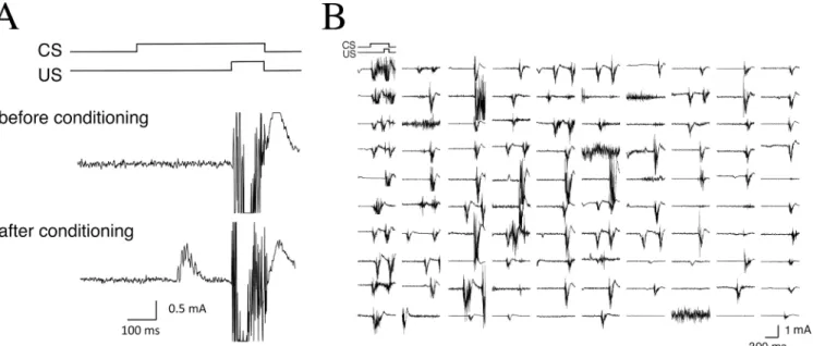
We examined delay conditioned eyeblink responses in monkeys with OO-EMG measurement methods.Fig 2Ashows typical EMG recordings from monkeys during the CS-US presenta-tions. Although CRs could not be elicited before conditioning, repeated paired presentation of the CS and US induced obvious CR expression before US onset.Fig 2Bshows all EMG data from one monkey from 1 day (100 trials on day 5).
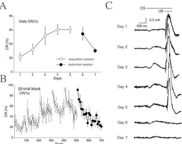
Fig 3Ashows the learning curve (daily CR%) for the delay eyeblink conditioning averaged over all monkeys (n= 5). The CR% increased progressively up to 60.5% during the 5-day acqui-sition phase. Following the acquiacqui-sition phase, the CR% decreased to 30.6% by the end of the 2-day extinction phase.Fig 3Bshows the development of CR% calculated on every 10-trial block, showing within-session variations. CR% tended to be higher in the first 10 trials than in the last 10 trials during both the acquisition and extinction phases.Fig 3Cshows the rectified and averaged EMG waveform on days 1–7, clearly indicating a gradual increase and decrease of the CR component over the acquisition and extinction sessions, respectively.
Delay eyeblink conditioning in monkeys by high-speed video analysis
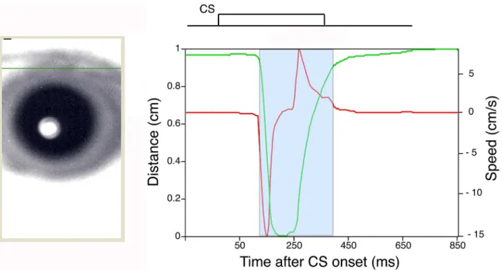
Delay eyeblink conditioning in monkeys was next evaluated with a novel high-speed camera-based tracking system.Fig 4shows an example of monkey eyelid movement analyzed with the high-speed camera-based tracking system (Fig 1A). The green solid line inFig 4shows an ex-ample of the position of the upper eyelid calculated as described in the Methods section.
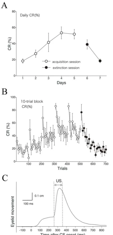
series of acquisition sessions. In the extinction sessions, CR% decreased to 18.6% in 2 days.Fig 5Bshows the development of CR% calculated for every 10-trial block. The result shows intra-day variation of CR%, indicating a tendency for a higher CR% in the first 10 trials compared to the last 10 trials during both the acquisition and extinction phases, as in the case of EMG-based analysis (Fig 3C).Fig 5Cshows the averaged eyelid movement distance against time within a session (100 trials) for all monkeys (n= 5), indicating an obvious CR component be-fore US onset.
Comparison of CR detection sensitivity between EMG analysis and
high-speed video sensing
We next compared CR detection sensitivities between EMG analysis (Figs2and3) and high-speed video analysis (Figs4and5). InFig 6A, the daily CR% as calculated by EMG analysis was compared with that determined with the high-speed video analysis. The EMG-based anal-ysis showed that the monkeys exhibited successful CR acquisition during 5 days of acquisition sessions, and rapid extinction of CRs during 2 subsequent days of extinction sessions. Data from the high-speed video analysis showed the monkeys exhibiting similar tendencies during both CR acquisition and extinction. However, in the acquisition phase, CR% detected by EMG-based analysis was somewhat higher than that by high-speed video analysis, even though ANOVA revealed no significant difference in the CR acquisition results calculated from these two channels (F1,8= 0.55,p= 0.48 for methodological effect;F4,32= 0.24,p= 0.92 for interac-tion effect). The same trend favoring the EMG channel appeared in the extincinterac-tion phase (F1,8= 4.59,p= 0.064 for methodological effect;F1,8= 0.18,p= 0.68 for interaction effect). Thus, EMG-based analysis is somewhat more sensitive for detecting eyeblink CR in monkeys. InFig 6B, we compared the results of the two different principal methods from the viewpoint of CR topographies. The averaged EMG waveform and average position of the eyelid as a time func-tion were plotted in a single graph. The CR peak given by the EMG method occurred at an ear-lier time than that given by high-speed video analysis.
Fig 2. Examples of eyelid EMG responses in monkeys.(A) Typical EMG recordings are shown in the CS-US timing diagram. Before conditioning, the conditioned response (CR) is not observed (upper panel). However, repeated paired presentation of the CS and US results during expression of significant CR before US onset (lower panel). (B) All EMG responses from the 100 trials of 1 typical session on day 5 are shown.
CR expression despite closed eyes
During conditioning, eye closures indicated that the monkeys often showed visible signs of drowsiness and behavioral microsleep. Rousing the subjects in such a situation would have in-troduced unacceptable artifacts into the data. Furthermore, in the process of assessing the rela-tionship between the actual state of eyelid closure and OO-EMG activity, we found that even when their eyes were nearly closed, the monkeys frequently showed obvious CRs that were seen as a slight, further closure (Fig 7A and 7BandS1andS2Videos).S1andS2Videos show eyelid movement during a CS-US paired trial or a CS-only trial with obvious CRs during eyelid closure. Accordingly, we quantitatively evaluated the correlation between the actual state of eyelid closure (Fig 8A) and CR expression.
First, we calculated the incidence of conditioning trials in which the subject’s eye was closed just before CS onset (closed-eye trial,Fig 8B). Throughout the entire experimental period, the ratio of closed-eye trials to all trials (100 trials) held steady between 15% and 20%. Second, we calculated the CR incidence in the closed-eye trials by EMG analysis (Fig 8C). The results showed a significant CR% increase during the 5-day acquisition phase and a significant CR% decrease during the 2-day extinction phase. Interestingly, even in the high-speed video analysis, the CR was expressed during the closed-eye trials, and the CR incidence in the closed-eye trials was significantly increased and decreased in the acquisition and extinction phases, respectively (Fig 8D).
Fig 3. Delay eyeblink CR incidence (%) calculated across trials in monkeys (EMG data).The CR incidence (CR%) for all monkeys (n= 5) calculated on a daily (A) and 10-trial-block (B) basis, with 10 such blocks being run per day. (C) The rectified and averaged EMG amplitudes from all monkeys on days 1–7. In the extinction sessions, presentation of the CS without the US results in loss of the CR.
Third, to test the effect of eye-open/closed status on CR expression, we compared the CR ex-pression incidence between closed- and open-eye trials (Fig 9). Using EMG-based analysis, no difference in CR% was found between closed- and open-eye trials during either acquisition (F1,8= 0.028,p= 0.87 for eye-condition effect;F4,32= 0.57,p= 0.69 for interaction effect) or ex-tinction (F1,8= 0.49,p= 0.51 for eye-condition effect;F1,8= 1.18,p= 0.31 for interaction effect; seeFig 9A).Fig 9Bshows the corresponding results obtained using video-based analysis. As with EMG analysis, no difference was detected between the closed- and open-eye trials during either acquisition (F1,8= 0.060,p= 0.81 for eye-condition effect;F4,32= 2.17,p= 0.095 for in-teraction effect) or extinction (F1,8= 0.040,p= 0.084 for eye-condition effect;F1,8= 0.88,
p= 0.38 for interaction effect). These results show that delay CR incidence is not influenced by eyelid closure just before CS onset.
Discussion
In the present study, we developed a monkey eyeblink conditioning system that simultaneously used an EMG method and 1-kHz high-speed camera-based tracking system [20,28]. Although a measuring system for trace eyeblink conditioning in monkeys based on an infrared camera has already been developed [16], the time and spatial resolutions were poor. Compared with trace eyeblink conditioning, delay eyeblink conditioning studies require greater time resolution because the interval between CS and US onsets is quite short. In the present study, the high-speed camera-based tracking and EMG analysis systems—both sampling at 1 kHz—enabled us to analyze the detailed incidence and latency of eyeblink CRs in monkeys. Furthermore,
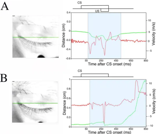
Fig 4. An example of eyelid movement obtained by high-speed video analysis.The eyelid movement data collected in a single CS-only trial after analysis with the Intelligent Vision Sensor. The green line plots the distance between the upper and lower eyelids versus time. The maximum eyelid distance (about 1 cm) represents the highest position of the upper eyelid (i.e., a completely open eye). Conversely, the status of the naturally close eyelid is defined as a distance of zero. The red line plots the velocity of the upper eyelid versus time. The blue area marks the period of time when the eyelid was moving, and the response within this area includes the CR. The left image showing the monkey’s eye represents the state of the eyelid immediately before CS onset. A green line was traced to mark the upper eyelid position. A schematic representation of CS timing for the delay eyeblink conditioning paradigm is shown above the plots of eyelid position and velocity.
because our system is a multi-measuring system that incorporates two different measuring principles, we were able for the first time to elucidate the relationship between the actual state of eyelid closure and OO-EMG activity in monkeys.
Fig 5. Delay eyeblink CR% in monkeys obtained by high-speed video analysis.The CR incidence (CR %) for all monkeys (n= 5) is calculated on a daily (A) and 10-trial-block (B) basis with data from the Intelligent Vision Sensor. Monkeys received five acquisition and two extinction sessions. (C) A plot of eyelid
displacement versus time during a session (average of all monkeys on day 5).
Fig 6. A comparison of the sensitivity of CR detection between EMG analysis and high-speed video analysis.(A) The all-subjects (n= 5) day-by-day average CR% is compared between the EMG and video-tracking channels. (B) Comparison of CR response timing analyzed by these two methods. The averaged EMG amplitude and video-recorded movement of the eyelid are compared in a single graph.
doi:10.1371/journal.pone.0129828.g006
Fig 7. A preliminary study of CR expression during eyes-closed periods.Examples of CR expression during eyes-closed periods in a CS-US paired trial (A) and CS-only trial (B). Monkeys in light sleep often exhibit delay CRs despite their eyelids being completely closed (S1andS2Videos). Note that in such cases, the monkeys exhibit more downward movement of the upper eyelid than is needed to close the eye, resulting in a negative distance measurement. A schematic representation of the CS and US is inset above the plots of eyelid movement and speed. Green and red lines show the position and velocity of the upper eyelid, respectively. The red arrow indicates robust CRs between CS and US onsets.
Using this novel system to record delay eyeblink conditioning in monkeys, we demonstrated successful CR acquisition and extinction (Figs3and5). Even though the attained level of CR% (approximately 60%) in the present study is lower than that observed in cats and rabbits [13, 30,31], it is comparable to or higher than the level reported in some human or mouse condi-tioning experiments [5,12,14,24]. The present study is the first attempt to evaluate eyeblink conditioning in monkeys and is significant in three ways: as the first report of monkey eyeblink conditioning by EMG, as the first by video processing, and as the first report of the delayed
Fig 8. Changes in CR expression in cases of closed eyes.(A) Representative images of an“open eye” and a“closed eye”as judged by the video-tracking system. (B) The percentage of all trials that were “closed-eye trials”during delay eyeblink conditioning in monkeys. (C, D) The CR incidence (CR%) in the“closed-eye trials”as detected by EMG analysis (C) and as detected by the Intelligent Vision Sensor (D). Data are presented as mean±S.E.M.
doi:10.1371/journal.pone.0129828.g008
Fig 9. Changes in CR expression over the experiment in cases of open vs. closed eyes.The CR incidence (CR%) averaged over all monkeys (n= 5) in cases of eyes open and closed just before CS onset are compared between the EMG channel (A) and the Intelligent Vision Sensor channel (B). Insets in (A): representative images of an“open eye”and a“closed eye”as judged by the video-tracking system. Data are presented as mean±S.E.M.
version of eyeblink conditioning in monkeys. The monkey eyeblink CRs detected by the high-speed camera-based tracking system were in good agreement with those detected by EMG, although the EMG-based analysis gave a percentage of trials with observed CRs that was somewhat higher than that measured by high-speed video analysis (Fig 6). EMG analysis occa-sionally detected a CR in a trial when the high-speed camera-based tracking system did not. The opposite also occurred, but with a lower incidence. Latency analysis showed that the CR peak for the EMG method preceded that for high-speed video analysis (Fig 6B). This is consis-tent with previous studies in other species and likely reflect the fact that the muscle EMG in-duces physical eyelid movement [32,33]. A detailed analysis also indicated that CR incidence tended to be higher in the first half of trials than in the latter half during each daily session (Figs3Band5B). This intraday variation in CR occurrence is comparable to that observed in mice [18]. The phenomenon is probably attributable to habituation to the stimulus or testing environment.
In the present conditioning procedure, each animal was placed in a monkey chair in a dimly lit room for more than 30 min every day. Thus, it often appeared that the monkeys dozed dur-ing conditiondur-ing, but we did not wake them up in such situations. The multi-measurdur-ing system could detect the actual state of eyelid closure and the monkey’s appearance, so we could sepa-rately evaluate performance in awake and light sleep states. As shown inS1andS2Videos, an interesting subsidiary finding was that the monkeys often exhibited obvious CRs even when their eyes had closed just before CS onset (Fig 7). Quantitative analysis confirmed that the probability of CR incidence was not influenced by whether or not the monkeys closed their eyelids just before CS onset. In fact, the incidence of both eyes-open and eyes-closed CRs rose and fell in parallel over the acquisition/extinction sequence (Fig 9). These results exclude the possibility that CR responses during closed-eye trials were startle responses caused by the tone CS.
Delay eyeblink conditioning has been tested in many animal species including human sub-jects, but few studies have evaluated this paradigm in monkeys [12,13]. This is somewhat surprising considering that one other major paradigm of cerebellar motor learning, the vesti-bulo-ocular reflex (VOR), has been extensively performed in monkeys [34,35]. Clark and Zola (1998) suggested several reasons for the absence of contemporary studies of eyeblink condi-tioning in monkeys prior to their research [16], though several premodern attempts had been made [36–38]. One of the reasons was that the expense of primate models is only justified by their special relevance to the cognitive aspects of human memory. Thus, it could be supposed that there was no perceived need to develop an eyeblink conditioning system for monkeys to supplement research on simple forms of human learning that are not especially evolved in hu-mans, such as eyeblink conditioning [16]. However, the context of monkey biology has greatly changed in recent years. Monkey research is embarking upon a new phase because of the devel-opment of genetically engineered nonhuman primates [39–41]. In the new era, the monkey eyeblink conditioning system could be a meaningful tool for elucidating the molecular basis of implicit and explicit memory [5,42,43]. Moreover, a positron-emission tomographic (PET) method for measuring brain functions in monkeys in a conscious state allows us to map brain functions and determine which regions are activated when the higher functions that are in-volved in learning, memory, or cognition are exercised [44,45]. In future, the combination of the conscious, nonanesthetized preparation with PET imaging and monkey eyeblink condi-tioning technology may more clearly elucidate the neural system(s) underlying delay eyeblink conditioning in higher primates, thus enabling direct, subtractive comparison with systems un-derlying higher functions.
and cerebellar deep nuclei [46,47] even though several studies have assumed a different role of the cerebellum in this type of associative learning [30,48]. At least in rabbits and rodents, delay conditioning is critically dependent on a brainstem-cerebellar circuit [4,8,46,49–51], whereas trace conditioning with a sufficiently long stimulus-free interval depends on the condition of the hippocampus [8]. Moreover, substantial research in humans suggests the importance of the cerebellum in the memory of delay eyeblink conditioning, which is consistent with the small-animal data [52–55]. However, a species difference in brain regions critical for delay-eyeblink memory formation may exist between higher mammals (including humans) and rodents. For example, in humans more than in rodents, the hippocampal contribution may be greater in the acquisition of delay eyeblink memory because delay eyeblink conditioning is a more sensitive test for cognitive deficit in patients with Alzheimer disease (AD) than the trace eyeblink condi-tioning paradigm [56], despite the fact that the cerebellum is a relatively spared area of the AD brain and that general motor learning is preserved in these patients [57,58]. In contrast, AD model mice exhibited significant impairment in long-trace but not delay eyeblink conditioning [23,25,59]. Delay eyeblink conditioning in monkeys could prove useful in identifying the brain regions that are critical for motor memory in higher mammals. An important issue is whether primates and rodents share common brain regions for implicit memory.
The observation of learning expressed as a behavioral output during microsleep or drowsiness is quite interesting and rare. Our present results suggest that delay eyeblink memory can be ex-pressed and might even be formed at low levels of arousal. This is consistent with the notion that delay eyeblink conditioning is a form of nondeclarative implicit memory [3]. Our present results from a novel multi-measuring system for eyeblink CRs in monkeys have important implications for elucidating the neural mechanisms underlying implicit/unconscious learning.
In conclusion, we have developed the first monkey eyeblink conditioning system using si-multaneous EMG and high-speed video analysis. The monkey eyeblink CRs detected by the high-speed camera-based tracking system were in good agreement with those detected by EMG analysis. This multi-measuring system incorporating two different measuring principles enabled us to elucidate the relationship between the actual presence of eyelid closure and OO-EMG activity during the experiment. Interestingly, the animal’s eyelid-open state, which is supposed to be related to the animal’s wakefulness and potential presence of cognitive process-ing [29], did not predict the probability of CR incidence. These results suggest that delay eye-blink memory can be expressed independently of behavioral state (degree of wakefulness or consciousness), confirming that this form of conditioning is implicit learning. An interesting problem for future studies is to determine whether the CR is observed in monkeys during trace eyeblink conditioning, which is characterized by a brief stimulus-free period between CS offset and US onset. Our system for cognitive assessment in monkeys based on eye closure will be a useful tool for elucidating the neural mechanisms underlying delay eyeblink conditioning—an acquisition process for implicit memory in higher mammals.
Supporting Information
S1 Video. Example of eyelid movement during a CS-US paired trial.An obvious CR is elic-ited before US onset even during closed-eye periods. Left: The physical movement of the eyelid is tracked by the Intelligent Vision Sensor; the green horizontal line illustrates the position of the upper eyelid over time. Right: time course of eyelid movement. The green line shows the position of the upper eyelid, and the red line shows velocity. The scale of the vertical axis is in-verted relative toFig 7A. The relationship between the CS and US is depicted above the time courses of eyelid movement.
S2 Video. Example of eyelid movement during a CS-only trial.An obvious CR is elicited be-fore US onset even during closed-eye periods. Left: The physical movement of the eyelid is tracked by the Intelligent Vision Sensor; the green horizontal line illustrates the position of the upper eyelid over time. Right: time course of eyelid movement. The green line shows the posi-tion of the upper eyelid, and the red line shows velocity. The CS timing is depicted above the time courses of eyelid movement.
(MP4)
Acknowledgments
We are grateful to Dr. Soichi Nagao for helpful discussions concerning this work.
Author Contributions
Conceived and designed the experiments: Y. Kishimoto MK Y. Kirino. Performed the experi-ments: Y. Kishimoto SY KS. Analyzed the data: Y. Kishimoto SY Y. Kirino. Contributed re-agents/materials/analysis tools: SY KS H. Toyoda H. Tsukada. Wrote the paper: Y. Kishimoto Y. Kirino. Designed the software used in analysis: SY KS H. Toyoda.
References
1. Thompson RF, Krupa DJ. Organization of memory traces in the mammalian brain. Annu Rev Neurosci. 1994; 17: 519–549. PMID:8210186
2. Thompson RF, Bao S, Chen L, Ciproano B, Grethe J, Kim JJ, et al. Associative learning. In: Schmah-mann JD, editor. The cerebellum and cognition. San Diego: Academic Press, 1997. pp. 152–189. 3. Thompson RF, Kim JJ. Memory systems in the brain and localization of a memory. Proc Natl Acad Sci
U S A. 1996; 93: 13438–13444. PMID:8942954
4. McCormick DA, Thompson RF. Cerebellum: essential involvement in the classically conditioned eyelid response. Science. 1984; 223: 296–299. PMID:6701513
5. Clark RE, Squire LR. Classical conditioning and brain systems: the role of awareness. Science. 1998; 280: 77–81. PMID:9525860
6. Kim JJ, Thompson RF. Cerebellar circuits and synaptic mechanisms involved in classical eyeblink con-ditioning. Trends Neurosci. 1997; 20: 177–181. PMID:9106359
7. Manns JR, Clark RE, Squire L. Single-cue delay eyeblink conditioning is unrelated to awareness. Cogn Affect Behav Neurosci. 2001; 1: 192–198. PMID:12467114
8. Attwell PJ, Cooke SF, Yeo CH. Cerebellar function in consolidation of a motor memory. Neuron. 2002; 34: 1011–1020. PMID:12086647
9. Aiba A, Kano M, Chen C, Stanton ME, Fox GD, Herrup K, et al. Deficient cerebellar long-term depres-sion and impaired motor learning in mGluR1 mutant mice. Cell. 1994; 79: 377–388. PMID:7954803
10. Andrews SJ, Freeman JH Jr, Carter CS, Stanton ME. Ontogeny of eyeblink conditioning in the rat: audi-tory frequency and discrimination learning effects. Develop Psychobiol. 1995; 28: 307–320. PMID:
7589817
11. Gruart A, Blazquez P, Delgado-Garcia JM. Kinematics of spontaneous, reflex, and conditioned eyelid movements in the alert cat. J Neurophysiol. 1995; 74: 226–248. PMID:7472326
12. Woodruff-Pak DS, Steinmetz JE. Eyeblink classical conditioning, volume 1: Applications in humans. Boston: Kluwer Academic Publishers; 2000.
13. Woodruff-Pak DS, Steinmetz JE. Eyeblink classical conditioning, volume 2: Animal models. Boston: Kluwer Academic Publishers; 2000.
14. Kishimoto Y, Kano M. Endogenous cannabinoid signaling through the CB1 receptor is essential for cer-ebellum-dependent discrete motor learning. J Neurosci. 2006; 26: 8829–8837. PMID:16928872
15. Keifer J, Armstrong KE, Houk JC. In vitro classical conditioning of abducens nerve discharge in turtles. J Neurosci. 1995; 15: 5036–5048. PMID:7623132
17. Leal-Campanario R, Delgado-García JM, Gruart A. The rostral medial prefrontal cortex regulates the expression of conditioned eyelid responses in behaving rabbits. J Neurosci Methods. 2013; 33: 4378– 4386. doi:10.1523/JNEUROSCI.5560-12.2013PMID:23467354
18. Kishimoto Y, Nakazawa K, Tonegawa S, Kirino Y, Kano M. Hippocampal CA3 NMDA receptors are cru-cial for adaptive timing of trace eyeblink conditioned response. J Neurosci. 2006; 26: 1562–1570. PMID:16452679
19. Skosnik PD, Edwards CR, O'Donnell BF, Steffen A, Steinmetz JE, Hetrick WP. Cannabis use disrupts eyeblink conditioning: evidence for cannabinoid modulation of cerebellar-dependent learning. Neurop-sychopharmacol. 2008; 33: 1432–1440.
20. Takumi M, Toyoda H, Mukozaka N. 1kHz smart camera with image processing feature. Proc IAPR Work Mach Vis. 2002; 8–37: 384–387.
21. Takechi H, Onoe H, Imamura K, Onoe K, Kakiuchi T, Nishiyama S, et al. Brain activation study by use of positron emission tomography in unanesthetized monkeys. Neurosci Lett. 1994; 182: 279–282. PMID:7715827
22. Miyata M, Kishimoto Y, Tanaka M, Hashimoto K, Hirashima N, Murata Y, et al. A role for myosin Va in cerebellar plasticity and motor learning: a possible mechanism underlying neurological disorder in myo-sin Va disease. J Neurosci. 2011; 31: 6067–6078. doi:10.1523/JNEUROSCI.5651-10.2011PMID:
21508232
23. Kishimoto Y, Oku I, Nishigawa A, Nishimoto A, Kirino Y. Impaired long-trace eyeblink conditioning in a Tg2576 mouse model of Alzheimer's disease. Neurosci Lett. 2012; 506: 155–159. doi:10.1016/j. neulet.2011.10.071PMID:22085694
24. Kishimoto Y, Hirono M, Atarashi R, Sakaguchi S, Yoshioka T, Katamine S, et al. Age-dependent im-pairment of eyeblink conditioning in prion protein-deficient mice. PLoS One. 2013; 8: e60627. doi:10. 1371/journal.pone.0060627PMID:23593266
25. Kishimoto Y, Kirino Y. Presenilin 2 mutation accelerates the onset of impairment in trace eyeblink condi-tioning in a mouse model of Alzheimer's disease overexpressing human mutant amyloid precursor pro-tein. Neurosci Lett. 2013; 538: 15–19. doi:10.1016/j.neulet.2013.01.025PMID:23370287
26. Sugiyama Y, Takumi M, Toyoda H, Mukozaka N, Ihori A, Kurashina T, et al. A high-speed CMOS image sensor with profile data acquiring function (in Japanese). IEEE J Solid State Circuits. 2005; 40: 2816–2823.
27. Adachi K, Hamada T, Nakano T, Yamamoto S. Blink measurement to detect a driver’s drowsy state by moving image processing (in Japanese). IEEJ Trans. 2004; 124: 776–783.
28. Nakamura Y, Matsuda J, Suzuki K, Toyoda H, Hakamata N, Shimamoto T, et al. Measurement of spon-taneous blinks with a high-speed blink analyzing system. Nippon Ganka Gakkai Zasshi. 2008; 112: 1059–1067. PMID:19157026
29. Malla AM, Davidson PR, Bones PJ, Green R, Jones RD. Automated video-based measurement of eye closure for detecting behavioral microsleep. In: Annual International Conference of the IEEE Engineer-ing in Medicine and Biology Society. 2010;6741–6744.
30. Sánchez-Campusano R, Gruart A, Delgado-García JM. Dynamic associations in the cerebellar-moto-neuron network during motor learning. J Neurosci. 2009; 29: 10750–10763. doi:10.1523/JNEUROSCI. 2178-09.2009PMID:19710326
31. Carretero-Guillén A, Pacheco-Calderón R, Delgado-García JM, Gruart A. Involvement of hippocampal inputs and intrinsic circuit in the acquisition of context and cues during classical conditioning in behav-ing rabbits. Cereb Cortex. 2015; 25: 1278–1289. doi:10.1093/cercor/bht321PMID:24243618
32. Koekkoek SK, Den Ouden WL, Perry G, Highstein SM, De Zeeuw CI. Monitoring kinetic and frequency-domain properties of eyelid responses in mice with magnetic distance measurement technique. J Neu-rophysiol. 2002; 88: 2124–2133. PMID:12364534
33. Koekkoek SK, Hulscher HC, Dortland BR, Hensbroek RA, Elgersma Y, Ruigrok TJ, et al. Cerebellar LTD and learning-dependent timing of conditioned eyelid responses. Science. 2003; 301: 1736–1739. PMID:14500987
34. Ito M. Historical review of the significance of the cerebellum and the role of Purkinje cells in motor learn-ing. Ann N Y Acad Sci. 2002; 978: 273–288. PMID:12582060
35. Lisberger SG. Internal models of eye movement in the floccular complex of the monkey cerebellum. Neuroscience. 2009; 162: 763–776. doi:10.1016/j.neuroscience.2009.03.059PMID:19336251
36. Hilgard ER, Marquis DG. Conditioned eyelid responses in monkeys with a comparison of dog, monkey, and man. Psychol Monogr. 1936; 47: 186–198.
38. Pennypacker HS, King FA, Achenbach KE, Roberts L. An apparatus and procedure for conditioning the eyeblink reflex in the squirrel monkey. J Exp Anal Behav. 1966; 9: 601–604. PMID:4960570
39. Sasaki E, Suemizu H, Shimada A, Hanazawa K, Oiwa R, Kamioka M, et al. Generation of transgenic non-human primates with germline transmission. Nature. 2009; 459: 523–527. doi:10.1038/ nature08090PMID:19478777
40. Inoue K, Koketsu D, Kato S, Kobayashi K, Nambu A, Takada M. Immunotoxin-mediated tract targeting in the primate brain: selective elimination of the cortico-subthalamic“hyperdirect”pathway. PLoS One. 2012; 7: e39149. doi:10.1371/journal.pone.0039149PMID:22761729
41. Tachibana M, Sparman M, Ramsey C, Ma H, Lee HS, Penedo MC, et al. Generation of chimeric rhesus monkeys. Cell. 2012; 148: 285–295. doi:10.1016/j.cell.2011.12.007PMID:22225614
42. Squire LR, Zola SM. Structure and function of declarative and nondeclarative memory systems. Proc Natl Acad Sci U S A. 1996; 93: 13515–13522. PMID:8942965
43. Lovibond PF, Liu JC, Weidemann G, Mitchell CJ. Awareness is necessary for differential trace and delay eyeblink conditioning in humans. Biol Psychol. 2011; 87: 393–400. doi:10.1016/j.biopsycho. 2011.05.002PMID:21586313
44. Yamamoto S, Nishiyama S, Kawamata M, Ohba H, Wakuda T, Takei N, et al. Muscarinic receptor occu-pancy and cognitive impairment: a PET study with [11C](+)3-MPB and scopolamine in conscious mon-keys. Neuropsychopharmacology. 2011; 36: 1455–1465. doi:10.1038/npp.2011.31PMID:21430646
45. Yamamoto S, Ohba H, Nishiyama S, Harada N, Kakiuchi T, Tsukada H, et al. Subanesthetic doses of ketamine transiently decrease serotonin transporter activity: a PET study in conscious monkeys. Neu-ropsychopharmacology. 2013; 38: 2666–2674. doi:10.1038/npp.2013.176PMID:23880871
46. Mauk MD. Roles of cerebellar cortex and nuclei in motor learning: contradictions or clues? Neuron. 1997; 18: 343–346. PMID:9115728
47. Hesslow G, Yeo C. Cerebellum and learning: a complex problem. Science. 1998; 280: 1817–1819. PMID:9669927
48. Sánchez-Campusano R, Gruart A, Delgado-García JM. The cerebellar interpositus nucleus and the dy-namic control of learned motor responses. J Neurosci. 2007; 27: 6620–6632. PMID:17581949
49. Krupa DJ, Thompson JK, Thompson RF. Localization of a memory trace in the mammalian brain. Sci-ence. 1993; 260: 989–991. PMID:8493536
50. Yeo CH, Hesslow G. Cerebellum and conditioned reflexes. Trends Cogn Sci. 1998; 2: 322–330. PMID:
21227228
51. Attwell PJ, Rahman S, Ivarsson M, Yeo CH. Cerebellar cortical AMPA-kainate receptor blockade pre-vents performance of classically conditioned nictitating membrane responses. J Neurosci. 1999; 19: RC45.
52. Daum I, Channon S, Canavan AG. Classical conditioning in patients with severe memory problems. J Neurol Neurosurg Psychiatry. 1989; 52: 47–51. PMID:2496204
53. Topka H, Valls-Sole J, Massaquoi SG, Hallett M. Deficit in classical conditioning in patients with cere-bellar degeneration. Brain. 1993; 116: 961–969. PMID:8353718
54. Gerwig M, Hajjar K, Dimitrova A, Maschke M, Kolb FP, Frings M, et al. Timing of conditioned eyeblink responses is impaired in cerebellar patients. J Neurosci. 2005; 25: 3919–3931. PMID:15829644
55. Gerwig M, Esser AC, Guberina H, Frings M, Kolb FP, Forsting M, et al. Trace eyeblink conditioning in patients with cerebellar degeneration: comparison of short and long trace intervals. Exp Brain Res. 2008; 187: 85–96. doi:10.1007/s00221-008-1283-2PMID:18253726
56. Woodruff-Pak DS, Papka M. Alzheimer's disease and eyeblink conditioning: 750 ms trace vs. 400 ms delay paradigm. Neurobiol Aging. 1996; 17: 397–404. PMID:8725901
57. Eslinger PJ, Damasio AR. Preserved motor learning in Alzheimer's disease: implications for anatomy and behavior. J Neurosci. 1986; 6: 3006–3009. PMID:3760945
58. Larner AJ. The cerebellum in Alzheimer's disease. Dement Geriatr Cogn Disord. 1997; 8: 203–209. PMID:9213064