Attention Deficit/Hyperactivity Disorder and
Urinary Nonylphenol Levels: A Case-Control
Study in Taiwanese Children
Ching-Jung Yu1, Jung-Chieh Du2, Hsien-Chih Chiou3, Shang-Han Yang1, Kai-Wei Liao1, Winnie Yang4, Ming-Yi Chung5, Ling-Chu Chien6, Betau Hwang2, Mei-Lien Chen1*
1Institute of Environmental and Occupational Health Sciences, School of Medicine, National Yang Ming University, Taipei, Taiwan,2Department of Pediatrics, Taipei City Hospital, Zhongxiao branch, Taipei, Taiwan,3Department of Child and Adolescent Psychiatry, Taipei City Hospital, Songde branch, Taipei, Taiwan,4Department of Pediatrics, Taipei City Hospital, Yangming branch, Taipei, Taiwan,5Department of Life Sciences and Institute of Genome Sciences, National Yang Ming University, Taipei, Taiwan,6School of Public Health, Taipei Medical University, Taipei, Taiwan
Abstract
Objective
Nonylphenol (NP) belongs to the family of endocrine disruptors, and it is widely used in industrial applications and is ubiquitous in daily foods. Animal studies have suggested that NP exposure might promote motor hyperactivity, likely by causing deficits in dopaminergic neurons. However, research assessing NP exposure and epidemiology studies on human populations are limited. The aim of this study was to explore the association between child NP exposure and ADHD while considering particular covariants, such as lead levels and dopamine-related gene variations.
Methods
A case-control study was conducted on patients with clinically diagnosed ADHD; the Swan-son, Nolan and Pelham, Fourth Revision (SNAP-IV) questionnaire was used to identify nor-mal controls aged 4–15 years. Participants were examined for urinary NP concentrations, blood lead levels, and select single-nucleotide polymorphisms of two dopamine-related genes (D4 dopamine receptor,DRD4, and dopamine transporter,DAT1). Socio-demo-graphic variables, maternal lifestyle factors during pregnancy and family medical history were obtained using a questionnaire.
Results
A total of 97 children with doctor-diagnosed ADHD and 110 normal controls were enrolled. The blood lead levels in both groups were similar (1.57±0.73 vs. 1.73±0.77μg/dL,p= 0.15).
No significant difference in urinary NP concentration was found between the children with ADHD and the control subjects (4.52±3.22μg/g cr. vs. 4.64±2.95μg/g cr.,p= 0.43). ADHD
was significantly more prevalent among males in this study (male to female ratio: 5:1 for the
OPEN ACCESS
Citation:Yu C-J, Du J-C, Chiou H-C, Yang S-H, Liao K-W, Yang W, et al. (2016) Attention Deficit/ Hyperactivity Disorder and Urinary Nonylphenol Levels: A Case-Control Study in Taiwanese Children. PLoS ONE 11(2): e0149558. doi:10.1371/journal. pone.0149558
Editor:Manuel Portolés, Hospital Universitario LA FE, SPAIN
Received:June 2, 2015
Accepted:February 2, 2016
Published:February 18, 2016
Copyright:© 2016 Yu et al. This is an open access article distributed under the terms of theCreative Commons Attribution License, which permits unrestricted use, distribution, and reproduction in any medium, provided the original author and source are credited.
Data Availability Statement:All relevant data are within the paper.
ADHD group and 1.3:1 for the control group,p<0.01). The analysis was repeated after excluding the females, but this had no effect on the association between NP and ADHD. The regression model, including or excluding females, indicated no increased odds of hav-ing ADHD in the context of NP exposure after adjusthav-ing for covariants.
Conclusion
This study indicated that NP exposure might not promote ADHD in children, even though children in Taiwan had relatively high levels of NP compared to those reported previously and those in developed nations.
Introduction
Nonylphenol (NP) and its parent compound, nonylphenol polyethoxylate (NPnEO), are used in various manufacturing processes and in a wide range of products to which humans are exposed [1–5]. The annual global production of alkylphenol polyethoxylate (APnEO) is approximately 650,000 tons, and NPnEO represents approximately 80% of this amount [6,7]. NP is prevalent in the environment and is a known endocrine-disrupting chemical with estro-gen-like effects [8–10]. Estrogen participates in various steps of cellular differentiation and affects the maturation of dopamine neurons [11]. After exposure, NP binds to estrogen recep-tors and can influence estrogen functions in the developmental stages of animals and humans [5,12–15].
Humans may be exposed to NP through drinking water; the intake of contaminated foods; the inhalation of air; and absorption via the skin [6,16]. NP is widespread in Taiwanese food, and the average daily intake of NP is 4- to 8.5-fold higher than that in other countries [17]. In addition, biological monitoring has revealed significant levels of NPs in the adipose tissue, plasma and urine of neonates, children, pubertal students, pregnant mothers, women, and workers in the textile and housekeeping industries [13,14,18–21]. However, a limited number of NP exposure assessments and epidemiology studies on human populations have been con-ducted [22]. Lopez-Espinosa et al. determined the NP concentrations in 20 non-occupationally exposed women in Spain; 100% of these women had detectable levels of NP. Body mass index (BMI) was associated with NP concentration [21]. NP exposure was also found to be associated with an increased risk of low neonatal weight and was shown to disturb pubertal development in Taiwan [13,14]. Infants and children in Taiwan are constantly exposed to NP, and the health implications of NP exposure for vulnerable children should be monitored.
Attention deficit hyperactivity disorder (ADHD) is one of the most common childhood neurobehavioral conditions, with a prevalence ranging from 5.9%-7.1% worldwide and from 7.5%-9.9% in Taiwan [23,24]. ADHD interferes with learning and social development; this condition typically begins during the preschool years and often persists into adulthood. The etiology of ADHD remains unclear; however, it is believed to involve individual genetic and environmental factors as well as interactions between these factors [25–27]. The heritability of ADHD has been estimated to be as high as 76% [28], and various polymorphisms in dopa-mine-related genes have been found to increase the risk of ADHD [29,30]. The associations between polymorphisms in the dopamine 4 receptor (DRD4) and dopamine transporter (DAT1) genes and ADHD have been most frequently verified [31]. Lead is one of the most well-studied environmental pollutants [32]. Low-level lead exposure has been associated with a clinical diagnosis of ADHD in several recent studies [31,33–35].
interpretation of data; in the writing of the report; and in the decision to submit the article for publication.
Competing Interests:The authors have declared
Animal studies have revealed that the chronic application of NP sensitizes mouse brains and causes neuronal apoptosis [36]. Intracisternal NP injection might promote motor hyperac-tivity, likely by causing deficits in dopaminergic neurons [37,38]. These animal investigations suggested that NP exposure was associated with adverse neurodevelopmental outcomes, such as hyperactivity. Several studies have focused on the health implications of ADHD and expo-sure to potential environmental pollutants, such as lead, phthalates and pesticides, in young and school-age children [35,39–41]. To the best of our knowledge, no studies have assessed the possible risks of ADHD associated with childhood exposure to NP. The purpose of this study was to explore the associations between NP exposure and ADHD by measuring the uri-nary concentrations of NP in both ADHD and non-ADHD children. We also considered known potential variables in the etiology of ADHD, such as blood lead levels (BLLs), genetic variations, parental psychosocial factors, family history of nervous system diseases and prenatal exposure to tobacco and alcohol.
Materials and Methods
Study subjects
The study protocol was approved by the Taipei City Hospital institutional review board. All participants gave verbal or written assent. Written informed consent was obtained from the participant’s parents or guardians. We recruited subjects between 4 and 15 years of age in out-patient waiting rooms at Taipei City Hospital. The case subjects in this study were children with ADHD identified by board-certified pediatricians or psychiatrists after at least a three-visit clinical assessment. Consecutive children admitted for initial or follow-up ADHD treat-ment were recruited as cases during the study period, with the exclusion criteria of neurological deficits or mental retardation. The diagnosis of ADHD was performed in accordance with the criteria of the Diagnostic and Statistical Manual of Mental Disorders, 4th edition, revised crite-ria (DSM-IV-TR) [42].
Control subjects were recruited by randomly selecting healthy children without ADHD aged 4–15 years who utilized Taipei City Hospital for non-ADHD-related visits during the same study period. The same exclusion criteria for the cases were applied to the controls. Con-trols were screened for the absence of ADHD symptoms based on assessments of their behavior at home and in the classroom by the parent(s) and teachers, respectively, according to the Chi-nese version of the Swanson, Nolan and Pelham, Fourth Revision (SNAP-IV) questionnaire [43–46]. The rating results were computed by pediatricians and examined for the absence of ADHD symptoms. The SNAP-IV Parent and Teacher Form uses the same format and directly adopts the DSM-IV symptoms; this form has been translated into Chinese and has been found to be a reliable and valid tool for screening for ADHD for clinical and research purposes in Tai-wan [45,46]. The 26-item SNAP-IV is based on a four-point (0–3) scale. It consists of the DSM-IV criteria for inattention (items 1–9), hyperactivity/impulsivity (items 10–18) as the symptoms for ADHD, and oppositional symptoms (items 19–26) characteristic of oppositional defiant disorder. If at least six of the nine inattention items or hyperactivity/impulsivity items were scored at 2 (quite a bit) or 3 (very much) in either the parent or teacher form, the children were defined as having the possible presence of ADHD syndromes [47]. Children scored as having possible ADHD were referred to pediatricians or psychiatrists for further assessment. All the cases were also assessed by SNAP-IV scoring at the initial visit.
during pregnancy and family history of nervous system diseases. Subjects provided a spot urine sample and blood (or saliva) sample during the clinic visit.
Sample preparation and analysis
Reagents. 4- Nonylphenol (p-isomers>85%) was obtained from Fluka (Buchs,
Switzer-land). Methanol, n-hexane, acetone, acetonitrile, hydrochloric acid, acetic acid, and ammo-nium acetate were LC grade, and all were obtained from Merck (Darmstadt, Germany).β -Glucuronidase (type H-2) was purchased from Sigma-Aldrich (USA). Deionized water, obtained using a Millipore water purification system (Bedford, MA, USA), was prepared before use and was collected in a glass container.
Instrumentation. High-performance liquid chromatograph (HPLC) coupled with a fluo-rescence detector (Hitachi, Tokyo) was used for NP analysis. The reverse-phase column was a Luna C18 (250 x 4.6 mm) with a 5-μm particle size. The isocratic mobile phase was a mixture
of acetonitrile and water (75:25, v/v) with a 1.0 mL/min flow rate. The fluorescence detector was operated at an excitation wavelength of 275 nm and an emission wavelength of 300 nm. The samples were injected at volumes of 20μL.
Sample pretreatment and analysis. This study adopted the same NP analytical method used previously in this lab [20]. Before testing, the urine samples were thawed and homoge-nized using a vortex mixer. The pH values of all the urine samples were adjusted to 5.5 with 1 M acetic acid, and the samples were mixed with 1 mL of 1 M ammonium acetate solution. The urine samples were deconjugated by adding 125μL ofβ-glucuronidase to detect total NP (free
and conjugated forms). It was followed by incubation at 37°C for 15 h in a shaker bath. The mixture was acidified to pH 3.0 using a 1 M HCl solution. Samples were cleaned using pH solid-phase extraction (SPE) cartridges (3 mL, RP, Supelco, USA), which had been precondi-tioned with 20 mL of methanol, followed by 3 mL of pure water that was acidified with 1 M HCl solution to pH 3.0. After sample application, each cartridge was washed with 5 mL of pure water. The absorbed compounds on the cartridge were eluted with 3 mL of methanol and were evaluated by HPLC. The NP recovery rate was in the range of 81%-107%, with a CV (coeffi-cient of variance) of 6.7%. The LOD (limit of detection) for NP was 1.6μg/L.
To avoid contamination of NP, glassware was used for the urine collection for both the case and control groups. Urine samples were sealed, chilled, and transported to the lab imme-diately. No NPnEO (which is biodegraded to NP)-containing detergents and plastics were used during sample collection, preparation and analysis. NP concentrations were adjusted using creatinine concentrations to correct for variable urine dilutions in the spot urine sam-ples. Urinary creatinine concentrations were determined using a commercially available diagnostic enzyme method (Eagle Diagnostic, USA) that is based on a modified Jaffe reaction [48]. The concentrations of urinary NP were expressed asμg per gram creatinine (μg/g cr.).
We excluded one child, an ADHD case, with extremely dilute urine (creatinine levels
<30 mg/dL).
Peripheral blood was drawn using a syringe or venoclysis needle, which was then sealed in a heparin-containing vacuum tube and immediately transported at 4°C to the laboratory. One milliliter of whole blood was frozen separately and stored for lead analysis. The BLLs were mea-sured using inductively coupled plasma-mass spectrometry (Thermo, USA). Trace Elements Serum L-2 (SeronormTM, Norway) was used to verify the precision and accuracy of the analyti-cal measurements. For the BLL measurements, the LOD was 0.001μg/dL with a CV of 3.2%.
The results are reported as the mean of two replicate measurements. Moreover, quality control samples were analyzed in each batch.
In the situation in which individuals were not able to supply a blood sample, DNA was extracted from a saliva sample. Saliva was spat into the Saliva DNA Collection and Preservation Kit (Norgen Biotek Corporation, Canada) and stored at room temperature until analysis.
Gene polymorphisms. Genotype analyses with haplotype-tagging SNPs ofDRD4and
DAT1were selected by screening the CHB (Han Chinese in Beijing) panel from the HapMap database (http://hapmap.ncbi.nlm.nih.gov/). To avoid redundancy and to obtain complete genetic coverage, we evaluated linkage disequilibrium patterns and set a maximum r2threshold of 0.8 for all SNPs that possessed a minor allele frequency (MAF)0.05. The non-synonymous SNPs were further inspected using the University of California at Santa Cruz (UCSC) genome browser (http://genome.ucsc.edu/). For the tagged SNPs, MAF values0.1 were used to check the genetic variations in the CHB by browsing the 1000 GENOME database (http://www. 1000genomes.org/).
Genomic DNA from peripheral blood leukocytes was extracted by using a DNA mini kit (Geneaid, CA, USA). DNA from saliva was isolated using a QIAamp DNA Mini Kit (Qiagen, Hilden, Germany) according to the manufacturer’s instructions. Genotyping of theDRD4and
DAT1SNPs was performed using the MassARRAY Platform (Sequenom, CA, USA) at the VYM Genome Research Center, National Yang-Ming University. Amplifications were per-formed in a 384-well polymerase chain reaction system at a minimum concentration of 10 ng/
μL DNA. The genotypes were tested for deviation from Hardy-Weinberg equilibrium, and
associations were analyzed using standard chi-square goodness-of-fit tests (http://ihg.gsf.de/ cgi-bin/hw/hwa1.pl).
Covariates. We examined covariates and potential confounders for the association between NP exposure and ADHD. Predictors were chosen based on their association with ADHD in previous studies. The following variables were considered as potential confounders: BLLs, dopamine-related gene variations, age, gender, BMI, maternal age at childbirth, gesta-tional age at birth (<37 weeks or37 weeks), parental education (high school education and below or college or advanced training), maternal smoking during pregnancy (yes or no) and maternal drinking during pregnancy (yes or no) [15,39,49–53]. In addition to environmental risk factors, we also included family history of nervous system disease as a covariate. The family history of nervous system diseases listed in the questionnaire includes Parkinson's disease, Alz-heimer's disease, ADHD, mental retardation, cerebral palsy, autism, epilepsy, developmental delay, multiple sclerosis and peripheral neuromuscular disease in grandparents, parents or sib-lings of the subject. BLLs and genetic variations inDRD4andDAT1were measured in this study, and the other variables were obtained from clinical records or questionnaires completed by the parents.
Statistical analysis. SPSS version 17.0 was used for the statistical analysis. Measurements below the LOD were given a value corresponding to LOD/2. We assessed the significance of differences between the case and control groups using the 2-sided nonparametric statistical Mann-Whitney U test for consecutive data and chi-squared tests or Fisher’s exact test for cate-gorical data, where appropriate [54]. The statistical significance was set atp<0.05. Logistic
regression analyses of NP concentrations and ADHD with and without adjusting for covariant factors were conducted to investigate the odds ratio. Covariates were included in the multivari-ate analyses if they were relmultivari-ated to ADHD atp<0.1. A covariate was controlled if the adjusted
Results
Demographic characteristics of subjects
Table 1lists the demographic characteristics of the 207 participating children. Of these partici-pants, 97 were diagnosed with ADHD (81 boys [83.5%] and 16 girls [16.5%]; M:F = 5:1), and 110 were control subjects (63 boys [57.3%] and 47 girls [42.7%]; M:F = 1.3:1). ADHD was sig-nificantly more likely to be diagnosed in boys than in girls (χ2= 16.7,p<0.01). The mean ages
(±SD) of cases and controls were 8.9±2.8 years and 8.9±2.0 years, respectively. Children with and without ADHD did not differ in terms of age, BMI, maternal age at birth, and maternal smoking during pregnancy. Age-related differences were examined, and no differences in the effect estimates were observed.
Control children had significantly higher paternal and maternal education levels compared to children with ADHD. This study also found an association between maternal drinking dur-ing pregnancy and ADHD. ADHD cases were significantly associated with a family history of nervous system diseases compared with the reference subjects. Among those variables with
p<0.1, only maternal education levels and maternal drinking during pregnancy had a greater
than 10% coefficient difference in the unadjusted and adjusted regression analyses, and these two variables were adjusted in the logistic regression model.
Table 1. Demographic and exposure characteristics of the study participants (N = 207).
Variables Controls ADHD p-value
N = 110 N = 97
Gender (%) <0.01*
Male 63 (57.3%) 81 (83.5%)
Female 47 (42.7%) 16 (16.5%)
Age (years) 8.9±2.0 8.9±2.8 0.86
BMI (kg/m2) 17.3±3.5 17.9±3.6 0.31
Maternal age at childbirth 30.3±4.2 30.4±5.5 0.71
Gestational age at birth (%) 0.11
<37 weeks 11 (10.0%) 17 (17.5%)
37 weeks 99 (90.0%) 80 (82.5%)
Paternal education levels (%) 0.01*
High School and below 26 (23.6%) 44 (45.4%) College or advanced training 84 (76.4%) 53 (54.6%)
Maternal education levels (%) 0.01*
High School and below 29 (26.9%) 42 (43.3%) College or advanced training 81 (73.1%) 55 (56.7%)
Maternal smoking during pregnancy (%) 0.16
No 106 (96.4%) 89 (91.8%)
Yes 4 (3.6%) 8 (8.2%)
Maternal drinking during pregnancy (%) <0.01*
No 106 (96.4%) 81 (83.5%)
Yes 4 (3.6%) 16 (16.5%)
Family history of nervous system diseases (%) <0.01*
No 91 (82.7%) 64 (66.0%)
Yes 19 (17.3%) 33 (34.0%)
*p<0.05
Genetic variation influences
Table 2lists the 10 SNPs that were related to genetic variations inDRD4andDAT1among the study participants. We observed a nominal significant difference in aDRD4gene polymor-phism (rs752306) between the case and control groups. However, the difference may have resulted from the limited number of subjects. Logistic regression models of NP and ADHD were estimated after including dopaminergic gene variations (those withp<0.1) to determine
whether genetic variation influences the risk of having ADHD. Variations in dopaminergic genes did not modify the association between NP exposure and ADHD in this study.
Lead exposure
There were challenges involved in obtaining blood during participant recruitment, especially for school-aged ADHD participants, and the difference in BLLs between the case and control groups was not significant. Therefore, we discontinued the blood collection after we had recruited 146 subjects (44 cases and 102 controls). The BLLs of these 146 subjects ranged from 0.44–4.71μg/dL (Table 3), and lead was detected in 100% of the participants because of the low
LOD at 0.001μg/dL. The mean (± SD) BLLs in the control and ADHD groups were 1.73
±0.77μg/dL and 1.57±0.73μg/dL, respectively. There was no significant difference in BLL
between children with and without ADHD (p= 0.15). There was a small difference (<10%,
data not shown) in the association between NP exposure and ADHD after adjusting for BLLs compared to the unadjusted association.
Urinary NP levels and association with ADHD
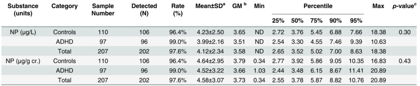
Urinary NP was detected in 97.6% of the participants (Table 4). The concentrations of NP ran-ged from ND (not detectable, less than 1.6μg/L) to 18.38μg/L. After creatinine adjustment, the
mean (± SD) concentrations of NP in the control and ADHD groups were 4.64±2.95 and 4.52 ±3.22μg/g cr., respectively. We did not observe a statistically significant relationship between
Table 2. Polymorphisms in dopamine-related genes (DRD4/DAT1) in the study participants (N = 207).
Gene SNP Genotypes
Control N (%) (N = 110) Case N (%) (N = 97) p-value
11 12 22 11 12 22
DRD4 rs7395429 53 (48.2%) 46 (41.8%) 11 (10.0%) 60 (61.9%) 30 (30.9%) 7 (7.2%) 0.07**
rs3758653 44 (40.0%) 55 (50.0%) 11 (10.0%) 48 (49.5%) 45 (46.4%) 4 (4.1%) 0.09** rs11246228 34 (30.9%) 51 (46.4%) 25 (22.7%) 18 (18.6%) 50 (51.5%) 29 (29.9%) 0.05** rs752306a 67 (60.9%) 38 (34.5%) 5 (4.5%) 75 (77.3%) 21 (21.6%) 1 (1.0%) <0.01
*
DAT1 rs6347 81 (73.6%) 28 (25.5%) 1 (0.9%) 78 (80.4%) 19 (19.6%) 0 (0%) 0.21
rs2975292 87 (79.1%) 22 (20.0%) 1 (0.9%) 70 (72.2%) 27 (27.8%) 0 (0.0%) 0.33
rs37022 30 (27.3%) 55 (50.0%) 25 (22.7%) 16 (16.5%) 63 (64.9%) 18 (18.6%) 0.47 rs40358 46 (41.8%) 48 (43.6%) 16 (14.3%) 37 (38.1%) 50 (51.5%) 10 (10.3%) 0.95
rs10040882 87 (79.1%) 22 (20.0%) 1 (0.9%) 73 (75.3%) 24 (24.7%) 0 (0.0%) 0.63
rs464049 50 (45.5%) 44 (40.0%) 16 (14.5%) 43 (44.3%) 47 (48.5%) 7 (7.2%) 0.5
a
The significant difference in this polymorphism (rs752306) between the case and control groups may have resulted from the limited number of participants.
*p<0.05 **p<0.1
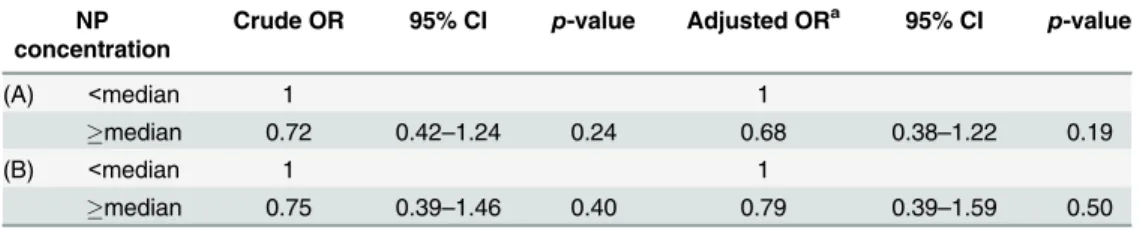
urinary NP levels and the presence of ADHD (p= 0.43). Moreover, logistic regression analyses, with females included and excluded, suggested that children with creatinine-adjusted NP con-centrations higher than the median may not present with a greater risk of having ADHD com-pared to children with concentrations below the median (Table 5). The odds ratios were essentially the same after adjusting for maternal education level and maternal alcohol exposure.
We detected a significant association between maternal drinking during pregnancy and ADHD (p<0.01), with few positive maternal drinking subjects reported in the control group.
After exclusion, the limited number of participants with in utero alcohol exposure did not affect the adjusted odds ratio in the logistic regression analysis.
Discussion
In this study, ADHD was significantly male predominant (male to female ratio: 5:1 in the ADHD group and 1.3:1 in the control group,p<0.01); ADHD was approximately three-fold
more likely to be diagnosed in boys than in girls [56]. Our finding was in accord with previous studies [56,57]. Biederman et al. suggested that girls with ADHD were less likely than boys to develop disruptive behavior disorder, a comorbid of ADHD, which may result in referral to specialists. The observation suggested that cases of girls with ADHD may be under-reported, which requires further attention [58,59]. Because of the limited number of females with ADHD in this study (N = 16), we examined different analytical approaches to evaluate
gender-Table 3. Distribution of BLLs in control and ADHD participants (N = 146). Substance
(units)
Category Sample Number
Detected (N)
Rate (%)
Mean±SDa GMb Min Percentile Max p-valuec
25% 50% 75% 90% 95%
Lead (μg/dL) Controls 102 102 100% 1.73±0.77 1.56 0.44 1.10 1.69 2.09 2.69 3.14 4.71 0.15
ADHD 44 44 100% 1.57±0.73 1.43 0.64 0.97 1.44 1.98 2.52 3.46 3.81
Total 146 146 100% 1.68±0.76 1.52 0.44 1.07 1.63 2.05 2.62 3.19 4.71
astandard deviation bgeometric mean c
2-sided Mann-Whitney U test.
doi:10.1371/journal.pone.0149558.t003
Table 4. Distribution of urinary NP (creatinine unadjusted and adjusted) in control and ADHD participants (N = 207). Substance
(units)
Category Sample Number
Detected (N)
Rate (%)
Mean±SDa GMb Min Percentile Max p-valuec
25% 50% 75% 90% 95%
NP (μg/L) Controls 110 106 96.4% 4.23±2.50 3.65 ND 2.72 3.76 5.45 6.88 7.66 18.38 0.30
ADHD 97 96 99.0% 3.99±2.16 3.51 ND 2.54 3.30 4.55 7.46 9.39 10.63
Total 207 202 97.6% 4.12±2.34 3.58 ND 2.65 3.52 5.02 7.00 8.63 18.38 NP (μg/g cr.) Controls 110 106 96.4% 4.64±2.95 3.79 0.34 2.77 3.92 5.86 9.05 10.35 16.83 0.43
ADHD 97 96 99.0% 4.52±3.22 3.66 1.03 2.44 3.48 6.15 8.67 11.41 20.89 Total 207 202 97.6% 4.58±3.07 3.73 0.34 2.55 3.78 5.87 8.82 10.76 20.89
astandard deviation bgeometric mean
c2-sided Mann-Whitney U test; ND: NP LOD = 1.6μg/L, measurements below the LOD were assigned a value corresponding to LOD/2.
related effects. A sensitivity analysis was performed by including and excluding females. The mean urinary NP levels in ADHD (N = 83) and non-ADHD (N = 61) boys in this study were 4.60±3.34 and 4.49±2.71μg/g cr., respectively (p= 0.67, data not shown). Higher urinary NP
levels did not increase the odds ratios between NP and ADHD when females were either included or excluded (Table 5). These data suggested that gender may not modify the relation-ship between urinary NP concentration and ADHD.
Several lines of evidence revealed that children with a family history of ADHD were more likely to be diagnosed with ADHD [32,39,49]. Our study found family history of nervous sys-tem diseases, including ADHD, was associated with higher risk for ADHD. Although the etiol-ogies of various nervous system diseases are still unclear and may be different, there might be deficits in the neuroprotection function against assorted risk factors for the probands, and these conditions are consequently transmitted within families.
We found that high maternal and paternal educational attainment was associated with a decreased susceptibility for ADHD. This finding is in agreement with previous results indicating that low parental education levels are an important adverse risk factor for ADHD [32,49,60]. In this study, a statistically significant association was observed between maternal drinking and ADHD. Our finding is consistent with several studies that have linked prenatal alcohol exposure to ADHD [61–63]. These studies support the inclusion of alcohol as a risk factor for ADHD.
Lead exposure of children in Taiwan
Lead exposure alters the dopamine system, which is relevant to the pathogenesis of ADHD [64,
65]. Low-level lead exposure has been associated with a clinical diagnosis of ADHD in several recent studies, even at concentrations much lower than the previous action level of 10μg/dL
(currently lowered to 5μg/dL) [31,34,35,66]. No significant difference in BLLs between the
control and case groups was observed (1.73±0.77μg/dL vs. 1.57±0.73μg/dL,p= 0.15). The
BLLs in this study were lower than those in the other two studies in Taiwan, which reported measurements of 5.50±1.86μg/dL for 934 primary school children in 2002 and a range from
1.97 to 2.49μg/dL for children 5–9 years of age in 2012 [67,68]. Leaded gasoline has been
banned in Taiwan since 2000. Meanwhile, 97.1% of the participants in the present study live in the greater Taipei area, which has few industrial emissions and tighter controls. These observa-tions may explain the lower BLL measurements in this study compared to those in previous studies that were performed in 2002 and 2012.
NP levels and detection rate compared to other studies
A previous study in this lab assessed the NP exposure of pubertal students aged 10.9–13.6 years and reported a geometric mean (GM) urinary NP level of 1.27μg/g cr.; 30% of the urine
Table 5. Odds ratios for ADHD according to the creatinine-adjusted NP concentration. NP
concentration
Crude OR 95% CI p-value Adjusted ORa 95% CI p-value
(A) <median 1 1
median 0.72 0.42–1.24 0.24 0.68 0.38–1.22 0.19
(B) <median 1 1
median 0.75 0.39–1.46 0.40 0.79 0.39–1.59 0.50
(A): all participants, N = 207; (B): boys, N = 144.
aAdjusted covariates: maternal education levels and maternal drinking during pregnancy.
samples were positive for NP [14]. In this current study of children aged 4–15 years, the urinary NP concentration GM was 3.73μg/g cr. (Table 4), and the detection frequency was 97.6%. The
NP levels in our samples were three-fold higher than those in pubertal students in Taiwan. Com-pared to studies from other nations, our measurements were approximately tenfold higher than reported levels in American adults (50% detection rate) and much higher than the readings in Belgium [69,70]. NP was not detected in the urine samples of 131 subjects in a general Belgian population (LOD, 0.23μg/L) [71]. However, the NP levels in the participants in our study (GM,
3.73μg/g cr.) were lower than those measured in China, in which a GM of 15.92μg/g cr. (100%
detected) was reported for 287 children and students aged 3 to 24 years [72].
The differences in NP levels between our study and other reference values reported in the Bel-gian and American data may be caused by several factors. First, NPnEO, which are biodegraded to NP, are massively produced and widely used in Taiwan [73–75]. Second, NP is not effectively removed due to insufficient wastewater treatment in Taiwan [76,77]. Third, detectable NP migrates into food from various plastic food containers and wrappings, which are widely used in Taiwanese daily activities [2,78]. The relatively high levels of NP exposure and the high detection frequency in this study and in research performed in China indicate that children in Asian nations are consistently exposed to high levels of NP, which represents a significant concern.
Association between NP exposure and the presence of ADHD
We report an absence of association between urinary NP concentrations and ADHD and no increased odds of ADHD for children with higher NP levels. The regression analyses, with and without excluding females, suggested that higher NP exposure may not promote the risk of having ADHD. Furthermore, no dose-response effects were observed.
According to Muller’s pharmacokinetic study, a bioavailability of 20% and a half-life of 2–3 h in blood were reported for NP. Moreover, only 10% of the applied dose is excreted in the urine [1]. Assuming that the volume of daily urine excretion is between 1000 and 1500 mL, the average daily body exposure of NP was estimated to be ~1.65–2.48μg/day/Kg body weight (b.w.) for
schoolchildren in Taiwan (data not shown). The daily NP exposure was calculated as follows: Estimated daily exposure of NP (μg/day/Kg b.w.) = urinary NP levels (μg /L) x urine
excre-tion per day (L/day) /10% NP exposure excreted through urine /body weight of participants (Kg)
The estimated daily exposure to NP in this study was lower than the tolerable daily intake (TDI) of 5μg/day/Kg b.w. proposed by the Danish Institute of Food Safety and Toxicology
[79]. Moreover, in Masuo’s animal study, young mice with a mean body weight of ~10 grams received an intracisternal injection of 87 nmol, which corresponds to 1.91 mg/Kg b.w. of NP exposure (NP has a molecular weight of 220 g/mole), approximately three orders of magnitude higher than the daily NP exposure in our study. An uncertainty factor of 1,000 typically was applied when deriving a no observed adverse effect level from experiments in laboratory ani-mals to a reference dose for children [80]. However, for an intracisternal injection animal study, the adverse effects of a dose applied to the brain will be much more severe than those after oral intake. Thus, the daily NP exposures of children in our study were much lower com-pared to the intra-brain dose in mice that triggered spontaneous motor activity. This relatively low-dose exposure probably explains why our data do not support the hypothesis that exposure to endocrine-disrupting NP increases the risk of ADHD.
Strengths and limitations
Taiwan. Moreover, the SNAP-IV questionnaire, a tool with acceptable reliability, was used to screen children with suspected ADHD. The diagnosis of ADHD was confirmed through an extensive evaluation based on the DSM-IV-TR clinical diagnosis performed by pediatricians or psychiatrists. Chances of misclassification between ADHD and non-ADHD were therefore minimal. In addition, we examined or adjusted for various important factors, including dopa-mine-related gene polymorphisms, exposure to lead, several socioeconomic indicators and maternal lifestyle factors.
However, this study also has several limitations. The limited sample size was related to the challenge of recruiting school-age children, especially those with ADHD, to participate in the study. The limited participation rate may be another limitation. Taipei City Hospital is a met-ropolitan hospital that serves the local population and accepts referrals from all the administra-tive districts in the city. In addition, 97.1% of the participants in the present study live in this metropolitan area. The cases and controls were recruited from the general pediatric clinics, not specific ADHD clinics, at the same hospital. Because attendance at Taipei City Hospital and the chance of being selected for the study were not dependent on NP levels, the chance of selec-tion bias was probably low [81]. In addition, there was the potential for recall bias in our study, particularly regarding prenatal tobacco and alcohol exposure and family history of nervous dis-eases. The recall bias was minimized by (1) objectively measuring the variables of interest (uri-nary NP levels, BLLs and gene polymorphisms) and by (2) asking for information that did not depend heavily on memory or subjective interpretation, such as parental education levels and maternal age at birth.
Conclusion
This case-control study found that NP exposure, at the estimated dosage of ~1.65–2.48μg/day/
kg b.w., was not a risk factor for ADHD in school-age children in Taiwan after adjusting for confounding factors. No differences were observed in BLLs between children with and without ADHD. More studies are warranted to further confirm the lack of a relationship between NP exposure and ADHD in children.
Acknowledgments
We would like to thank the Department of Health, Taipei City Government, and Aiming for the Top University Plan from the Ministry of Education of the Republic of China, Taiwan, for financially supporting this research. The work was also supported by the National Science Council of Taiwan. The authors also acknowledge the High-throughput Genome Analysis Core Facility of National Core Facility Program for Biotechnology, Taiwan, for SNP genotyp-ing. This manuscript has been edited by American Journal Experts.
Author Contributions
Conceived and designed the experiments: CJY JCD WY MYC BH MLC. Performed the experi-ments: CJY JCD HCC SHY KWL WY. Analyzed the data: CJY JCD SHY KWL MYC. Contrib-uted reagents/materials/analysis tools: JCD LCC MLC. Wrote the paper: CJY MLC.
References
1. Muller S, Schmid P, Schlatter C. Pharmacokinetic behavior of 4-nonylphenol in humans. Environ Toxi-col PharmaToxi-col. 1998; 5(4):257–65. Epub 1998/06/02. doi: S1382-6689(98)00009-X [pii]. PMID:
21781872.
3. John DM, White GF. Mechanism for biotransformation of nonylphenol polyethoxylates to xenoestro-gens in Pseudomonas putida. Journal of bacteriology. 1998; 180(17):4332–8. PMID:9721266 4. Lu J, Jin Q, He Y, Wu J, Zhang W, Zhao J. Anaerobic degradation behavior of nonylphenol
polyethoxy-lates in sludge. Chemosphere. 2008; 71(2):345–51. PMID:17936332
5. Soares A, Guieysse B, Jefferson B, Cartmell E, Lester J. Nonylphenol in the environment: a critical review on occurrence, fate, toxicity and treatment in wastewaters. Environment international. 2008; 34 (7):1033–49. doi:10.1016/j.envint.2008.01.004PMID:18282600
6. Guenther K, Heinke V, Thiele B, Kleist E, Prast H, Raecker T. Endocrine disrupting nonylphenols are ubiquitous in food. Environmental science & technology. 2002; 36(8):1676–80.
7. White R, Jobling S, Hoare S, Sumpter J, Parker M. Environmentally persistent alkylphenolic com-pounds are estrogenic. Endocrinology. 1994; 135(1):175–82. PMID:8013351
8. Markey CM, Rubin BS, Soto AM, Sonnenschein C. Endocrine disruptors: from Wingspread to environ-mental developenviron-mental biology. The Journal of steroid biochemistry and molecular biology. 2002; 83 (1):235–44.
9. Brooke L, Thursby G. Ambient aquatic life water quality criteria—nonylphenol. US Environmental
Pro-tection Agency (EPA), Office of Water Office of Science and Technology, Washington, DC Available: http://water.epa.gov/scitech/swguidance/standards/criteria/aqlife/nonylphenol/upload/2006_5_18_ criteria_nonylphenol_final-doc.pdf. Accessed: 19 November 2013]. 2005.
10. Laws SC, Carey SA, Ferrell JM, Bodman GJ, Cooper RL. Estrogenic activity of octylphenol, nonylphe-nol, bisphenol A and methoxychlor in rats. Toxicological Sciences. 2000; 54(1):154–67. PMID:
10746942
11. Küppers E, Ivanova T, Karolczak M, Lazarov N, Föhr K, Beyer C. Classical and nonclassical estrogen action in the developing midbrain. Hormones and behavior. 2001; 40(2):196–202. PMID:11534982 12. Tan BL, Kassim NM, Mohd MA. Assessment of pubertal development in juvenile male rats after
sub-acute exposure to bisphenol A and nonylphenol. Toxicology letters. 2003; 143(3):261–70. PMID:
12849686
13. Chang C-H, Chen M-L, Liao K-W, Tsai Y-A, Mao I, Wang T-H, et al. The association between maternal nonylphenol exposure and parity on neonatal birth weight: A cohort study in Taiwan. Chemosphere. 2013; 93(6):1145–52. doi:10.1016/j.chemosphere.2013.06.048PMID:23871597
14. Chen M-L, Lee H-Y, Chuang H-Y, Guo B-R, Mao I. Association between nonylphenol exposure and development of secondary sexual characteristics. Chemosphere. 2009; 76(7):927–31. doi:10.1016/j.
chemosphere.2009.04.054PMID:19476970
15. Kajta M, Wójtowicz AK. Impact of endocrine-disrupting chemicals on neural development and the onset of neurological disorders. Pharmacological Reports. 2013; 65(6):1632–9. PMID:24553011
16. Huang Y-F, Wang P-W, Huang L-W, Yang W, Yu C-J, Yang S-H, et al. Nonylphenol in pregnant women and their matching fetuses: Placental transfer and potential risks of infants. Environmental research. 2014; 134:143–8. doi:10.1016/j.envres.2014.07.004PMID:25127525
17. Lu Y-Y, Chen M-L, Sung F-C, Wang PS-G, Mao I-F. Daily intake of 4-nonylphenol in Taiwanese. Envi-ronment international. 2007; 33(7):903–10. PMID:17512594
18. Tsai M-S, Chang C-H, Tsai Y-A, Liao K-W, Mao I-F, Wang T-H, et al. Neonatal outcomes of intrauterine nonylphenol exposure—A longitudinal cohort study in Taiwan. Science of the total environment. 2013;
458:367–73. doi:10.1016/j.scitotenv.2013.04.039PMID:23680990
19. Chen M-L, Chang C-C, Shen Y-J, Hung J-H, Guo B-R, Chuang H-Y, et al. Quantification of prenatal exposure and maternal-fetal transfer of nonylphenol. Chemosphere. 2008; 73(1):S239–S45. doi:10.
1016/j.chemosphere.2007.04.091PMID:18442845
20. Chen M-L, Lee W-P, Chung H-Y, Guo B-R, Mao I-F. Biomonitoring of alkylphenols exposure for textile and housekeeping workers. International Journal of Environmental Analytical Chemistry. 2005; 85(4–
5):335–47.
21. Lopez-Espinosa M, Freire C, Arrebola J, Navea N, Taoufiki J, Fernandez M, et al. Nonylphenol and octylphenol in adipose tissue of women in Southern Spain. Chemosphere. 2009; 76(6):847–52. doi:10.
1016/j.chemosphere.2009.03.063PMID:19409598
22. Asimakopoulos AG, Thomaidis NS, Koupparis MA. Recent trends in biomonitoring of bisphenol A, 4-t-octylphenol, and 4-nonylphenol. Toxicology letters. 2012; 210(2):141–54. doi:10.1016/j.toxlet.2011.
07.032PMID:21888958
23. Huang HL, Chao CC, Tu CC, Yang PC. Behavioral parent training for Taiwanese parents of children with attention‐deficit/hyperactivity disorder. Psychiatry and clinical neurosciences. 2003; 57(3):275–81.
PMID:12753567
25. Thapar A, Cooper M, Eyre O, Langley K. Practitioner review: what have we learnt about the causes of ADHD? Journal of Child Psychology and Psychiatry. 2013; 54(1):3–16. doi:10.1111/j.1469-7610.2012.
02611.xPMID:22963644
26. Millichap JG. Attention deficit hyperactivity disorder handbook: a physician's guide to ADHD: Springer Science & Business Media; 2009.
27. Banerjee TD, Middleton F, Faraone SV. Environmental risk factors for attention‐deficit hyperactivity
dis-order. Acta paediatrica. 2007; 96(9):1269–74. PMID:17718779
28. Faraone SV, Perlis RH, Doyle AE, Smoller JW, Goralnick JJ, Holmgren MA, et al. Molecular genetics of attention-deficit/hyperactivity disorder. Biological Psychiatry. 2005; 57(11):1313–23. PMID:15950004 29. DiMaio S, Grizenko N, Joober R. Dopamine genes and attention-deficit hyperactivity disorder: a review.
Journal of Psychiatry and Neuroscience. 2003; 28(1):27. PMID:12587848
30. Hechtman L. Families of children with attention deficit hyperactivity disorder: a review. Canadian Jour-nal of Psychiatry. 1996; 41(6):350–60. PMID:8862854
31. Aguiar A, Eubig PA, Schantz SL. Attention deficit/hyperactivity disorder: a focused overview for chil-dren's environmental health researchers. Environmental Health Perspectives. 2010; 118(2):1646–53. 32. Wang H-L, Chen X-T, Yang B, Ma F-L, Wang S, Tang M-L, et al. Case-control study of blood lead levels
and attention deficit hyperactivity disorder in Chinese children. Environ Health Perspect. 2008; 116 (10):1401–6. doi:10.1289/ehp.11400PMID:18941585
33. Needleman H. The current status of childhood low-level lead toxicity. Neurotoxicology. 1992; 14(2–
3):161–6.
34. Chiodo LM, Jacobson SW, Jacobson JL. Neurodevelopmental effects of postnatal lead exposure at very low levels. Neurotoxicology and Teratology. 2004; 26(3):359–71. PMID:15113598
35. Eubig PA, Aguiar A, Schantz SL. Lead and PCBs as risk factors for attention deficit/hyperactivity disor-der. Environmental Health Perspectives. 2010; 118(2):1654–67.
36. Mao Z, Zheng Y-L, Zhang Y-Q, Han B-P, Chen L-T, Li J, et al. Chronic application of nonylphenol-induced apoptosis via suppression of bcl-2 transcription and up-regulation of active caspase-3 in mouse brain. Neuroscience letters. 2008; 439(2):147–52. doi:10.1016/j.neulet.2008.05.006PMID:
18514416
37. Masuo Y, Ishido M, Morita M, Oka S. Effects of neonatal treatment with 6-hydroxydopamine and endo-crine disruptors on motor activity and gene expression in rats. Neural plasticity. 2004; 11(1–2):59–76.
PMID:15303306.
38. Masuo Y, Morita M, Oka S, Ishido M. Motor hyperactivity caused by a deficit in dopaminergic neurons and the effects of endocrine disruptors: a study inspired by the physiological roles of PACAP in the brain. Regulatory peptides. 2004; 123(1):225–34.
39. Bouchard MF, Bellinger DC, Wright RO, Weisskopf MG. Attention-deficit/hyperactivity disorder and uri-nary metabolites of organophosphate pesticides. Pediatrics. 2010; 125(6):e1270–e7. doi:10.1542/
peds.2009-3058PMID:20478945
40. De Cock M, Maas YG, van de Bor M. Does perinatal exposure to endocrine disruptors induce autism spectrum and attention deficit hyperactivity disorders? Review. Acta paediatrica. 2012; 101(8):811–8.
doi:10.1111/j.1651-2227.2012.02693.xPMID:22458970
41. Kim B-N, Cho S-C, Kim Y, Shin M-S, Yoo H-J, Kim J-W, et al. Phthalates exposure and attention-defi-cit/hyperactivity disorder in school-age children. Biological Psychiatry. 2009; 66(10):958–63. doi:10.
1016/j.biopsych.2009.07.034PMID:19748073
42. Association AP. Diagnostic and statistical manual-text revision (DSM-IV-TRim, 2000): American Psy-chiatric Association; 2000.
43. Swanson J. SNAP-IV Scale. Irvine, CA: University of California Child Development Center. 1995.
44. Bussing R, Fernandez M, Harwood M, Hou W, Garvan CW, Eyberg SM, et al. Parent and teacher SNAP-IV ratings of attention deficit hyperactivity disorder symptoms psychometric properties and nor-mative ratings from a school district sample. Assessment. 2008; 15(3):317–28. doi:10.1177/
1073191107313888PMID:18310593
45. Gau SS-F, Lin C-H, Hu F-C, Shang C-Y, Swanson JM, Liu Y-C, et al. Psychometric properties of the Chinese version of the Swanson, Nolan, and Pelham, version IV scale-Teacher Form. Journal of pedi-atric psychology. 2009; 34(8):850–61. doi:10.1093/jpepsy/jsn133PMID:19074488
46. Gau SSF, Shang CY, Liu SK, Lin CH, Swanson JM, Liu YC, et al. Psychometric properties of the Chi-nese version of the Swanson, Nolan, and Pelham, version IV scale–parent form. International journal of
methods in psychiatric research. 2008; 17(1):35–44. doi:10.1002/mpr.237PMID:18286459 47. Swanson JM, Kraemer HC, Hinshaw SP, Arnold LE, Conners CK, Abikoff HB, et al. Clinical relevance
the end of treatment. Journal of the American Academy of Child & Adolescent Psychiatry. 2001; 40 (2):168–79.
48. Heinegård D, Tiderström G. Determination of serum creatinine by a direct colorimetric method. Clinica chimica acta. 1973; 43(3):305–10.
49. Millichap JG. Etiologic classification of attention-deficit/hyperactivity disorder. Pediatrics. 2008; 121(2): e358–e65. doi:10.1542/peds.2007-1332PMID:18245408
50. Cheuk DK, Wong V. Attention-deficit hyperactivity disorder and blood mercury level: a case-control study in Chinese children. Neuropediatrics. 2006; 37(4):234–40. Epub 2006/12/21. doi:
10.1055/s-2006-924577PMID:17177150.
51. Linnet KM, Dalsgaard S, Obel C, Wisborg K, Henriksen TB, Rodriguez A, et al. Maternal lifestyle factors in pregnancy risk of attention deficit hyperactivity disorder and associated behaviors: review of the cur-rent evidence. American Journal of Psychiatry. 2003; 160(6):1028–40. PMID:12777257
52. Linnet KM, Wisborg K, Agerbo E, Secher N-J, Thomsen PH, Henriksen TB. Gestational age, birth weight, and the risk of hyperkinetic disorder. Archives of Disease in Childhood. 2006; 91(8):655–60.
PMID:16754656
53. Brookes K, Xu X, Chen W, Zhou K, Neale B, Lowe N, etc. The analysis of 51 genes in DSM-IV com-bined type attention deficit hyperactivity disorder:association signals in DRD4, DAT1 and 16 other genes. Molecular Psychiatry. 2006b; 11:934–5.
54. Mundry R, Fischer J. Use of statistical programs for nonparametric tests of small samples often leads to incorrectPvalues: examples fromAnimal Behaviour. Animal behaviour. 1998; 56(1):256–9. PMID:
9710485
55. Maldonado G, Greenland S. Simulation study of confounder-selection strategies. American journal of epidemiology. 1993; 138(11):923–36. PMID:8256780
56. Emond V, Joyal C, Poissant H. [Structural and functional neuroanatomy of attention-deficit hyperactivity disorder (ADHD)]. L'encéphale. 2009; 35(2):107–14. doi:10.1016/j.encep.2008.01.005PMID:
19393378
57. Biederman J. Attention-deficit/hyperactivity disorder: a selective overview. Biol Psychiatry. 2005; 57 (11):1215–20. Epub 2005/06/14. doi: S0006-3223(04)01100-X [pii] doi:10.1016/j.biopsych.2004.10.
020PMID:15949990.
58. Biederman J, Kwon A, Aleardi M, Chouinard V- A, Marino T, Cole H, et al. Absence of gender effects on attention deficit hyperactivity disorder: findings in nonreferred subjects. American Journal of Psychiatry. 2014.
59. Biederman J, Mick E, Faraone SV, Braaten E, Doyle A, Spencer T, et al. Influence of gender on atten-tion deficit hyperactivity disorder in children referred to a psychiatric clinic. American Journal of Psychia-try. 2002; 159(1):36–42. PMID:11772687
60. Sauver JLS, Barbaresi WJ, Katusic SK, Colligan RC, Weaver AL, Jacobsen SJ, editors. Early life risk factors for attention-deficit/hyperactivity disorder: a population-based cohort study. Mayo Clinic Pro-ceedings; 2004: Elsevier.
61. Aronson M, Hagberg B, Gillberg C. Attention deficits and autistic spectrum problems in children exposed to alcohol during gestation: a follow‐up study. Developmental Medicine & Child Neurology.
1997; 39(9):583–7.
62. Mick E, Biederman J, Faraone SV, Sayer J, Kleinman S. Case-control study of attention-deficit hyper-activity disorder and maternal smoking, alcohol use, and drug use during pregnancy. Journal of the American Academy of Child & Adolescent Psychiatry. 2002; 41(4):378–85.
63. Knopik VS, Heath AC, Jacob T, Slutske WS, Bucholz KK, Madden PA, et al. Maternal alcohol use disor-der and offspring ADHD: disentangling genetic and environmental effects using a children-of-twins design. Psychological medicine. 2006; 36(10):1461–71. PMID:16734942
64. Cory-Slechta D. Relationships between lead-induced learning impairments and changes in dopaminer-gic, cholinerdopaminer-gic, and glutamatergic neurotransmitter system functions. Annual review of pharmacology and toxicology. 1995; 35(1):391–415.
65. Schneider J, Huang F, Vemuri M. Effects of low-level lead exposure on cell survival and neurite length in primary mesencephalic cultures. Neurotoxicology and Teratology. 2003; 25(5):555–9. PMID:
12972068
66. CDC. Centers for Disease Control and Prevention: CDC's Childhood Lead Poisoning Prevention Pro-gram, Atlanta, GA. Available:http://www.cdc.gov/nceh/lead/acclpp/blood_lead_levels.htm. Accessed: 3 October 2015.
68. Huang P-C, Su P-H, Chen H-Y, Huang H-B, Tsai J-L, Huang H-I, et al. Childhood blood lead levels and intellectual development after ban of leaded gasoline in Taiwan: a 9-year prospective study. Environ-ment international. 2012; 40:88–96. doi:10.1016/j.envint.2011.10.011PMID:22280932
69. Calafat AM, Kuklenyik Z, Reidy JA, Caudill SP, Ekong J, Needham LL. Urinary concentrations of bis-phenol A and 4-nonylbis-phenol in a human reference population. Environmental Health Perspectives. 2005:391–5. PMID:15811827
70. Shen Y, Xu Q, Ren M, Feng X, Cai Y, Gao Y. Measurement of phenolic environmental estrogens in women with uterine leiomyoma. PloS one. 2013; 8(11):e79838. doi:10.1371/journal.pone.0079838 PMID:24255718
71. Pirard C, Sagot C, Deville M, Dubois N, Charlier C. Urinary levels of bisphenol A, triclosan and 4-nonyl-phenol in a general Belgian population. Environment international. 2012; 48:78–83. doi:10.1016/j.
envint.2012.07.003PMID:22885664
72. Li X, Ying G-G, Zhao J-L, Chen Z-F, Lai H-J, Su H-C. 4-Nonylphenol, bisphenol-A and triclosan levels in human urine of children and students in China, and the effects of drinking these bottled materials on the levels. Environment international. 2013; 52:81–6. doi:10.1016/j.envint.2011.03.026PMID:
21794921
73. Cheng C-Y, Ding W-H. Determination of nonylphenol polyethoxylates in household detergents by high-performance liquid chromatography. Journal of Chromatography A. 2002; 968(1):143–50.
74. Chen HW, Liang CH, Wu ZM, Chang EE, Lin TF, Chiang PC, et al. Occurrence and assessment of treatment efficiency of nonylphenol, octylphenol and bisphenol-A in drinking water in Taiwan. Sci Total Environ. 2013; 449C:20–8. Epub 2013/02/14. doi: S0048-9697(13)00057-0 [pii] doi:10.1016/j.
scitotenv.2013.01.038PMID:23403099.
75. Pan YP, Tsai SW. Determination and residual characteristic of alkylphenols in household food deter-gents of Taiwan. Chemosphere. 2009; 76(3):381–6. Epub 2009/04/21. doi:10.1016/j.chemosphere.
2009.03.034S0045-6535(09)00349-X [pii]. PMID:19375146.
76. Ding W-H, Fann JC. Application of pressurized liquid extraction followed by gas chromatography–mass
spectrometry to determine 4-nonylphenols in sediments. Journal of Chromatography A. 2000; 866 (1):79–85. PMID:10681011
77. Shue M-F, Chen F-A, Chen T-C. Total estrogenic activity and nonylphenol concentration in the Dong-gang River, Taiwan. Environmental monitoring and assessment. 2010; 168(1–4):91–101. doi:10.1007/
s10661-009-1093-4PMID:19609692
78. Loyo-Rosales JE, Rosales-Rivera GC, Lynch AM, Rice CP, Torrents A. Migration of nonylphenol from plastic containers to water and a milk surrogate. Journal of Agricultural and Food Chemistry. 2004; 52 (7):2016–20. PMID:15053545
79. Nielsen E, Ostergaard G, Thorup I, Ladefoged O, Jelnes JE. Toxicological Evaluation and Limit Values for Nonylphenol, Nonylphenol Ethoxylates, Tricresyl Phosphates and Benzoic Acid. The Institute of Food Safety And Toxicology. Available:http://www2.mst.dk/Udgiv/publications/1999/87-7909-566-6/ pdf/87-7909-565-8.pdf[Accessed 19 Nov. 2013], 1999 0105–3094.
80. Frumkin H. Environmental health: from global to local: John Wiley & Sons; 2005.
![Table 1 lists the demographic characteristics of the 207 participating children. Of these partici- partici-pants, 97 were diagnosed with ADHD (81 boys [83.5%] and 16 girls [16.5%]; M:F = 5:1), and 110 were control subjects (63 boys [57.3%] and 47 girls [42](https://thumb-eu.123doks.com/thumbv2/123dok_br/18275441.345004/6.918.305.865.497.1011/demographic-characteristics-participating-children-partici-diagnosed-control-subjects.webp)