Leticia Vilaça Willeman Bastos*, Ricardo de Souza Tesch**, Odilon Victor Porto Denardin***
Cephalometric deviations present in
children and adolescents with
temporomandibular joint disorders
Introduction: Temporomandibular disorders (TMD) have proved to be a risk factor for
developing hyperdivergent facial growth patterns. Objective: The aims of this study were: (1) Assess differences between the cephalometric measurements in children with articular TMD and a control group, before and after mandibular growth peak according to cervical vertebral maturation; and (2) Identify a predictive model capable of differ-entiating patients with TMD and control group patients based on early cephalometric
characteristics. Method: The study included children and adolescents with maximum
age of 17 years, divided into experimental group (n=30) diagnosed with articular TMD— according to the Research Diagnostic Criteria for Temporomandibular Disorders (RDC/ TMD) for children and adolescents—subdivided according to growth stage, called pre-peak (n=17) and post-pre-peak (n=13) and control group (n = 30), matched by gender, skel-etal maturity stage of the cervical vertebrae and classification of malocclusion. Lateral cephalometric and craniofacial structures were traced and their relations divided into: Cranial base, maxilla, mandible, intermaxillary relations, vertical skeletal relations and dental relations. Differences between the means for each variable were evaluated by applying the statistical Student t test for independent samples. Results: The means of the variables analyzed in the pre-peak showed no statistically significant differences. However, analysis of post-peak showed that the experimental group displayed decreased SNA and SNB and increased SN.Gn and 1.NB (p<0.05). Conclusion: It was possible to identify a predictive model able to differentiate patients with TMD and asymptomatic controls from early cephalometric characteristics.
Abstract
Keywords: Facial growth. TMD. Malocclusion.
* MSc in Health Sciences, TMD and Orofacial Pain, Heliopolis Hospital (Hosphel).
** MSc in Health Sciences, TMD and Orofacial Pain, Hosphel. Head of TMD and Orofacial Pain Department, Petrópolis Medical School. *** PhD in Endocrinology, São Paulo Federal University (UNIFESP). Head of Statistics Department, Hosphel.
How to cite this article: Bastos LVW, Tesch RS, Denardin OVP.
Cephalo-metric deviations present in children and adolescents with temporoman-dibular joint disorders. Dental Press J Orthod. 2012 Jan-Feb;17(1):74-84.
IntROduCtIOn
Although the treatment need for temporo-mandibular disorders (TMD) is directly related to pain and functional disability produced by these conditions, the presence of clinical signs and symptoms arising from changes in mor-phology and/or function of the temporoman-dibular joint (TMJ), such as joints sounds, are much more frequent.
Currently, the Research Diagnostic Crite-ria for Temporomandibular Disorders (RDC/ TMD),10 represents the most studied diagnostic
tool regarding validity and accuracy of TMD’s diagnostic classification based on specific diag-nostic criteria. This system, through its physical axis, allows the possibility of multiple diagno-ses for one individual, being the joint disorders divided into two broad categories not mutually exclusive: The disc displacements, with or with-out reduction; and arthralgia, osteoarthritis and osteoarthrosis of the temporomandibular joint.10
The RDC/TMD was not originally designed for use in children and adolescents, but was modified and validated for this purpose by Wahlund et al33 and was later applied to this
population group by several researchers. Among the articular TMD, disc displace-ment with reduction may be clinically recog-nized by the occurrence of a clicking sound during opening and closing mouth movements, eliminated when the mouth is open sustain-ing maximum protrusion. In the diagnosis of articular disc displacement without reduction although there may be a history of joint clicks, the same must be absent at the time of diag-nosis, associated with limitations and/or uncor-rected deviation of mandibular movement.10
Degenerative changes of the TMJ are char-acterized by the presence of clinical signs of continuous joint soundsin the form of crepitus. According to RDC/TMD, crepitation can be ac-companied by arthralgia, known as osteoarthri-tis or, in the absence of pain, osteoarthrosis.10
The temporomandibular joint arthralgia is char-acterized by pre-auricular spontaneous pain or pain induced by palpation and/or function.10
Population studies about the prevalence of articular disorders’ signs described above, in chil-dren and adolescents, showed that 8% to 29% of the assessed population showed clicking sounds in the TMJ, while only 1% displayed crepitus.11
Several factors distinguish the stomato-gnathic system of adults, children and ado-lescents, for example, the components of the masticatory system, which undergo different growth and development patterns.32 During this
development process, the craniofacial struc-tures can be influenced by several factors such as congenital abnormalities,15 uncontrolled
hor-monal levels,13 arthritis,17 condylar hypoplasia,4
deleterious breathing habits,34 trauma,3
infec-tions,19 and orthopedic forces.24 Such factors
can alter the tissue adaptive capacity during growth and thus alter facial morphology.25,26
The studies that correlate articular TMD and facial growth patterns were conducted in adult patients,1,2,6,16 as well as in children and
adolescents.24,25,26,31
Thus, the involvement ofTMJ by
degenera-tive changes or disc displacements proved to be an important risk factor for identification of individuals with hyperdivergent facial growth patterns. However, the pathophysiological pro-cesses responsible for this relationship are not yet fully established.
Ahn et al2 evaluated the presence or absence
of anterior disc displacement diagnosis in 134 women, using magnetic resonance image (MRI). The means of 36 cephalometric variables were compared and the discriminant analysis mod-el was conducted to determine which are the most important factors for identifying patients with anterior disc displacement.
overjet intensified gradually with the progres-sion of internal derangement of TMJ, and sub-jects with bilateral joint disorders showed the largest dentofacial morphological changes. Dis-criminant analysis was able to correctly classify 79% of subjects and demonstrated that those with the smallest angle between the long axis of the lower central incisor and the Frankfort plan and greater overjet had an increased risk to develop articular TMD.
The lack of knowledge regarding when the pa-tient was affected by these disorders, which are more prevalent in ages subsequent to completion of facial growth, becomes a potential bias in estab-lishing the real power of this association, which can be further important if early identified.
It seems therefore, important to identify, during the different stages of skeletal matu-rity that makes up the facial growth, signs of articular TMD, evaluating the real impact of these disorders on the facial growth pattern. It is expected therefore to show that the awaited changes in cephalometric measurements occur mainly in the later stages of facial development after the mandibular growth peak (stages III and IV of cervical vertebral maturation).
The aims of this study were: (1) To evaluate differences between the cephalometric vari-ables selected for identification of facial growth pattern in children and adolescents with articu-lar TMD and control group free of that disorder before and after the mandibular growth peak, according to cervical vertebral maturation (CVM) and (2) Identify a predictive model able to differentiate patients with TMJ disor-ders and asymptomatic controls based on early skeletal cephalometric characteristics.
MAteRIAl And MethOds
This study followed an observational and cross-sectional protocol, performed at the Preven-tive Orthodontics Clinic of the Petrópolis Medi-cal School (Rio de Janeiro State, Brazil), including
patients aged up to 17 years old of both genders. It was approved by the Scientific and Research Eth-ics Committee of the mentioned school.
Before performing any procedure all pa-tients received detailed information about the research and signed a consent form.
The experimental group comprised 30 pa-tients diagnosed with articular TMD, subdivided into two groups according to the corresponding growth period, named pre-peak of growth spurt (n=17) and post-peak of growth spurt (n=13). The inclusion criteria were: (1) Articular TMD Group II or III, according to Axis I of RDC/ TMD for children and adolescents—the Portu-guese version; (2) Good quality lateral radio-graphs obtained prior to orthodontic treatment, carried out in the same cephalostat (Cerdo Ra-diology Clinic, Petrópolis, RJ).
The following exclusion criteria were used: (1) Diagnosis of juvenile rheumatoid arthritis or other systemic joint diseases; (2) Orthodon-tic treatment prior to examination.
The control group consisted of 30 volunteers without TMD, matched by gender, cervical ver-tebral maturation index and Angle’s classifica-tion of malocclusion. Inclusion criteria were the same adopted for the study group except for the need of articular TMD diagnosis, Groups II and III, according to Axis I of RDC/TMD for chil-dren and adolescents, Portuguese version, which becomes an exclusion criteria in addition to the others previously related to the study group.
The clinical examination conducted, part of the Axis I diagnosis protocol of RDC/TMD for children and adolescents, Portuguese version,27
has been applied and validated in adolescents in previous studies,33 comprising 10 question.
were divided into the following categories for analysis: Cranial base, maxilla, mandible, inter-maxillary relations, vertical skeletal and dental relations (Table 1, Figs 1, 2, 3 and 4).
The angular and linear measurements were taken from the analysis previously described by Downs,9 Jarabak14 and McNamara.21
The measures found were tabulated. Means and standard deviations for each variable in the patient sample were calculated using the statis-tical software SPSS (SPSS Inc, Chicago, USA).
The differences between the means for each of the variables selected for the group of patients with articular TMD and the control group were evaluated by applying Student t test for independent samples and the discriminant analysis with stepwise entry for patients in post-peak of pubertal growth spurt. The differences
that had less than a 5% chance of having oc-curred by chance (p<0.05) were considered sta-tistically significant.
To test the magnitude of the measurement error for the cephalometric variables in this study, the lateral radiographs of 15 patients and volunteers were randomly chosen and mea-sured again. By applying Dahlberg’s8 formula,
the error found was between 0.31 and 0.79 mm for linear measurements and 0.30 and 0.98º for angular measurements.
Results
Patients in the experimental group—articu-lar TMD (n=30)—had a mean age of 9 years old (range 5-17 years old), being 12 individuals (40%) males and 18 (60%) females. Of these, 7 were in stage I of cervical vertebral matura-tion (23%), 10 in stage II (33%), 12 in stage III (40%) and 1 in stage IV (4%).
In the control group—no articular TMD (n=30)—the average age was 10 years old (range 5-14 years old), and presented the same distribution for gender and cervical vertebral maturation index found in the experimental group due to the pairing of the samples.
When the total experimental and total con-trol groups were subdivided according to the stage of skeletal maturity, assessed by the cer-vical vertebral maturation index, into the sub-groups pre-peak of pubertal growth spurt and post-peak of pubertal growth spurt, the means of the analyzed variables in the pre-peak of pu-bertal growth spurt period showed no statisti-cally significant differences (Table 2).
However, the analysis of post-peak of pu-bertal growth spurt showed that the experi-mental group had mean values for SNA and SNB angles decreased, and the facial axis angle (SN.Gn) and lower incisor inclination (1.NB) increased in relation to the mean values found in the control group, revealing statistically sig-nificant differences (Table 3).
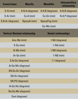
TABLE 1 - Linear and angular cephalometric measurements. Cranial base Maxilla Mandible Intermaxillary
relationship
S-N (mm) S.N.A (degrees) S.N.B (degrees) A.N.B (degrees)
S-Ar (mm) Co.A (mm) Co-Gn (mm) N.A.P (degrees)
S.N.Ar (degrees) NperpA (mm) NperpPog (mm)
Go-Me (mm)
Vertical Skeletal relationship Dental relationships
Ans-Me (mm) 1.NA (degrees)
S-Go (mm) 1-NA (mm)
N-Me (mm) 1.NB (degrees)
Ar-Go (mm) 1-NB (mm)
S.Ar.Go (degrees) 1.1 (degrees)
Ar.Go.Me (degrees)
SN.Go.Gn (degrees)
SN.Gn (degrees)
SN.PO (degrees)
Ar.Go.Na (degrees)
Na.Go.Me (degrees)
3
3 1
1
2
3 4
5
1 2
3 4
5
6 7
8 9
10 1
2
2
4
4 5
5
7
7
8
8
9 9
10 11
11 12
12 13
10
6
6
The discriminant analysis, with stepwise vari-able input, was applied only to varivari-ables whose dif-ferences between experimental and control groups had previously shown to be statistically significant. When performed in patients in the post-peak of pubertal growth spurt, as demonstrated by the cer-vical vertebral maturation index, it were selected
two variables: SNA and lower incisor inclination. The discriminant function nonstandard coeffi-cients led to the following equation, which gives each new subject an individual score to classify them as a patient with articular TMD or control.
Individual score = 0.829 X (S.N.A) – 0.645 X (1.NB)
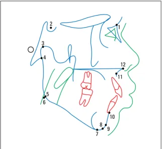
FIGURE 1 - Points used in this study: 1= N; 2= S; 3= Co; 4= Ar; 5= anatomic Go; 6= cephalometric Go; 7= Me; 8= Gn; 9= Pog; 10= B; 11= A; 12= Ans.
FIGURE 2 - Lines used in this study: 1= S-N; 2= S-Ar; 3= Ar-Go; 4= Go-Me; 5= S-Go; 6= Co-A; 7= Co-Gn; 8= N-Me; 9= NPog-P; 10= NA-P; 11= 1-NA; 12= 1-NB; 13= Ans-Me.
FIGURE 3 - Angles used in this study: 1= S.N.Ar; 2= S.Ar.Go; 3= Ar.Go.Me; 4= Ar.Go.Na; 5= Na.Go.Me.
Cephalometric variables
Experimental Group
Control Group
p
(mean± DP) (mean± DP)
Cranial base
S-N (mm) 67.35±3.53 66.44±5.04 0.546
S-Ar (mm) 32.37±3.04 30.56±4.44 0.176
S.N.Ar (degrees) 123.45±5.23 124.58±6.35 0.575
Maxilla
S.N.A (degrees) 82.05±3.03 81.39±4.34 0.611
Co.A (mm) 84.17±6.25 83.91±8.35 0.921
NperpA (mm) 1.19±3.70 1.03±3.69 0.898
Mandible
S.N.B (degrees) 4.30±1.91 3.98±4.63 0.799
Co-Gn (mm) 108.35±7.95 109.76±7.61 0.600
NperpPog (mm) -5.52±6.38 -2.44±4.67 0.117
Go-Me (mm) 66.87±5.34 69.21±5.68 0.224
Intermaxillary relationship
A.N.B (degrees) 4.30±1.91 3.98±4.63 0.799
N.A.P 8.57±4.53 6.71±7.21 0.375
Vertical skeletal relationship
Ans-Me (mm) 64.31±63.79 63.79±4.65 0.788
S-Go (mm) 68.25±6.10 67.23±5.68 0.619
N-Me (mm) 110.40±7.96 110.24±7.69 0.952
Ar-Go (mm) 39.22±3.86 39.46±3.50 0.853
S.Ar.Go (degrees) 144.92±6.86 145.38±5.93 0.836
Ar.Go.Me (degrees) 129.66±6.75 126.15±5.37 0.104
SN.GoGn (degrees) 35.52±4.82 36.08±5.27 0.748
SN.Gn (degrees) 67.97±3.69 68.29±3.19 0.791
SN.Po (degrees) 18.17±4.59 18.15±4.19 0.990
Ar.Go.N (degrees) 53.77±5.43 52.27±2.76 0.316
N.Go.Me (degrees) 75.87±4.47 73.94±4.33 0.211
Sum 398.12±4.50 396.12±3.73 0.167
Dental relationships
1.NA (degrees) 24.48±6.04 22.45±8.65 0.434
1-NA (mm) 4.38±1.77 4.62±2.30 0.737
1.NB (degrees) 31.25±7.24 26.56±8.54 0.094
1-NB (mm) 6.24±2.14 6.77±5.34 0.710
1.1 (degrees) 122.57±11.30 128.07±15.26 0.241
Cephalometric variables
Experimental Group
Control Group
p (mean± DP) (mean± DP)
Cranial base
S-N (mm) 70.85±3.47 67.81±5.23 0.094
S-Ar (mm) 33.93±3.20 33.58±3.74 0.801
S.N.Ar (degrees) 125.26±6.25 125.53±6.31 0.914
Maxilla
S.N.A (degrees) 78.25±3.55 82.90±4.53 0.008
Co.A (mm) 86.19±5.73 87.93±3.69 0.368
NperpA (mm) -0.13±4.04 2.79±3.63 0.065
Mandible
S.N.B (degrees) 74.69±3.63 79.26±4.75 0.011
Co-Gn (mm) 114.51±7.80 115.94±3.46 0.552
NperpPog (mm) -7.08±8.77 -2.52±7.95 0.178
Go-Me (mm) 73.03±5.84 72.97±3.35 0.974
Intermaxillary relationship
A.N.B (degrees) 3.55±2.98 3.71±2.89 0.891
N.A.P 6.45±6.57 6.92±6.03 0.852
Vertical skeletal relationship
Ans-Me (mm) 68.88±6.83 67.93±4.46 0.678
S-Go (mm) 73.42±6.42 72.99±4.36 0.842
N-Me (mm) 121.26±9.21 115.37±7.58 0.088
Ar-Go (mm) 42.18±3.53 43.12±3.99 0.532
S.Ar.Go (degrees) 146.10±7.19 141.92±7.34 0.155
Ar.Go.Me (degrees) 125.76±5.59 128.02±4.42 0.265
SN.GoGn (degrees) 38.59±7.15 34.35±4.87 0.090
SN.Gn (degrees) 72.08±5.13 67.85±4.12 0.029
SN.Po (degrees) 18.85±6.32 15.19±4.47 0.101
Ar.Go.N (degrees) 50.06±4.24 53.39±4.87 0.075
N.Go.Me (degrees) 75.82±6.25 74.66±4.31 0.588
Sum 397.18±7.90 395.45±4.41 0.499
Dental relationships
1.NA (degrees) 27.38±5.05 23.90±7.83 0.190
1-NA (mm) 6.67±3.59 5.44±3.00 0.355
1.NB (degrees) 30.50±4.07 26.41±5.52 0.042
1-NB (mm) 6.41±4.85 5.64±2.02 0.603
1.1 (degrees) 119.26±6.34 124.86±10.29 0.108
TABLE 2 - Distribution of means and standard deviations for cephalo-metric variables in patients with pre-peak vertebral maturity.
The score found for the experimental group was 45.20 and for the control group was 51.96. The critical score, calculated as the average of the scores found for the experimental group and control group was 48.58. Thus the percent-age of subjects correctly classified was 80.76%, leading to misclassification of 2 subjects in the experimental group (15.39%) and 3 control subjects (23.08%).
dIsCussIOn
The condyle plays an active role in man-dibular growth, which varies according to its cartilage primary potential. Children and ado-lescents have high adaptive capacity, so that the components of their TMJ and dentoalveolar re-gions undergo shape and size changes during growth.12 Such changes tend to be more
rel-evant during the pubertal growth spurt, which corresponds to the period of greatest relative mandibular growth.
The displacement of the articular disc has the ability to change the joint function, possibly exceeding the adaptive capacity of the growing tissues.32 During mandibular growth, when
pro-liferation of the condylar cartilage provides the joint fibrocartilaginous tissue, the articular disc displacement can result in rupturing of the cell proliferation layer. This disruption can reduce the formation of extracellular collagen and teoglycan matrix, which contribute to the pro-cess of condylar growth.20
Experiments with animals, in which ante-rior displacements of the articular disc were surgically obtained in rabbits, and the posterior ligament kept intact over the condyle, demon-strated that the jaw of these animals became significantly shorter on the same side of the disc displaced, which resulted in a midline shift to the affected side. The mandibular asymme-try was not observed in the control group, with no displacement of the articular disc.18
These results suggest that the articular disc
displacement precede the development of asymmetry of the jaw and can therefore be considered as risk factors for these conditions. However, if this sequence of events is relevant to human mandibular growth and develop-ment, it has not been determined yet, although it has been previously suggested.25,26
Mandibular asymmetries were also observed by Kambylafkas et al16 in patients with
unilat-eral osteoarthritis who had mandibular devia-tion to the affected side. The cephalometric measurements of patients revealed decreased condylar height and increased antegonial depth on the same side of the diagnosis of degenera-tive articular changes.
In this study, when the total experimental group and control group were subdivided ac-cording to the time of skeletal maturity, as as-sessed by cervical vertebral maturation index, into subgroups pre-peak and post-peak of pu-bertal growth spurt, the mean variables ana-lyzed in the pre-peak of pubertal growth spurt showed no statistically significant differences.
However, the analysis of post-peak of pubertal growth spurt showed that the experimental group had greater maxillary and mandibular retrusion, increased facial convexity and labial inclination of upper incisors when compared to the control group. These results suggest that the compensa-tory skeletal and dentoalveolar changes resulting from TMJ disorders manifest themselves prefer-entially after the mandibular growth peak, a fact that has not been previously demonstrated.
Recently, several studies published, used mag-netic resonance images (MRI) for the diagnosis of positional changes of the articular disc and de-generative diseases of the TMJ trying to correlate those diagnoses with changes on the craniofacial measurements of growing patients.24,25,26,31 The
However, the relatively high cost of MRI precludes its routine clinical indications for confirmation of the diagnosis of articular TMD, which can be detected clinically with accept-able levels of validity and accuracy.22
The compensatory proclination of lower in-cisors is probably related to increased overjet, arising from the posterior positioning of the mandible and its clockwise rotation. This rota-tion is associated with decreased height of the mandibular ramus found in patients with TMD in different studies.1,25,26
A relative maxillary retrusion, evaluated through the SNA angle, was observed in patients of the experimental group in the post-peak of puber-tal growth stage when compared with the control group. Although it has been previously described in studies evaluating cephalometric characteristics of patients with TMD,2,6 it has not been discussed
satisfactorily, probably due to the absence of ob-vious relationship between intra-articular changes and maxillary growth. However, the SNA measure uses as reference point A, a dentoalveolar land-mark and therefore capable of suffering the influ-ence of possible dentoalveolar compensation on the superior arch due to mandibular changes.
Most published studies relating cephalomet-ric variables and TMD suggest that the lateral cephalometric radiograph could be used to iden-tify patients potentially suffering from internal disorders and/or degenerative changes of the TMJ.1,2,6 Recently, certain variables selected from
panoramic radiographs also demonstrated to be associated with the diagnosis of TMD,7 opening
the field for further research in this area.
However, in a study published by Bósio et al,5
56% of symptomatic women with bilateral disc displacement and Angle Class I malocclusion, dis-played a mean value for the SNB angle (75.6°) significantly lower than the pattern observed in normal individuals. However, when women with similar articular diagnoses, but presenting Class II malocclusions were divided into Division 1 and
Division 2 sub-groups, no statistically significant difference was found for the angle SNB when compared to normal individuals.
Thus, according to the authors, it would only be possible to diagnose—with accept-able degree of validity—the articular disc dis-placement based on lateral cephalometric ra-diographs when the patient develops typical signs or symptoms from TMD, Angle Class I malocclusion and SNB below the average cal-culated in the above described study (75.6°). This would make the prediction possible, but not very practical and definitely not diagnostic.
Therefore, the identification of certain cephalometric characteristics in specific patient groups can be considered an indicating factor for the necessity of diagnostic tools aimed at early identification of articular changes in chil-dren and adolescents, many times neglected.
The discriminant analysis was used previously to establish a predictive formula capable of clas-sifying patients with TMD, with greater reliabil-ity.2 The two variables selected by the stepwise
process were: the inclination of lower incisors in relation to the Frankfurt horizontal plane and the amount of overjet. Thus, patients with internal derangement of the TMJ showed a lower incisor angle and greater overjet than in control subjects with normal joints. The discriminant analysis showed a higher validity in predicting patients with TMD (93.3%) than in the prediction of controls with normal joints (65.9%).
The results of this study demonstrate a rela-tionship between TMJ disorders in children and adolescents and the presence of a hyperdiver-gent pattern of facial growth. Even though the direction of this relationship can be assumed, it could not be determined due to the cross-sec-tional nature of this study, where the supposed risk factors and outcomes were assessed at the same time point. Therefore, the hypothesis of these disorders influencing on facial growth pattern must be confirmed by a longitudinal follow up to adulthood, from a growing cohort of patients, with and without TMJ disorders.
Future studies in this direction should also, ideally, consider the different sub-groups of pa-tients with articular disc displacement,
accord-ing to the RDC/TMD,10 in the cephalometric
evaluation of structural characteristics since, according to Bósio et al,5 the position of the
articular disc can influence the condylar
posi-tion and thus the jaw posiposi-tion and their cepha-lometric measurements.
Ren, Isberg and Westesson29 demonstrated
that joints with disc displacement have more posterior condylar position than that found in normal joints. Likewise, Ronquillo et al30 and
Pullinger28 suggested that patients with
1. Ahn SJ, Kim TW, Nahm DS. Cephalometric keys to internal derangement of temporomandibular joint in women with Class II malocclusion. Am J Orthod Dentofacial Orthop. 2004;126(4):486-94.
2. Ahn SJ, Baek SH, Kim TW, Nahm DS. Discrimination of internal derangement of temporomandibular joint by lateral cephalometric analysis. Am J Orthod Dentofacial Orthop. 2006;130(3):331-9.
3. Amaratunga NA. Mandibular fractures in children: a study of clinical aspects, treatment needs, and complications. J Oral Maxillofac Surg. 1988;46:637-40.
4. Bjork A. Facial growth in bilateral hypoplasia of the mandibular condyles. In: Kraus BS, Reidel RA. Vistas in Orthodontics. Philadelphia: Saunders; 1962. p.104-40. 5. Bósio JA, Burch JG, Tallents RH, Wade DB, Beck FM.
Lateral cephalometric analysis of asymptomatic volunteers and symptomatic patients with and without bilateral temporomandibular joint disk displacement. Am J Orthod Dentofacial Orthop. 1998;114(3):248-55.
6. Brand JW, Nielson KJ, Tallents RH, Nanda RS, Currier GF, Owen WL. Lateral cephalometric analysis of skeletal patterns in patients with and without internal derangement of the temporomandibular joint. Am J Orthod Dentofacial Orthop. 1995;107(2):121-8. 7. Ahn SJ, Kim TW, Lee DY, Nahm DS. Evaluation of internal
derangement of the temporomandibular joint by panoramic radiographs compared with magnetic resonance imaging. Am J Orthod Dentofacial Orthop. 2006;129(4):479-85. 8. Dahlberg G. Statistical methods for medical and biological
students. New York: Interscience Publication; 1940. 9. Downs WB. Variation in facial relationship: their
significance in treatment and prognosis. Am J Orthod. 1948;34:812-40.
10. Dworkin SF, LeResche L. Research diagnostic criteria for temporomandibular disorders: review, criteria, examinations and speciications. Critique J Craniomand Disord Facial Oral Pain. 1992;6:302.
11. Egermark-Eriksson I, Carlsson GE, Ingervall B. Prevalence of mandibular dysfunction and orofacial parafunction in 7-, 11- and 15-year-old Swedish children. Eur J Orthod. 1981;3(3):163-72. 12. Enlow DH. Facial growth. 3rd ed. Philadelphia: WB Saunders; 1990. 13. Hoskin WE, Asling CW. Inluence of growth hormone and
thyroxine and endochondral osteogenesis in the mandibular condyle and proximal tibial epiphysis. J Dent Res. 1977; 56:509-17.
RefeRenCes
14. Jarabak JR, Fizzel JA. Technique and treatment with light wire appliances. St. Louis: Mosby;1972.
15. Kaban LB, Mulliken JB, Murray JE. Three-dimension approach to analysis and treatment of hemifacial microsomia. Cleft Palate J. 1981;18:90-9.
16. Kambylafkas P, Kyrkanides S, Tallents RH. Mandibular asymmetry in adult patients with unilateral degenerative joint disease. Angle Orthod. 2005;75(3):305-10. 17. Larhcim TA, Haanacs HR, Ruud AF. Mandibular growth,
temporomandibular joint changes and dental occlusion in juvenile rheumatoid arthritis: a 17-year follow-up study. Seand J Rheumatol. 1981;10:225-33.
18. Legrell PE, Reibel J, Nylander K, Horstedt P, Isberg A. Temporomandibular joint condyle changes after surgically induced non-reducing disk displacement in rabbits: a macroscopic and microscopic study. Acta Odontol Scand. 1999;57(5):290-300.
19. Leighty SM, Spach DH, Myall RW, Burns JL. Septic arthritis of the temporomandibular joint: review of the literature and report of two cases in children. Int J Oral Maxilofac Surg. 1993;22:292-7.
20. Macher DJ, Westesson PL, Brooks SL, Hicks DG, Tallents RH. Temporomandibular joint: surgically created disk displacement causes arthrosis in rabbit. Oral Surg Oral Med Oral Pathol. 1992;73:645-9.
21. McNamara JA Jr, Howe RP, Dischinger TG. A comparison of Herbst and Frankel treatment in Class II malocclusion. Am J Orthod Dentofacial Orthop. 1990;98:134-44.
22. John MT, Dworkin SF, Mancl LA. Reliability of clinical
temporomandibular disorder diagnoses. Pain. 2005;118(1-2):61-9. 23. Meikle MC. Remodeling the dentofacial skeleton: the
biological basis of orthodontics and dentofacial orthopedics. J Dent Res. 2007;86(1):12-24.
24. Nebbe B, Major PW, Prasad NG. Adolescent female craniofacial morphology associated with advanced bilateral TMJ disk displacement. Eur J Orthod. 1998; 20:701-12. 25. Nebbe B, Major PW, Prasad NG. Female adolescent
facial pattern associated with TMJ disk displacement and reduction in disk length: part I. Am J Orthod Dentofacial Orthop. 1999;116:168-78.
26. Nebbe B, Major PW, Prasad NG. Male adolescent facial pattern associated with TMJ disk displacement and reduction in disk length: part II. Am J Orthod Dentofacial Orthop. 1999:116:301-7.
COnClusIOns
According to the study’s findings, it can be concluded that:
1.
Differences were found betweencepha-lometric variables SNA, SNB, SN.Gn and 1.NB in children with articular TMD and control group free of that disorder, in the
stages of cervical vertebral maturation before the mandibular growth peak. 2. It was possible to identify a predictive
Contact address
Leticia Vilaça Willeman Bastos Rua Ajuratuba, 121 bloco A/403 – Méier Zip code: 20.735-050 – Rio de Janeiro / RJ, Brazil E-mail: [email protected]
Submitted: June 14,2007
Revised and accepted: November 21, 2008 27. Pereira MGM, Tesch RS, Denardin OVP, Favilla EE. Versão para
a língua portuguesa do questionário das características da dor, suas conseqüências e sintomas associados com Disfunção Temporomandibular em criança e adolescentes e do exame de suas condições físicas segundo o RDC/TMD. Trabalho apresentado no 8º Congresso Brasileiro de Dor; Centro de Convenções; 2008 out 18-15; Goiânia, GO: SEBD; 2008. 28. Pullinger AG. The signiicance of condyle position in normal
and abnormal temporomandibular joint function. In: Clark G, Solberg W, editors. Perspectives in temporomandibular disorders. Chicago: Quintessence; 1987. p. 89-103. 29. Ren YF, Isberg A, Westesson PL. Condyle position in the
temporomandibular joint. Oral Surg Oral Med Oral Pathol Oral Radiol Endod. 1995 ;80(1):101-7.
30. Ronquillo HI, Guay J, Tallents RH, Katzberg RW, Murphy W. Tomographic analysis of mandibular condyle position as compared to arthrographic indings of the temporomandibular joint. J Craniomandib Disord. 1988;2(2):59-64.
31. Schellhas KP, Pollei SR, Wilkes CH. Pediatric internal derangements of the temporomandibular joint: effect on facial development. Am J Orthod Dentofacial Orthop. 1993;104(1):51-9.
32. Stegenga B, de Bont LG, Boering G, van Willigen JD. Tissue responses to degenerative changes in the temporomandibular joint: a review. J Oral Maxillofac Surg. 1991;49(10):1079-88.
33. Wahlund K, List T, Dworkin SF. Temporomandibular disorders in children and adolescents: Reliability of a questionnaire, clinical examination, and diagnosis. J Orofac Pain. 1998;12(1):42-51.