Francisco Marcelo Paranhos Pinto*, Luciana Baptista Pereira Abi-Ramia**, Andrea Sasso Stuani***, Maria Bernadete Sasso Stuani****, Flavia Artese*****
Introduction: Rapid maxillary expansion (RME) for the treatment of maxillary deficiency
and posterior crossbite may induce changes in the vertical dimension. Expanders with
occlusal splints have been developed to minimize unwanted vertical effects. Objective:
This preliminary study used cephalometric radiographs to evaluate the vertical effects of
RME using a Hyrax appliance in children with maxillary deficiency. Method:
Twenty-six patients (11 boys; mean age = 8 years and 5 months) with maxillary deficiency and posterior crossbite were treated using a Hyrax appliance with an acrylic occlusal splint. Radiographs and cephalometric studies were performed before the beginning of the treat-ment (T1) and after RME active time (T2), at a mean interval of 7 months. Results were
compared with normative values. Results and Conclusions: At the end of treatment, there
were no statistically significant changes, and measurements were similar to the normative values. Data showed that there were no significant effects on vertical growth, which sug-gests that appliances with occlusal splints may be used to correct transverse deficiencies regardless of the patient’s growth pattern.
Abstract
Keywords: Palatal expansion techniques. Vertical dimension. Interceptive orthodontics.
How to cite this article: Pinto FMP, Abi-Ramia LBP, Stuani AS, Stuani MBS, Artese F. Vertical growth control during maxillary expansion using a bonded Hyrax appliance. Dental Press J Orthod. 2012 Jan-Feb;17(1):101-7.
» The authors report no commercial, proprietary, or inancial interest in the
products or companies described in this article.
Vertical growth control during
maxillary expansion using a bonded
Hyrax appliance
* Specialist in Orthodontics, School of Dentistry, State University of Rio de Janeiro.
** Specialist in Orthodontics, School of Dentistry, State University of Rio de Janeiro. Student of the Masters Program in Dentistry, concentration area in Orthodontics, School of Dentistry, State University of Rio de Janeiro.
*** MSc in Orthodontics, School of Dentistry, Federal University of Rio de Janeiro. PhD in Orthodontics, School of Dentistry, State University of Rio de Janeiro.
**** MSc and PhD in Orthodontics, School of Dentistry, Federal University of Rio de Janeiro. Professor of Orthodontics, Ribeirão Preto Dental School, University of São Paulo.
IntROduCtIOn
Rapid maxillary expansion (RME) has been used to correct cases of maxillary deficiency and posterior crossbite in growing patients. The incidence of this type of malocclusion among children ranges from 7 to 23%, and RME has become a usual procedure in orthodontics.
Conventional RME results in midpalatal su-ture opening,16,29 increase of basal bone width
and dental arch perimeter,1,13 and decrease of
airflow resistance.10,15,19 However, treatment of
transverse maxillary deficiencies using palatal expanders may have unwanted effects on the sagittal and vertical planes.5
Studies that evaluated radiographs and den-tal casts found that the maxilla moves forward and downward,10,12,16,29 the maxillary molars
ex-trude,4,11,26,27 the mandibular plane angle4,12,13,26,29
and, consequently, the total facial height increas-es.4,10,14,27 However, other studies demonstrated
that, although there were no statistical differences regarding the forward movement of the maxilla, the mandible and the palatal plane rotated clock-wise.1,10,11,26 A systematic review of the long-term
effects of RME18 concluded that there were no
significant anteroposterior or vertical changes in the maxilla or mandible.
Several methods have been developed for RME,15,19,21,28 such as the bonded Hyrax
devel-oped by McNamara and Brudon19 (a Hyrax with
an acrylic splint bonded to the occlusal surface of posterior teeth) to minimize the vertical ef-fects of conventional expansion appliances.
The comparison of banded and bonded ex-panders revealed less forward and downward movement of the maxilla when the bonded ap-pliance was used, which suggests that it should be used to minimize the unwanted vertical ef-fects of RME.3,25 According to Garib et al,14 the
side effects of RME with no vertical control are not significant in the long term, which do not contraindicate this treatment for patients with vertical growth pattern.
Despite the several studies in this area1,3, 4,8,9,10,13,14,18,25,26,29, there is still a controversy if
bonded expanders provide vertical control. The aim of this preliminary study was to evaluate the cephalometric vertical effects of RME with a bonded Hyrax in children with transverse maxillary deficiency. Results were compared with normative follow-up values of dentofacial growth and development in untreated children.
MAteRIAl And MethOds sample selection
Twenty-six children (11 boys), with mean age of 8 years and 5 months (range = 6 years and 11 months to 10 years and 11 months) were in-cluded in this study. They all sought treatment in the Department of Orthodontics of the Ri-beirão Preto Dental School, São Paulo University (FORP-USP). The inclusion criteria were good general and oral health, no caries or periodontal disease, erupted permanent maxillary and man-dibular first molars, transverse maxillary deficien-cy and uni- or bilateral posterior crossbite. This study was approved by the Ethics Committee of the São Paulo University.
Occlusal radiographs were obtained to con-firm the opening of the palatal suture after expansion, and lateral cephalograms were ob-tained before and after treatment for the analy-sis of vertical effects.
Modified hyrax appliance
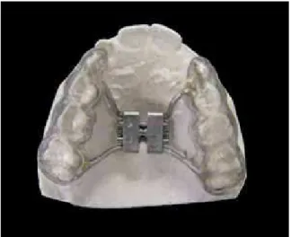
A bonded Hyrax (a rapid palatal expander with an acrylic occlusal splint) was used for RME, similar to the one described by McNamara and Brudon19 (Fig 1). The appliance was adjusted
to the patient’s mouth to obtain the highest pos-sible number of occlusal contact points.
15 seconds. A dual-cure resin cement (Rely X, Unitek/3M, St. Paul, MN, USA) was then applied on the internal surface of the acrylic splint and immediately placed in the patient’s mouth. After removing the excess, the resin was light-cured for 40 seconds, as recommended by the manufacturer.
Patients and parents received hygiene and appliance activation instructions (2 turns per day). The expansion phase was weekly moni-tored up to the point of crossbite overcorrec-tion. The opening of midpalatal suture was confirmed through occlusal radiographs. The appliance was then fixed using acrylic resin and remained as a retainer for 107 days.
Cephalometric analysis
The cephalograms were obtained and scanned by only one technician in the Laboratory of Dental Radiographic Analysis and Control (LACIRO) of FORP-USP at two time points: before treatment (T1) and immediately after the removal of the ex-pansion appliance (T2). The mean time interval
between T1 to T2 was 7 months. All cephalograms were traced by the same examiner. Based on con-ventional cephalometric analysis (Steiner, Downs, Tweed, Ricketts, USP, Unicamp,) available in Ra-diocef software (Radio Memory, Belo Horizonte/ BH, Brazil), we developed our own analytical method using 9 angles and 3 linear measurements (Fig 2) to evaluate the vertical bone changes and the facial height after the treatment.
statistical analysis
To evaluate the error of method, 12 cephalo-grams were traced twice by the same examiner at an interval of at least 3 hours. The Pearson (r) cor-relation test and a t-test for r were used to statisti-cally compare the values. Results were described and compared as means and standard deviations using a paired t-test. Statistical significance was tested at p
≤
0.05. To define whether treatment af-fected the vertical dimension in our experimental group, the values obtained for this sample were compared to a group of untreated patients.7,24 FIGURE 1 - Bonded Hyrax expander with acrylic occlusal splint used inthis study.
FIGURE 2 - Cephalogram showing the landmarks, lines, planes and cephalometric measurements used in this study.
S N
ANS
Pr Or
MeGn Pog Go
Results
There was a high correlation (r
≥
0.99) be-tween repeated measurements to evaluate the error of method, with a great significance for r (p<0.001).Comparison of angular and linear cepha-lometric measurements in the vertical plane between T1 and T2 revealed that the changes were not statistically significant (Table 1), and that there was no change in vertical growth pattern. Although there was no significance, the greatest variation was found in the anterior facial height (AFH), with the mean difference between T1 and T2 of 0.5958 mm.
According to the Bolton’s7 dentofacial growth
and development standards, FMA and SN-GoGn values for children of 8 years of age are 26.2º and 31.6º, respectively, with a small reduction of these angles every year. The mandibular plane angle
(FMA) established by Ricketts24 is about 28º at 3
years of age, with a 1º reduction every 3 years up to maturity. Therefore, at 8 years of age (a compat-ible phase with the mean initial age of this sample), FMA should be about 26º. The mean FMA angle in our sample was 26º at T1 and 26.2º at T2. Al-though there was an increase in the mean value of SN-GoGn angle when compared to the Bolton standards (37.06º at T1 and 36.80º at T2), there were no significant changes in this angle. Taken all together, the data suggested that there were no changes in the vertical growth of the mandible.
Moreover, the linear values used in this sam-ple (AFH, UpAFH and LoAFH) were greater
than the Bolton’s7 standards. However, they
were balanced between them, with a small but not statistically significant increase between T1 and T2, which is in agreement with the varia-tions described by that author.
TABLE 1 - Descriptive statistics of cephalometric variables before and after treatment.
Measurements
Before treatment (T1)
After treatment (T2)
Difference (T2-T1)
Paired t test
mean SD mean SD mean SD p values
SNOcl 20.0415 4.0931 20.1308 4.3207 0.0893 1.5746 0.775
SNGoMe 39.1738 5.2802 38.8958 5.2642 -0.2781 1.7458 0.424
GoGnOcl 17.0535 2.7608 16.7096 2.4922 -0.3439 1.7366 0.322
FMA 26.0965 4.4902 26.2550 4.5948 0.1585 1.4388 0.579
SNGn 69.0262 4.5551 68.7873 4.5124 -0.2389 1.1776 0.311
SNGoGn 37.0612 5.2509 36.8019 5.2126 -0.2593 1.7869 0.466
SNPP 6.8831 2.7199 6.7908 2.8008 -0.0923 1.7113 0.786
PPGoGn 30.1196 4.0914 30.0346 4.1125 -0.0850 2.1920 0.845
PPPoOr 4.1850 2.7565 3.8612 2.7186 -0.3238 1.4011 0.250
AFH 108.5315 4.8511 109.1273 5.1055 0.5958 1.8994 0.122
UpAFH 45.6777 2.8383 46.0577 3.0975 0.3800 1.2889 0.145
dIsCussIOn
Different expanders with occlusal splints
have been proposed19,21,28 to avoid changes in
the vertical dimension when using banded ap-pliances10,11,14,16,17,27,29 and the unfavorable effect
on patient’s facial height.3,11,19,25,28 The
appli-ance described by McNamara and Brudon19 was
chosen for this study because it is widely used in orthodontics, it is easy to manufacture and it is more hygienic.
The cephalometric measurements used in this study evaluated the vertical growth pat-tern of the patients. The focus was in the an-terior facial height, and maxillary and occlusal plane tipping, relatively to the cranial base and the mandible, respectively. There was no con-trol group in this study, and some of the results were compared with normative follow-up val-ues for growth without orthodontic treatment. The lack of statistical significance in the vertical measurements between T1 and T2 demonstrated the maintaining of the vertical pattern in the evaluated group. These results are in agreement with those reported by previ-ous studies,3,19,23,25 which did not find any side
effects on the vertical plane when bonded ex-panders were used, one of the great advantages of this type of treatment. These findings might be associated with the fact that appliances with an acrylic occlusal splint may prevent alveolar growth, excessive eruption of anchor-age teeth, tilting of the alveolar processes and, consequently, buccal movement of maxillary posterior teeth.3,19,21,25,28 In contrast, clockwise
mandibular rotation in response to RME was seen in other studies3,6 using banded and
bond-ed appliances. In both cases, the explanation was in the buccal movement of the anchorage teeth. Such mandibular rotation was not con-firmed in this study, and may be assigned to the skeletal resistance between the maxillary segments that did not allow a symmetrical ex-pansion of the maxilla.3,8
It can be suggested that the decrease of the palatal plane angle in relation to the cra-nial base (SN-PP) might be resulted from the downward displacement of the posterior region of the maxilla,3,10,25 rather than from the
up-ward displacement of the anterior region. The downward and forward maxillary movement
after RME, also reported by Haas,16 resulted
from the fact that this procedure is traumatic and causes fractures along the maxillary tuber-osity, which facilitated such movements.20
Although several studies found statistically and clinically significant short-term changes in the vertical dimension of patients with banded expan-sion appliances,10,11,14,16,17,27,29 we believe that these
changes are not significant in the long run, and the procedure should not be contraindicated for patients with a vertical growth pattern.14
The results of this study suggested that verti-cal changes after RME with occlusal splint also have no statistical and clinical significance. As the means found in this study were similar to normative values,7,24 we believe that the bonded
Hyrax might be a good option for the correction of transverse maxillary deficiency, regardless of vertical dimension or facial pattern.
This is a preliminary study of the vertical effects of palatal expansion using an expand-er with occlusal splints. As it is a longitudinal study, other patients, as well as a control group, are still being followed up. This sample selec-tion was based only on maxillary transverse di-mension and did not take into consideration as-pects associated with growth pattern. Further studies with a standardized sample should be conducted to clarify the benefits of expansion appliances with occlusal splints for patients with a vertical growth pattern.
COnClusIOns
1. Akkaya S, Lorenzon S, Uçem TT. A comparison of sagittal and vertical effects between bonded rapid and slow maxillary expansion procedures. Eur J Orthod. 1999; 21(2):175-80.
2. Almeida RR. Ortodontia preventiva e interceptadora: mito ou realidade? Rev Dental Press Ortod Ortop Facial. 1999;4(6):87-108.
3. Asanza S, Cisneros GJ, Nieberg LG. Comparison of Hyrax and bonded expansion appliances. Angle Orthod. 1997;67(1):15-22.
4. Basciftci FA, Karaman AI. Effects of a modiied acrylic
bonded rapid maxillary expansion appliance and vertical chin cap on dentofacial structures. Angle Orthod. 2002; 72(1):61-71.
5. Bishara SE, Staley RN. Maxillary expansion clinical implications. Am J Orthod Dentofacial Orthop. 1987;91(1):3-14.
6. Bramante FS. Estudo cefalométrico em norma lateral das alterações dentoesqueléticas produzidas por três tipos de expansores: colado, tipo Haas e Hyrax. [dissertação]. Bauru (SP): Universidade de São Paulo; 2000.
7. Broadbent BH, Broadbent BH Jr, Golden WH. Bolton standards of dentofacial developmental growth. 1st ed. Saint
Louis: C.V. Mosby; 1975.
8. Brossman RE, Bennett CG, Merow WW. Facioskeletal remodelling resulting from rapid palatal expansion in the monkey (Macaca cynomolgus). Arch Oral Biol. 1973;18(8):987-94.
RefeRenCes
9. Chang JY, McNamara JA Jr, Herberger TA. A longitudinal study of skeletal side effects induced by rapid maxillary expansion. Am J Orthod Dentofacial Orthop. 1997; 112(3):330-7.
10. Chung CH, Font B. Skeletal and dental changes in the sagittal, vertical, and transverse dimensions after rapid palatal expansion. Am J Orthod Dentofacial Orthop. 2004;126(5):569-75.
11. Cozza P, Giancotti A, Petrosino A. Rapid palatal expansion in mixed dentition using a modified expander: a cephalometric investigation. J Orthod. 2001;28(2):129-34. 12. Davis WM, Kronman JH. Anatomical changes induced by
splinting of the midpalatal suture. Angle Orthod. 1969; 39(2):126-32.
13. Garib DG, Henriques JFC, Hanson GP. Longitudinal cephalometric appraisal of rapid maxillary expansion effects. Rev Dental Press Ortod Ortop Facial. 2001;6(6):17-30.
14. Garib DG, Henriques JFC, Carvalho PEG, Gomes SC. Longitudinal effects of rapid maxillary expansion. A retrospective cephalometric study. Angle Orthod. 2007;77(3):442-8.
15. Haas AJ. The treatment of maxillary deficiency by opening the midpalatal suture. Angle Orthod. 1965;35:200-17. 16. Haas AJ. Palatal expansion: Just beginning of dentofacial
orthopedics. Angle Orthod. 1970;50(3):189-217.
changes in the vertical plane are not statisti-cally significant, which suggests that the verti-cal growth of patients that undergo this type of treatment does not change due to the use
Contact address
Flávia Artese
Rua Santa Clara, 75 sala 1110 – Copacabana Zip code: 22.041-010 – Rio de Janeiro/RJ, Brazil E-mail: flaviaartese@gmail.com
24. Ricketts RM. Perspectives in the clinical application of cephalometrics. Angle Orthod. 1981;51(2):115-50. 25. Sarver DM, Johnston MW. Skeletal changes in vertical and
anterior displacement of the maxilla with bonded rapid palatal expansion appliances. Am J Orthod Dentofacial Orthop. 1989;95(6):462-6.
26. Silva Filho OG, Boas MC, Capelozza Filho L. Rapid maxillary expansion in the primary and mixed dentitions: A cephalometric evaluation. Am J Orthod Dentofacial Orthop. 1991;100(2):171-9.
27. Silva Filho OG, Caricati, JAP, Capelozza Filho L, Cavassan AO. Expansão rápida da maxila na dentadura permanente: avaliação cefalométrica. Ortodontia. 1994; 27(2):68-76. 28. Spolyar JL. The design, fabrication, and use of a
full-coverage bonded rapid maxillary expansion appliance. Am J Orthod. 1984;86(2):136-45.
29. Wertz RA. Skeletal and dentofacial changes accompanying rapid midpalatal suture opening. Am J Orthod.
1970;58(1):41-66.
Submitted: December 2, 2008 Revised and accepted: August 22,2009
17. Kawakami RY. Comparação dos efeitos dentoesqueléticos produzidos por dois tipos de disjuntores palatinos, por meio de análise cefalométrica em norma lateral [dissertação]. Bauru (SP): Universidade de São Paulo; 1995.
18. Lagravere MO, Major PW, Flores-Mir C. Long-term skeletal changes with rapid maxillary expansion: A systematic review. Angle Orthod. 2005;75(6):1046-52.
19. McNamara JA Jr, Brudon WL. Bonded rapid maxillary expansion appliances. In: McNamara JA Jr, Brudon WL. Orthodontic and orthopedic treatment in mixed dentition. 5th
ed. Ann Arbor: Needham Press; 1995. p. 145-69. 20. Melsen B, Melsen F. The postnatal development of the
palatomaxillary region studied on human autopsy material. Am J Orthod. 1982;82(4):329-42.
21. Mondro JF, Litt RA. An improved direct-bonded palatal expansion appliance. J Clin Orthod. 1977;11(3):203-6. 22. Pearson LE. Treatment of a severe open bite excessive vertical pattern with an eclectic non-surgical approach. Angle Orthod. 1991;61(1):71-6.