This report describes the treatment of a 15-year-old female patient with Angle Class I malocclusion, severe maxillary anterior crowding, maxillary midline shift to the left, and maxillary atresia associated with posterior crossbite. The treatment consisted of palatal expansion using a modified Haas expander and placement of a standard Edge-wise fixed appliance. Interproximal reduction was performed on mandibular incisors and canines to align anterior teeth, as well as to reduce Bolton discrepancy due to wide mandibular teeth. This case was submitted to the Committee of the Brazilian Board of Orthodontics and Facial Orthopedics (BBO) in the Free Case category as part of the requisites to obtain the BBO Diploma.
Abstract
Keywords: Angle Class I malocclusion. Atresia. Palatal expansion. Crowding.
* MSc and PhD in Orhtodontics, Federal University of Rio de Janeiro (UFRJ). Adjunct Professor of Orthodontics, UFRJ. Visiting Associate Professor - Case Western Reserve University, Cleveland/Ohio. Diplomate of the Brazilian Board of Orthodontics.
HISTORY AND ETIOLOGY
The main complaint of this 15-year-old fe-male patient in good general health was the position of her maxillary left canine. During history taking, her guardians reported that her adenoids had not been removed. The size of her palatine tonsils was normal. The patient showed
nasal breathing pattern, satisfactory oral hy-giene and no severe periodontal condition.
DIAGNOSIS
Facial examination did not reveal any evident asymmetries, but the lower third of the face was slightly longer than expected. The facial profile
How to cite this article: Nojima LI. The conservative treatment of Class I
malocclusion with maxillary transverse deiciency and anterior teeth crowding. Dental Press J Orthod. 2011 Sept-Oct;16(5):163-71.
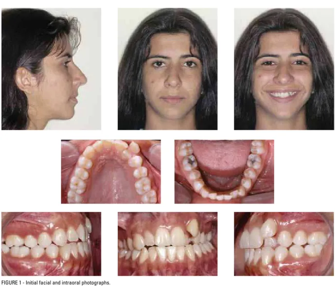
was straight, the nasolabial angle was obtuse, and the upper lip was retruded in relation to the Steiner S line (-1 mm). When smiling, max-illary midline shift to the left could be seen, and her smile was not pleasing because of the labioversion of tooth #23 and palatoversion of tooth #22 (Fig 1).
Intraoral examination revealed Angle Class I malocclusion and anterior crossbite of tooth #12, as well as posterior crossbite. Maxillary atresia resulted in anterior edge-to-edge bite and com-plete absence of space for the correct alignment
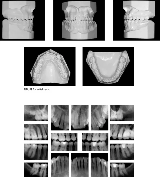
of tooth #23. Dentoalveolar discrepancy was -4 mm and, according to Bolton analysis, discrepancy of the anterior mandibular teeth was 2 mm. There was no overbite or overjet (Figs 1 and 2).
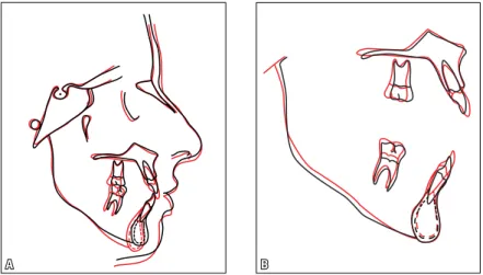
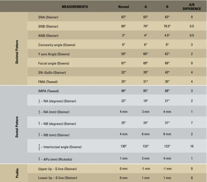
Periapical radiographs showed normal bone trabecula and agenesis of teeth #28, #38 and #48; all other permanent teeth were present (Fig 3). Cephalometric analyses showed a good relationship of maxillary and mandibular bones: ANB = 4° (SNA = 83° and SNB = 79°); and vertical growth pattern with GoGn-SN = 39° (Fig 4, Table 1).
A B
TREATmENT ObjEcTIvES
The purposes of the treatment were to ex-pand the maxillary arch to treat maxillary atresia and crossbite, to gain space to align the maxil-lary and mandibular anterior teeth, to create ad-equate overjet and overbite and to correct the maxillary midline. Further objectives were to maintain normal molar occlusion, improve the shape of dental arches, and avoid tooth extrac-tions during orthodontic treatment. For the fa-cial profile, the objective was to keep the bal-anced position of the upper lip.
TREATmENT PLAN
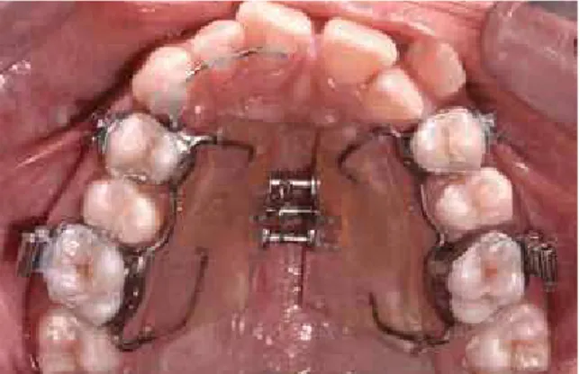
Palatal expansion was achieved with a modi-fied Haas expander with extension to the lin-gual surface of tooth #13, followed by the use of standard 0.022 x 0.028-in Edgewise fixed ap-pliance and alignment and leveling using round 0.016-in and 0.018-in stainless steel wires. At this stage, midline was corrected and spaces obtained to correct crossbite of tooth #22 and move tooth #23 to its correct position in the dental arch. Bands were cemented to teeth #36, #37, #46 and #47, and accessories bonded to the other teeth. To align mandibular incisor and canines, 2 mm interproximal reduction was performed and the triangular anatomic shape of
those teeth corrected. Round 0.014-in to 0.020-in wires were used to align and level the man-dibular teeth. For finishing, rectangular 0.019 x 0.025-in archwires were used, and first, second and third order bends individually prepared ac-cording to the need. A wraparound retainer was used for the maxilla, and a 0.028-in intercanine fixed retainer, for the mandible.
TREATmENT PROGRESS
The patient’s main complaint was associated with crowding and crossbite. She received ex-planations about the need of palatal expansion due to the atresia of the maxillary bone and pre-maxillary hypoplasia, which resulted in a facial profile with the upper lip positioned behind the esthetic line. The modified Haas expander with extension to tooth #13 was placed. The pur-pose of the extension was to anchor tooth #13, to which a coaxial 0.028-in wire was bonded on the lingual surface, extending to teeth #12 and #11 (Fig 5). The expander was activated 2/4 of a turn daily, in a total of 8 mm of screw activation. As the interincisal diastema was created, tooth #21 moved mesially towards the open space, and tooth #11 remained anchored to teeth #12 and #13, which avoided its mesial movement towards the diastema and corrected the midline shift.
A fixed 0.022 x 0.028-in standard Edgewise appliance was then used. Bands were adapted and bonded to teeth #36, #46, #37 and #47, and to the other mandibular teeth except the incisors. In addition, slight interproximal reduction of teeth #42, #41, #32 and #31 was performed. The ex-pander was removed five months after its stabi-lization, and a 0.045-in buccal archwire was an-chored to the 0.051-in buccal tubes, as a retainer for the expansion achieved. For maxillary align-ment and leveling, 0.014-in to 0.018-in round archwires were used, and a rectangular 0.016 x 0.022-in blue Elgiloy archwire was used for slight protrusion of maxillary incisors. Immediately af-ter that, the mandibular incisors brackets were bonded, and interproximal reduction of teeth #43 to #33 was performed. Adequate overbite was achieved using a 0.020 x 0.025-in stabilizing archwire with delta loops mesial to teeth #33 and #43, and a 0.019 x 0.025-in maxillary archwire with delta loops mesial to canines, associated with Class II and vertical anterior elastics. Finally, rect-angular 0.019 x 0.025-in archwires were manu-factured with first, second and third order bends according to the need. Maxillary retention was achieved with a removable wraparound appliance, and mandibular retention, with a bonded 0.028-in intercanine fixed retainer.
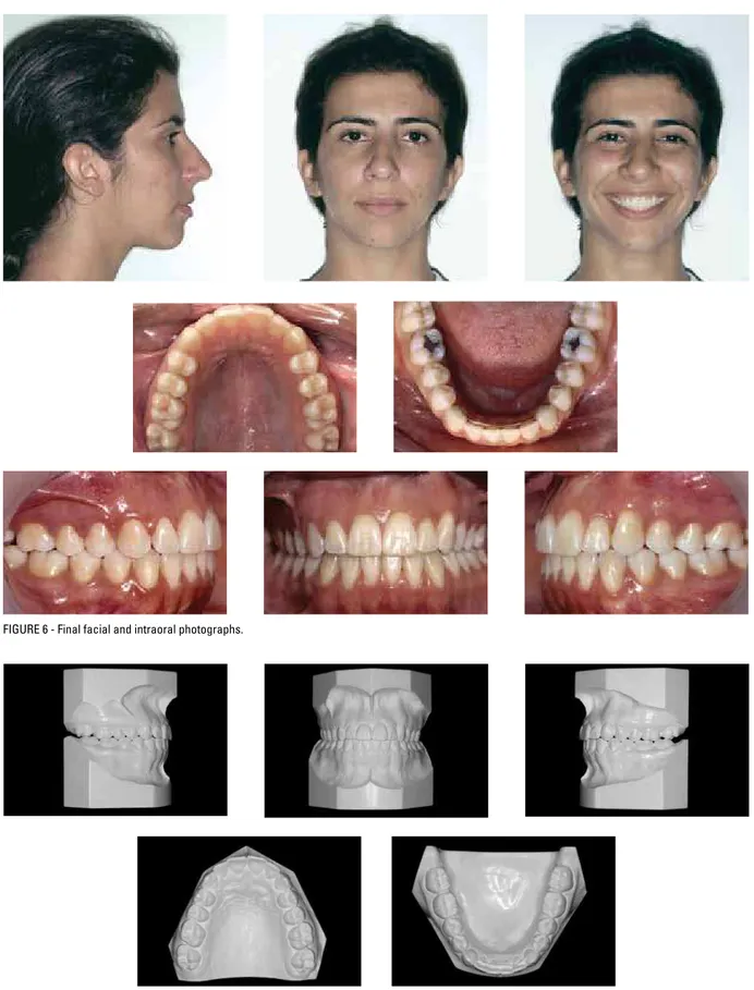
and a more adequate anatomic width (Table 2). This increase in the upper transverse dimensions affected the intermolar width in the mandibular arch, which resulted in a more vertical position of molars. How-ever, the mandibular plane was increased.3 The space gained in palatal expansion, associated with anchor-age of the expander to teeth #11, #12 and #13, re-sulted in adequate correction of maxillary midline shift and alignment of teeth #22 and #23 (Fig 6).
To align the dental arches, resilient stainless steel archwires were used to define the direc-tion of the forces applied and to move teeth to the desired positions, as well as to obtain a better periodontal response from support tissues. The protrusive movement of maxillary incisors was achieved by the use of mild forces applied by
rect-angular 0.016 x 0.022-in blue Elgiloy archwires.5,6
To align mandibular teeth, a 2 mm space was created by interproximal reduction of anterior teeth. This procedure promoted the stability of the final position as it increased the contact be-tween these teeth, which had originally an exces-sively triangular shape (Figs 6 and 7).7
Normal molar occlusion was achieved, as well as a good relationship of maxillary and mandib-ular teeth in static occlusion. The mandibmandib-ular movements presented normal excursion, with right and left lateral group function, and
protru-sion with posterior guidance.8
Maxillary retention was prescribed for 12 months with a removable appliance to be used 24 hours a day and 12 more months of night use only. Mandibular retention was performed with a 0.028-in stainless steel intercanine fixed retainer.
A
A
B
B
FIGURE 8 - Final periapical radiographs.
FIGURE 9 - Final cephalometric profile radiograph (A) and cephalometric tracing (B).
CAst trAnsverse meAsures A B
Mandibular intercanine width (mm) 24.5 24.5
Mandibular intermolar width (mm) 42.5 44.0
Maxillary intermolar width (mm) 46.5 50 TABLE 1 - Summary of cephalometric measurements.
TABLE 2 - Cast models linear measurements. FINAL cONSIDERATIONS
Facial esthetics became harmonious with slight protrusion of upper lips. The treatment of the maxillary arch was more complex due to the skeletal problem of maxillary atresia as-sociated with the vertical growth pattern, in addition to other problems, such as the
maxil-meAsurements normal A B A/B
DIFFerenCe
skeletal Pattern
SNA(Steiner) 82° 83° 83° 0
SNB(Steiner) 80° 79° 78.5° 0.5
ANB(Steiner) 2° 4° 4.5° 0.5
Convexity angle(Downs) 0° 6° 9° 3
Y axis Angle(Downs) 59° 60° 62° 2
Facial angle(Downs) 87° 89° 89° 0
SN–GoGn(Steiner) 32° 39° 43° 4
FMA(Tweed) 25° 31° 35° 4
Dental Pattern
IMPA(Tweed) 90° 85° 88° 3
–
1 – NA (degrees) (Steiner) 22° 19° 21° 2
–
1 – NA (mm) (Steiner) 4 mm 3 mm 4 mm 1
–
1 – NB (degrees)(Steiner) 25° 24° 31° 7
–
1 – NB (mm)(Steiner) 4 mm 6 mm 8 mm 2
– 1
1 – Interincisal angle(Downs) 130° 133° 123° 10
–
1 – APo (mm)(Ricketts) 1 mm 3 mm 4 mm 1
Proile
Upper lip – S line(Steiner) 0 mm -1 mm -1 mm 0
1. Tweed CH. A philosophy of orthodontic treatment. Am J Orthod Oral Surg. 1945;31:74.
2. Burstone CJ. Lip posture and its signiicance in treatment
planning. Am J Orthod. 1967;53(4):262-84.
3. Baratieri C, Nojima LI, Alves M Jr, Souza MMG, Nojima MCG. Transverse effects of rapid maxillary expansion in Class II malocclusion patients: a Cone-Beam Computed Tomography study. Dental Press J Orthod. 2010;15(5):89-97. 4. Kokish VG, Nappen DL, Shapiro PA. Gingival contour and
clinical crown length: their effect on the esthetic appearance of maxillary anterior teeth. Am J Orthod. 1984;86(2):89-94.
REFERENcES
contact address Lincoln I. Nojima
Av. Professor Rodolpho Paulo Rocco, 325
CEP: 21.941-617 – Ilha do Fundão – Rio de Janeiro/RJ, Brazil E-mail: linojima@gmail.com
Submitted: August 2, 2011
Revised and accepted: August 26, 2011
5. Rickets RM, Bench RW, Gugino CF, Hilgers JJ, Schulof RJ, Bioprogressive therapy. Denver, CO: Rocky Mountain Orthodontics; 1979.
6. Burstone CJ. Deep overbite correction by intrusion. Am J Orthod. 1977;72(1):1-22.
7. Tuverson DL. Anterior interocclusal relations. Part II. Am J Orthod. 1980;78(4):371-93.