Neonatal meningitis according to
the microbiological diagnosis
A decade of experience in a tertiary center
Maria Regina Bentlin1, Gabriel Luís Ferreira2,
Ligia Maria Suppo de Souza Rugolo3, Geraldo Henrique Soares Silva4, Alessandro Lia Mondelli3, Antonio Rugolo Júnior1
ABSTRACT
The aim of this study was to evaluate the incidence of and mortality due to meningitis and compare data according to microbiological diagnosis. This was a ten-year retrospective study conducted at a neonatal intensive care unit (NICU). Newborns with meningitis confirmed by positive CSF culture were included; those with congenital infection or malformations that made lumbar puncture impossible were excluded. The variables investigated were birth weight, gestational and postnatal age, procedures, hematological and CSF parameters, and complications. Parametric and non-parametric tests were used (statistical value p<0.05). The incidence of meningitis was 0.6% and mortality was 27%. Of the 22 cases, 59% involved Gram-negative bacteria; 36% Gram-positive and 5% fungi. The groups did not differ in relation to birth weight, gestational and postnatal age, procedures or hematological and CSF parameters. Sepsis, convulsions and deaths were frequent in both groups, without statistical difference. Gram-negative cases showed abscesses and higher frequency of ventriculitis and hydrocephaly. Meningitis was infrequent, but presented high mortality and frequent complications.
Key words: meningitis, newborn, central nervous system, sepsis.
Meningite neonatal de acordo com diagnóstico microbiológico: uma década de experiência em centro terciário
RESUMO
O objetivo do estudo foi avaliar incidência e mortalidade da meningite e comparar dados de acordo com o diagnóstico microbiológico. Estudo retrospectivo, de 10 anos, em UTI Neonatal. Incluídos RNs com meningite confirmada por cultura de líquor positiva; RN com infecção congênita ou malformações que impedem punção lombar foram excluídos. Variáveis: peso ao nascimento, idades gestacional e pós natal, procedimentos, parâmetros hematológicos e liquóricos, complicações. Testes paramétricos e não paramétricos foram utilizados (valor estatístico p<0,05). A incidência de meningite foi de 0,6% e mortalidade de 27%. Dos 22 casos, 59% foram por bactérias negativas; 36% por bactérias Gram-positivas e 5% por fungos. Grupos não diferiram quanto ao peso ao nascimento, idades gestacional e pós-natal, procedimentos e por parâmetros hematológicos e liquóricos. Sepse, convulsões e óbitos foram frequentes e não diferiram entre os grupos. Gram-negativos causaram abscessos e mais frequentemente ventriculite e hidrocefalia. Meningite não foi freqüente, mas apresentou alta mortalidade e complicações.
Palavras-chave: meningite, recém-nascido, sistema nervoso central, sepse. Correspondence
Maria Regina Bentlin Departamento de Pediatria da Faculdade de Medicina de Botucatu UNESP - Distrito de Rubião Júnior s/nº 18618-970 Botucatu SP - Brasil E-mail: [email protected]
Received 30 November 2009 Received in final form 10 May 2010 Accepted 17 May 2010
Neonatal meningitis is still a disease with high mor-bidity, although advances in perinatal care over the last few decades have been able to reduce its mortality rates to approximately 10%. Nevertheless, 20 to 58% of survi-vors show neurological sequelae. Another worrisome is-sue is the association of sepsis with meningitis, which has remained constant and rather frequent, showing rates of approximately 25%1-3. A recent review study on
neona-tal infections has reported meningitis incidence ranging from 0.8 to 6.1 cases in every 1,000 live newborns4.
Meningitis occurring during the first week of life, particularly on the irst two to three days, suggests that maternal transmission may be involved. Such cases are more frequently caused by group-B streptococci, E. coli
and Listeria monocytogenes, whereas occurrences after this period suggest that nosocomial infection consist-ing of staphylococcal species and Gram-negative rods is the main etiological agent2,3,5. Studies in developed
coun-tries comparing neonatal meningitis data over the peri-ods 1985-1987 and 1996-1997 showed little variation be-tween etiological agents, and group-B streptococci were the agents most frequently isolated2,6,7. However, in
de-veloping countries, Gram-negative bacteria are the main causative agents of meningitis8.
he hypothesis that meningitis caused by Gram-nega-tive agents shows greater severity of progression, and the possibility that there might be characteristic indings in this group of patients that could help in the diagnosis, was the motivation for conducting this study. he aim was to evaluate the incidence of and mortality due to neonatal meningitis conirmed by culture, and to compare the clin-ical and laboratory characteristics of meningitis caused by Gram-negative and Gram-positive agents.
METHOD
A retrospective epidemiological study was conduct-ed in the Neonatal Intensive Care Unit (NICU) of a ter-tiary center from January 1997 to December 2006 fol-lowing approval from the institution’s Medicine Ethics Committee.
All newborns admitted to the NICU (either born at this hospital or born elsewhere) who developed meningitis with positive cerebrospinal luid (CSF) culture before the age of 28 days were included in the study. Newborns with clinical and serological evidence of congenital infection; central nervous system malformation that made lumbar puncture impossible; CSF contamination (more than one agent in the same culture); positive CSF culture for coagu-lase-negative staphylococci in the absence of concomitant positive blood culture for this agent); and cases in which it was not possible to obtain the protocol data were excluded. Meningitis was deined laboratorially as the presence of a positive CSF culture, regardless of cytological and
bio-chemical CSF abnormalities. Coagulase-negative staph-ylococci were considered to be the etiological agent of meningitis only when they was isolated in CSF and blood cultures at the same time. The indications for lumbar puncture followed the standard protocol at our service: suspected sepsis and/or meningitis, presence of positive blood culture and non-deined infectious focus. Among preterm infants older than 35 weeks that were born in situations of infection risk, and among newborns pre-senting fever, the puncture indication was individualized.
he patients were stratiied into two groups, according to the microbiological diagnosis of meningitis (Gram-neg-ative vs. Gram-positive), and were evaluated with regard to clinical and laboratory data. Because of the low inci-dence of fungal meningitis, such patients were considered only in relation to estimations of incidence and mortality. he study began with an assessment of all positive CSF cultures from newborns admitted to the NICU during the study period. he CSF cultures were obtained from the hospital’s microbiology laboratory. An automated method (BACTEC Peds Plus F) was used in this laboratory. After obtaining the culture registration numbers, the patients’ charts were selected for illing out the protocols.
Variables studied
Maternal and gestational: Age; prenatal follow-up; premature rupture of membranes ≥18 hours; chorioam-nionitis (maternal fever associated with at least two signs among the following: uterine hypotonia, fetid amniotic luid, maternal leukocytosis, maternal or fetal tachycar-dia); premature labor and delivery.
Neonatal: Gender, birth weight, gestational and post-natal age, Apgar<7 at the ifth minute and congenital mal-formation.
Care procedures: Vascular catheters, parenteral nu-trition, mechanical ventilation, previous antibiotic ther-apy and ventriculoperitoneal shunt.
Hematological parameters: Leukocyte count, plate-lets, total neutrophil ratio; immature-to-segmented neutrophil ratio, Rodwell scoring system9 and
positivity of C-reactive protein (CRP). In the initial years of the study, CRP levels were determined using a semi-quantitative method, but since the year 2000, it has been determined using turbidimetry. A CRP level >1 mg/dl is considered abnormal.
CSF parameters: Leukocyte count, red cells, glucose and protein.
Cultures: Agents isolated in blood and CSF.
Clinical course: Meningitis associated with sepsis or complicated with convulsions; ventriculitis; abscess; hy-drocephaly diagnosed by means of the imaging exami-nations routinely performed on all newborns with men-ingitis (preferably cranial ultrasound); and death during hospitalization.
Continuous data were described in terms of either means and standard deviations or as medians and inter-quartile ranges (percentiles 25 and 75), while categori-cal variables were expressed in terms of the number and proportion of events. Comparisons between the groups were performed using the t test or Mann Whitney test for numerical variables, and using the chi-square or Fisher’s test for categorical variables. Results were considered to be signiicant when p<0.05.
RESULTS
Among the 3,910 newborns (NB) admitted to the NICU of the University Hospital over the 10-year study period, 24 presented meningitis that was conirmed by positive CSF culture. Two patients were excluded (due to congenital infection and not obtaining the protocol vari-ables). Considering the 22 newborns included, the inci-dence of meningitis was 0.6% and the mortality rate was 27% (6 NB). he overall mortality rate at the NICU over this period was approximately 12%.
In only one case did meningitis occur within the irst three days of life. In this case, the agent isolated from CSF was group-B streptococci, and the patient died. he other episodes occurred after the irst week of life.
Among the 22 patients studied, meningitis due to Gram-negative bacteria predominated, with this inding in 59% of the cases (13 NB). Infection occurred particu-larly during the irst ive years of the study, and the bac-teria most frequently observed were Pseudomonas aerug-inosa (3 NB), Burkholderia cepacea (2 NB) and Klebsiel-la sp (2 NB). E. coli was not isolated in any of the cul-tures. In the cases of meningitis due to Gram-positive bacteria (36%), the most frequent agents were S. aureus
(2 NB), coagulase-negative staphylococci (2 NB) and group-B streptococci (2 NB). here was only one case (5%) of fungal meningitis (Candida sp), which was diag-nosed on the 22nd day of life of a septic 28-week
prema-ture infant with birth weight of 1,000 g. No cases of co-agulase-negative staphylococci meningitis were excluded due to contamination.
he demographic data and hematological and CSF indings from newborns with meningitis according to the microbiological diagnosis are shown in Tables 1, 2 and 3. It is noteworthy that most of the newborns were pre-mature, had low birth weight and were diagnosed with meningitis in the third week of life. Malformation in the central nervous system corresponded to two cases of
hy-drocephaly and one of Dandy-Walker cyst, which were not excluded because it was possible to perform the lum-bar puncture.
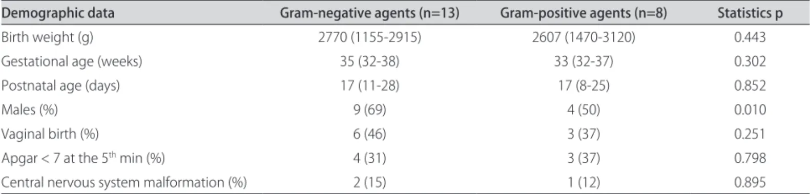
When comparing the groups based on etiological agents, no diference was observed in relation to mater-nal or neonatal variables, except for male gender, which was more frequent among the cases of meningitis due to Gram-negative agents.
Vascular catheters, parenteral nutrition, mechanical ventilation and previous antibiotic therapy were the most common procedures, but without diferences between the groups. he mean and median values of the hematologi-cal tests were found to be normal and also did not difer between the groups, except CRP positivity, which was sig-niicantly higher in cases of meningitis caused by Gram-negative agents (Table 2).
he high CSF values for leukocytes and proteins, as well as the low glucose concentrations in both groups, without diferences between positive and Gram-negative agents (Table 3), are also noteworthy.
Blood cultures were positive in 46% of the cases of Gram-negative meningitis and in 50% of the Gram-pos-itive cases.
he clinical course of Gram-negative meningitis was worse, with a larger number of patients developing ven-triculitis and hydrocephaly. Abscess occurred only in the Gram-negative meningitis group. Mortality was high, particularly among patients infected with Gram-positive agents; however, no diference was observed between the groups (Table 4). By analyzing deaths according to study periods (every ive years), it was observed that mortality dropped from 50% to 22% over the last ive years studied.
DISCUSSION
his study on conirmed cases of meningitis at a ter-tiary university center aimed to learn about the realities of this devastating disease, which is diicult to diagnose during the neonatal period and is responsible for high mortality rates. he hypothesis that there might be clini-cal and/or laboratory indings that could diferentiate be-tween meningitis infections due to Gram-negative and Gram-positive agents, along with the scarcity of data on this disease in Brazilian publications, was the motivation for conducting this study. Given the controversy regard-ing clinical and laboratory menregard-ingitis conirmations1,10,11,
it was decided to use a gold-standard-based diagnosis, i.e. positive CSF culture.
he incidence of meningitis conirmed by culture was not high (0.6%). An American multicenter study found a rate of 1%11, and another review has recently shown that,
in newborns with late-onset infection, meningitis preva-lence ranged from 1.3% to 3.5%12.
new-borns with meningitis, which was twice as high as the general rate for the NICU during this period. Howev-er, it is noteworthy that in the last ive years of the study, the mortality rate was reduced from 50% to 22%. he lit-erature reports a decrease in mortality rate, which went down from 50% in the 1950s to the current igures of ap-proximately 20%1,2. British studies comparing two
peri-ods, 1985-87 and 1996-97, showed a reduction in mortali-ty among conirmed meningitis cases from 29% to 10%6,7 .
he detection of only one case of early meningitis cor-roborates the inding of low rates of positive cultures due to early infection that was observed in this NICU by Sil-va. his author studied early sepsis over a ive-year pe-riod (1999 to 2003) and found positive blood cultures in only 1.5% of the cases13. Factors such as delay in
perform-ing lumbar puncture due to hemodynamic instability and use of antibiotics preceding the puncture may interfere with bacterial growth11.
Table 1. Demographic data from newborns with conirmed meningitis, according to microbiological diagnosis.
Demographic data Gram-negative agents (n=13) Gram-positive agents (n=8) Statistics p
Birth weight (g) 2770 (1155-2915) 2607 (1470-3120) 0.443
Gestational age (weeks) 35 (32-38) 33 (32-37) 0.302
Postnatal age (days) 17 (11-28) 17 (8-25) 0.852
Males (%) 9 (69) 4 (50) 0.010
Vaginal birth (%) 6 (46) 3 (37) 0.251
Apgar < 7 at the 5th min (%) 4 (31) 3 (37) 0.798
Central nervous system malformation (%) 2 (15) 1 (12) 0.895
Table 2. Hematological parameters of newborns with conirmed meningitis, according to microbiological diagnosis.
Hematological parameters Gram-negative agents (n=13) Gram-positive agents (n=8) Statistics p
Leukocytes (mm3 ) 15210±11980 13090±5090 0.646
Platelets (×103 /mm3) 244 (103-355) 293 (100-525) 0.483
Immature-to-segmented neutrophil ratio 0.03 (0.02-0.20) 0.10 (0.06-0.20) 0.684
Immature-to-total neutrophil ratio 0.02 (0.01-0,20) 0.09 (0.05-0.11) 0.717
Rodwell score 1.8±1.5 1.0±1.1 0.200
C-reactive protein (%) 88 64 <0.001
Table 3. CSF parameters of newborns with conirmed meningitis, according to microbiological diagnosis.
CSF parameters Gram-negative agents (n=13) Gram-positive agents (n=8) Statistics p
Leukocytes (mm3 ) 837 (207-3336) 111 (9-5680) 0.203
Red cells (mm3) 166 (13-1680) 50 (10-690) 0.563
Glucose (mg%) 10 (10-27) 10 (10-38) 0.817
Protein (mg%) 398±272 415±376 0.914
Table 4. Clinical course of newborns with conirmed meningitis, according to microbiological diagnosis.
Clinical course Gram-negative agents (n=13) Gram-positive agents (n=8) Statistics p
Association with conirmed sepsis;
positive blood culture (%) 6 (46) 4 (50) 0.671
Convulsions (%) 8 (61) 5 (62) 1.000
Ventriculitis (%) 10 (77) 5 (62) 0.032
Abscess (%) 2 (15) 0 0.015
Hydrocephaly (%) 10 (77) 5 (62) 0.032
he agents causing meningitis difer according to re-gional and care-provision characteristics and may be the same as those causing sepsis. In developed countries, it has been reported that 30 to 40% of meningitis cas-es are Gram-negative, and that E. coli is the most fre-quent bacterium1,2. In developing countries,
Gram-nega-tive meningitis is more frequent4,14. he predominance of
Gram-negative bacteria in the present study relected the bacteriological pattern in the NICU in the late 1990s and early 2000s13. From 2001 onwards, after implementation
of infection control measures, empirical therapy proto-cols for nosocomial infection and restrictions on the use of third-generation cephalosporin and vancomycin, coag-ulase-negative staphylococci became the causative agent of late sepsis in more than 60% of the cases13, a pattern
that is also found in CSF cultures.
In Gram-positive meningitis, group-B streptococci were not common. Staphylococci predominated among the Gram-positive agents and they are currently the main agents causing late-onset sepsis4,14. A multicenter study
involving 134 very low birth weight premature infants with conirmed meningitis showed that Gram-positive agents were responsible for 63% of the episodes, with oc-currences of coagulase-negative staphylococci in 29% of the cases. When the sepsis cases with meningitis were evaluated, coagulase-negative staphylococci were the agents responsible for 44% of the cases15 .
In the present study, the newborns were mostly pre-mature, but without low birth weight. The literature warns about occurrences of meningitis in premature in-fants with higher weight and older gestational age at birth, as documented in the study by Holt et al., in which only 15% of the newborns were premature and less than 33 weeks of gestational age, and 17% weighed less than 2,000 grams at birth7. Krebs and Taricco studied 50 newborns
with bacterial meningitis at the Children’s Institute in São Paulo and showed that the medians for gestational age and weight at birth were 36 weeks and 2,577 g respective-ly, with a postnatal age of 13 days at the time of meningitis diagnosis16. At present, there is great concern about
pre-mature infants of gestational age greater than or equal to 34 weeks that show physiological and developmental im-maturity and greater morbidity-mortality17-19. Studies on
ontogeny and maturation sequence are being developed in an attempt to explain these patients’ greater suscepti-bility to infection20,21 .
he hypothesis that Gram-negative meningitis would be more devastating and that there could be some char-acteristic inding in its development that would help in early identiication of this condition was the motivation for comparing the two groups of agents. he association of meningitis and sepsis with positive blood cultures was high: 46% for Gram-negative and 50% for
Gram-posi-tive agents. An association between sepsis and meningi-tis has been described in approximately 25% of the cas-es22, but these igures may vary. A multicenter study in the
National Institute of Child Health on 134 very low birth weight newborns with conirmed meningitis showed that the pathogens responsible for meningitis were similar to those associated with sepsis, and that one third of these newborns did not have positive blood cultures15.In a
mul-ticenter study, Ansong et al. showed that 20% of infants with group-B streptococcal meningitis had negative blood cultures23. In another multicenter study on 9,111
new-borns with gestational age greater than or equal to 34 weeks who underwent lumbar puncture, 95 patients with meningitis were detected, and of these, 62% showed pos-itive blood culture. he authors pointed out that menin-gitis frequently occurs in the absence of bacteremia and, for that reason, lumbar puncture is an important part of the diagnostic investigation in cases of suspected sepsis, since it may be the only positive test11.
Another study conducted in São Paulo on 87 new-borns with meningitis diagnosed by cytological and bio-chemical alterations reported that 39% of the cases had positive CSF cultures and only 17% showed positive blood cultures24.
Hematological tests have not been shown to be useful for diferentiating between the agents and are unspeciic in relation to infection and meningitis diagnosis22.
Concerning the analysis of cytological and biochem-ical CSF parameters, the expectation that they would be able to help with diagnosis based on the etiological agents was not conirmed. Increased leukocyte and protein lev-els and decreased glucose concentrations occurred in both groups. hese data difer from the results obtained by Smith et al.5 in a multicenter study, in which
leuko-cyte counts were signiicantly higher in cases of Gram-negative meningitis, without diferences in other param-eters. he larger number of patients included in that mul-ticenter study must be emphasized, as it could explain the diferences found. Garges et al.11 evaluated the
corre-lation between CSF culture, blood culture and CSF pa-rameters and observed that, for the meningitis cases con-irmed by culture, a leukocyte count greater than 0/mm3
showed sensitivity of 97% and speciicity of 11%. A leu-kocyte count greater than 21/mm3 reduced the
95 cases of meningitis. he accuracy obtained was 0.80 for leukocyte count, 0.63 for glucose and 0.72 for protein. hus, caution must be used in interpreting CSF values for meningitis diagnosis in premature infants, since the ac-curacy of these parameters is low25.
In agreement with data in the literature, complications such as ventriculitis, hydrocephaly and abscesses were more frequent in Gram-negative meningitis. Ventriculi-tis may occur in 40 to 90% of the cases, particularly when bacteria persist in the CSF, while hydrocephaly is usually associated with ventriculitis and hemorrhage24. he
fre-quency of abscesses was rather high (15%) in comparison with the rates of 1% to 3.4% reported in the literature24,26.
Abscesses may develop slowly, and they are not always easily seen, which hinders diagnosis. hey are often only detected in necropsies.
he high frequency of complications detected in this study points towards another worrisome aspect of neo-natal meningitis: the risk of sequelae over time. Although this was not the objective in the present study, the liter-ature shows that Gram-negative meningitis is especially responsible for sequelae such as delayed motor and men-tal development, auditory and visual deicits and behav-ioral problems, thus increasing the social and econom-ic costs and compromising patients’ and their relatives’ quality of life1,27.
Another relevant point was the high mortality rate among Gram-positive agents (one death caused by group-B streptococci, one by S. aureus and another by coagulase-negative staphylococci). his was also shown in the multicenter study by Heath et al., which report-ed death rates of 12 to 24% among meningitis cases due to group-B streptococci and of up to 36% among oth-er Gram-positive agents, whoth-ereas mortality caused by Gram-negative agents ranged from 0 to 50%2.
he present study has limitations, since it was retro-spective, only involved a single center and only included a small number of subjects. However, it relects the reali-ty of a reference center for caring for high-risk newborns over a period of one decade. It is the irst Brazilian study to focus on conirmed cases of meningitis by comparing groups of agents. It may contribute towards improving the knowledge on this severe disease, which despite tech-nological advances in perinatal care, continues to present high morbidity and sequelae rates worldwide.
In conclusion, neonatal meningitis is infrequent, but shows high morbidity and mortality rates, particularly among premature infants with low birth weight. CSF pa-rameters do not help in diferentiating between agents. he clinical course of Gram-negative meningitis is more severe and shows a higher frequency of ventriculitis, hy-drocephaly and abscesses.
REFERENCES
1. Polin RA, Harris MC. Neonatal bacterial meningitis. Semin Neonatol 2001;6: 157-172.
2. Heath PT, Nik Yusof NK, Baker CJ. Neonatal meningitis. Arch Dis Child Neo-natal Ed 2003;88:F173-F178.
3. Haussen DC, Brandalise LN, Praetzel FA, et al. Meningite neonatal. Aspectos associados. Arq Neuropsiquiatr 2005;63:625-631.
4. Thaver D, Zaidi AKM. Burden of neonatal infections in developing coun-tries: a review of evidence from community-based studies. Pediatr Infect Dis J 2009;28(Suppl):S3-S9.
5. Smith PB, Cotton CM, Garges HP, et al. A comparison of neonatal gram-negative rod and gram-positive cocci meningitis. J Perinatol 2006;26: 111-114.
6. de Louvois J, Blackbourn J, Hurley R, Harvey D. Infantile meningitis in England and Wales: a two year study. Arch Dis Child 1991;66:603-607. 7. Holt DE, Halket S, de Louvois J, Harvey D. Neonatal meningitis in England and
Wales: 10 years on. Arch Dis Child Fetal Neonatal Ed 2001;84:F85-F89. 8. WHO. Bacterial etiology of serious infections in young infants in
devel-oping countries: results of a multicenter study. The WHO young infants study group. Pediatr Infect Dis J 1999;18(Suppl):S17-S22.
9. Rodwell Rl, Leslie Al, Tudehope D. Early diagnosis of neonatal sepsis using a hematologic scoring system. J Pediatr 1988; 112:761-767.
10. Rodriguez AF, Kaplan SL, Mason EO. CSF values in the very-low-birth-weight infant. J Pediatr 1990;116:971-974.
11. Garges HP, Moody A, Cotton M, et al. Neonatal meningitis: What is the cor-relation among CSF cultures, blood cultures and CSF parameters? Pediatr 2006;117:1094-1100.
12. Malbon K, Mohan R, Nicholl R. Should a neonate with possible late onset in-fection always have a lumbar puncture? Arch Dis Child 2006;91:74-83. 13. Silva GHS. Análise clínica e laboratorial da sepse com hemocultura positiva
em recém-nascidos internados em Unidade de Terapia Intensiva Neonatal durante 5 anos. [dissertação] Botucatu: Faculdade de Medicina, Univer-sidade Estadual Paulista; São Paulo 2007.
14. Zaidi AKM, Thaver Dali AS, Khan TA. Pathogens associated with sepsis in newborn and young infants in developing countries. Pediatr Infect Dis J 2009; 28(Suppl):S10-S18.
15. Stoll BJ, Hansen N, Fanarof AA, et al. To tap or not to tap>high likelihood of meningitis without sepsis among very low birth weight infants. Pediatrics 2004;113:1181-1186.
16. Krebs VLJ, Taricco LD. Fatores de risco para meningite bacteriana no re-cém-nascido. Arq Neuropsiquiatr 2004;62:630-634.
17. Escobar GJ, Li D, Armstrong MA, et al. Neonatal sepsis workups in infants ≥2000g grams at birth: a population based study. Pediatr 2000;106:256-263. 18. Romero R, Espinoza J, Chaiworapongsa T, Kalache K. Infection and pre-maturity and the role of preventive strategies. Semin Neonatol 2002;7: 259-274.
19. Silva LPA, Cavalheiro LG, Queirós F, Vila Nova C, Lucena R. Prevalence of newborn meningitis and sepsis during the pregnancy period for public health care system participants in Salvador, Bahia, Brazil. BJID 2007;11: 272-276.
20. Clapp DW. Developmental regulation of the immune system. Semin Per-inatol 2006;30:69-72.
21. Raju TN, Riggins RD, Stark AR, Leveno KJ. Optimizing care and outcome for late preterm (near term) infants: a summary of workshop sponsored by National Institute of Child Health and Human Development. Pediatrics 2006;118:1207-1214.
22. Stoll BJ. Neonatal infections: a global perspective. In: Remington JS, Klein JO, (Eds). Infectious diseases of fetus, newborn and infants. 6th ed.
Phila-delphia, PA: WB Saunders; 2005:27-57.
23. Ansong AK, Smith PB, Benjamin DK , et al. Earlu Hum Dev 2009; 85:S5-S7. 24. Krebs VLJ, Costa GAM. Clinical outcome of neonatal bacterial meningitis
ac-cording to birth weight. Arq Neuropsiquiatr 2007;65:1149-1153. 25. Smith PB, Garges HP, Cotton CM, Walsh TJ, Clark RH, Benjamin DK Jr.
Men-ingitis ion preterm neonates: importance of CSF parameters. Am J Peri-natol 2008;25:421-426.
26. Feferbaum R, Diniz EM, Valente M, et al. Brain abscess by Citrobacter di-versus in infancy: case report. Arq Neuropsiquiatr 2000;58:736-740. 27. de Louvois J, Halket S, Harvey D. Neonatal meningitis in England and Wales: