151
REV. HOSP. CLÍN. FAC. MED. S.PAULO 54(5):151-154, 1999 SEPTEMBER-OCTOBER
From the Department of Pediatrics, University of São Paulo School of Medicine, São Paulo -Brazil.
INTRAVENTRICULAR HEMORRHAGE IN VERY LOW
BIRTH WEIGHT INFANTS: ASSOCIATED RISK
FACTORS AND OUTCOME IN THE NEONATAL
PERIOD
Monique Catache Mancini, Naila Elias Barbosa, Débora Banwart, Sandra Silveira, José Luiz Guerpelli and Cléa Rodrigues Leone
RHCFAP/2982
MANCINI, M. C. et al. - Intraventricular hemorrhage in very low birth weight infants: associated risk factors and outcome in the neonatal period. Rev. Hosp. Clín. Fac. Med. S. Paulo 54 (5):151-154,1999.
SUMMARY: Intraventricular hemorrhage (IVH) is a severe complication in very low birth weight (VLBW) newborns (NB). With the purpose of studying the incidence of IVH, the associated risk factors, and the outcomes for these neonates, we studied all the VLBW infants born in our neonatal unit. Birth weight, gestational age, presence of perinatal asphyxia, mechanical ventilation, length of hospitalization, apnea crisis, hydrocephalus, and periventricular leukomalacia were analyzed. The diagnosis of IVH was based on ultrasound scan studies (Papile’s classification) performed until the tenth day of life and repeated weekly in the presence of abnormalities. Sixty-seven/101 neonates were studied. The mortality rate was 30.6% (31/101) and the incidence of IVH was 29.8% (20/67) : 70% grade I, 20% grade III and 10% grade IV. The incidence of IVH in NB <1,000 g was 53.8% (p = 0.035) and for gestational age <30 weeks was 47.3% (p = 0.04), both considered risk factors for IVH. The length of hospitalization (p = 0.00015) and mechanical ventilation (p = 0.038) were longer in IHV NB. The IVH NB had a relative risk of 2.3 of developing apnea (p = 0.02), 3.7 of hydrocephalus (p = 0.0007), and 7.7 of periventricular leukomalacia (p < 0.00001). The authors emphasize the importance of knowing the risk factors related to IVH so as to introduce prevention schemes to reduce IVH and to improve outcomes of affected newborns.
DESCRIPTORS: Intraventricular hemorrhage. Newborns.
Intraventricular hemorrhage (IVH) is one of the most important neuro-logical complications in very low birth weight (VLBW) infants during the neonatal period. The incidence ranges from 5% to 90%, depending on the center, although it frequently oscillates between 30% and 40% 1,2,3,4.
It is important to detect this patho-logy as early as possible because of the associated high rate of mortality 5, as
well as the possibility of neurodeve-lopmental sequelae leading to dis-turbances in the neuropsychomotor development of those newborns.
Several factors have been implica-ted in IVH pathogenesis. Among the involved cardiocirculatory factors, any situation leading to an alteration in the cerebral blood flow and/or central nervous system (CNS) blood pressure may develop into IVH, such as mechanical ventilation, barotraumas, apnea crisis, congestive heart failure,
sepsis, etc. 3,4,6,7,8,9,10,11.
The peculiarities of the CNS micro-vasculature in VLBW infants favor the onset of IVH, due to the presence of a marked capillary fragility, especially in relation to the germinal matrix vessels. These capillaries are especially suscep-tible to ischemic lesions, promoting breakage and subsequent bleeding 3,4,6,7.
In addition to these factors, subs-tantial activity of fibrinolytic proteins has been observed in areas of he-morrhage, favoring the occurrence of capillary hemorrhages, which converge to the pre-existing hemorrhage 3,6.
Thus, it is important to identify associated risk factors linked to IVH, so as to intervene, seeking to diminish
its incidence and consequent mortality and associated neurological sequelae.
For the purpose of determining the incidence of IVH, identifying associa-ted risk factors in VLBW infants in a high-risk newborn center, and descri-bing their evolution in the neonatal period, a prospective study was carried out at the Nursery Annexed to the Maternity, in the Clinics Hospital Medicine School of São Paulo Univer-sity (NAM-HC).
CASES AND METHODS
152
REV. HOSP. CLÍN. FAC. MED. S.PAULO 54(5):151-154, 1999 SEPTEMBER-OCTOBER
The IVH diagnosis was based on USG examination performed up until the 10th postnatal day, always by the same team. Classification used was that of Papile et al. 12:
G-I: hemorrhage restricted to the germinal matrix;
G-II: intraventricular hemorrhage without ventricular dilatation; G-III: intraventricular hemorrhage with
ventricular dilatation; G-IV: parenchymal hemorrhage.
Follow-up examinations were re-peated weekly if the first exam showed alterations. The equipment utilized was an Aloka 630 and 5 MHz linear and sector transducers.
The following risk factors were analyzed:
• Birth weight;
• Gestational age, determined by the date of last menstruation. Whenever this differed by up to two weeks as indicated by the USG performed until the 20th week of gestation, Dubowitz or New Ballard methods were used as reference. Whenever mother infor-mation was absent or unconfirmed, definite gestational age was indi-cated by Dubowitz or New Ballard methods;
• Presence of acute perinatal asphyxia, considered as an Apgar score ≤3 at 1 minute and or ≤5 at 5 minutes of life;
• Period of mechanical ventilation longer than 21 days.
During the neonatal period there was follow-up of the following evolu-tion data:
• Apnea crisis, defined as breathing pauses >20 seconds, followed by bradycardia and/or cyanosis and/or oxygen saturation drop;
• Hydrocephalus and multicystic leukoencephalomalacia, diagnosed by USG;
• Length of hospitalization >45 days. The relative risk of each factor and the significance (p< 0.05) were deter-mined by the chi-square method with a Yates’ correction with a 95% confidence interval. Comparison of mean rates was carried out using the Student’s t test.
RESULTS
One hundred and one VLBW NB were admitted to the HAM-HC during the study period. The neonatal morta-lity rate during the period was 30.6% (31/101). Sixteen deaths occurred during the first 48 hours of life, and these were excluded from the study. Another 18 newborns were excluded because they did not meet the selection criteria. Of the 67 newborns studied, 19.4% (13/67) weighed <1000 g, and 13.4% (9/67) were <30 weeks of gestational age.
The incidence of IVH in this study reached 29.8% (20/67), and the distri-bution according to intensity was 70% grade I (14/20), 20% grade III (4/20), and 10% grade IV (2/20). Mortality rate of the IVH group was 20% (4/20), while in the group without IVH, it was 10.6% (5/47) (Table 1).
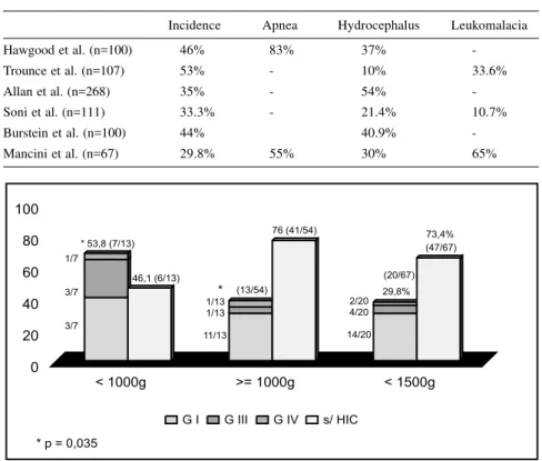
Within the group of newborns <1000 g, 53.8% (7/13) developed IVH: 42.8% (3/7) grade I, 42.8% (3/7) grade III, and 14.2% (1/7) grade IV (Figure 1). (No cases of grade II were obser-ved.)
Figure 2 shows IVH distribution in relation to gestational age: 47.3% (9/ 19) in newborns under 30 weeks of gestational age; 44.4% (4/9), 33.3% (3/ 9) and 22.2% (2/9) at grades I, III, and IV, respectively.
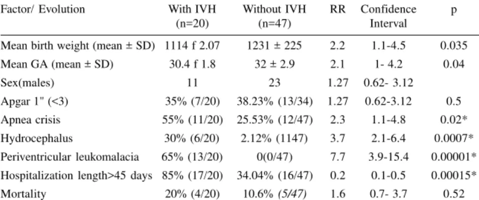
The newborn characteristics accor-ding to the occurrence of IVH are listed in table 1. The results indicate that IVH occurs with lower birth weight (p = 0.035), lower gestational age (p = 0.04), and longer hospita-lization length (p = 0.00015). The mean period of mechanical ventilation in the group with IVH was 27.7 + 36.9 days, compared to 12.78 + 14.34 in the group without IVH (p = 0.038).
Table 1 - NB characteristics according to IVH occurrence during the neonatal period.
Factor/ Evolution With IVH Without IVH RR Confidence p
(n=20) (n=47) Interval
Mean birth weight (mean ± SD) 1114 f 2.07 1231 ± 225 2.2 1.1-4.5 0.035
Mean GA (mean ± SD) 30.4 f 1.8 32 ± 2.9 2.1 1- 4.2 0.04
Sex(males) 11 23 1.27 0.62- 3.12
Apgar 1" (<3) 35% (7/20) 38.23% (13/34) 1.27 0.62-3.12 0.5
Apnea crisis 55% (11/20) 25.53% (12/47) 2.3 1.1-4.8 0.02*
Hydrocephalus 30% (6/20) 2.12% (1147) 3.7 2.1-6.4 0.0007*
Periventricular leukomalacia 65% (13/20) 0(0/47) 7.7 3.9-15.4 0.00001* Hospitalization length>45 days 85% (17/20) 34.04% (16/47) 0.2 0.1-0.5 0.00015*
Mortality 20% (4/20) 10.6% (5/47) 1.6 0.7- 3.7 0.52
* p < 0.05
153
REV. HOSP. CLÍN. FAC. MED. S.PAULO 54(5):151-154, 1999 SEPTEMBER-OCTOBER
IVH NB have a 2.3 risk of develo-ping apnea crisis (p = 0.02), 3.7 of hydrocephalus (p = 0.0007), and 7.7 of periventricular leukomalacia (p < 0.00001).
In relation to mortality, statistically significant differences were not detec-ted between the two groups analyzed.
DISCUSSION
A cranial ultrasound scan in the first week of life reveals the vast majority of IVH cases since 90% of these occur within the first 72 hours of life 6,4,13,14. Using equipment with high
frequency transducers permits optimal characterization of IVH in relation to severity, as well as early detection of sequelae, such as hydrocephalus and periventricular leukomalacia 7,15,
in-creasing examination specificity and sensitivity 10.
The current study revealed an incidence of 29.8% IVH in <1500 g newborns. These results are similar to those encountered in other institu-tions 1,2,3,4,6,15,16. Philip et al.15, studying
newborns <34 weeks, demonstrated a decrease in the incidence of IVH from 1979 to 1987 of 34% to 19%, and correlated these results to changes in the care of VLBW newborns 16. The
multi-institutional study conducted by the National Institute of Child Health and Human Development/Neonatal Research Network (NICHD) between 11/1987 and 01/1991 including <1500 g newborns, similar to this case study, indicated an incidence of 44%, making evident an important decrease in grades III and IV of IVH during this study 2.
Volpe et al., in a study conducted during the period between 1980 and 1989, also revealed the importance of detecting this pathology because of its repercussions, especially in the more immature NB, which are surviving in greater numbers now, increasing the number at risk 3.
The mortality rate was 20% and 10.6% in NB with and without IVH, respectively (no statistical significance
was found). However, Hawgood et al. 15 in 1984, found a higher mortality in NB with IVH, even higher in the severe cases. It is possible that we did not find statistical differences because of the higher incidence of grade I, which does not influence mortality rates, as shown by Thorburn et al. 17.
Regarding the severity of IVH, grade I hemorrhage occurs with varia-ble incidence. The NICHD study found 42.2% grade I 2, where as other studies
revealed an incidence around 60% 14,18.
It is important to notice that grades I and II have lower complication rates during follow-up, as previously de-monstrated by Allan et al.19, Soni et
al.14, and Burstein et al.18.
Gestational age under 30 weeks was an important risk factors for IVH development, as well as for the severity of the disease. These data are similar those of others, who found a higher incidence of IVH in earlier gestational ages 2,5.
Hawgood et al. 14 as well as
Partridge et al. 13 demonstrated an IVH
incidence of 61% and 67%, respec-tively, in NB under 1000 g, showing this low birth weight is a risk factor for IVH. Our results also indicate that birth weight less than 1000 g is an important risk factor for the more severe forms IVH.
Follow-up of IVH NB reveals complications such as apnea crisis (55%), hydrocephalus (30%), and periventricular leukomalacia (65%). Table 2 shows comparative data for IVH NB follow-up, highlighting the importance of follow-up. We found a higher incidence of complications in NB with IVH grades III and IV. Furthermore, in our study IVH NB remained under mechanical ventilation longer (p = 0.038) and were hospita-lized longer.
It is important for every neonatal unit to know the IVH incidence and re-lated risk factors so prevention sched-ules can be introduced. Ultrasound scan is a very important tool in the neonatal period for early detection of IVH and prevention of complications.
Table 2 - Incidence and evolution data of VLBW-NB with IVH on this study in relation to the literature data.
Incidence Apnea Hydrocephalus Leukomalacia
Hawgood et al. (n=100) 46% 83% 37%
-Trounce et al. (n=107) 53% - 10% 33.6%
Allan et al. (n=268) 35% - 54%
-Soni et al. (n=111) 33.3% - 21.4% 10.7%
Burstein et al. (n=100) 44% 40.9%
-Mancini et al. (n=67) 29.8% 55% 30% 65%
154
REV. HOSP. CLÍN. FAC. MED. S.PAULO 54(5):151-154, 1999 SEPTEMBER-OCTOBER
RESUMO RHCFAP/2982
MANCINI, M. C. e col. – Hemorragia intracranianana: evolução dos recém-nascidos de baixo peso no período neonatal. Rev. Hosp. Clín. Fac Med. S. Paulo 54 (5):1151-154, 1999.
A hemorragia intracraniana cons-tituí uma grave complicação na evo-lução dos recém-nascidos de muito baixo peso (RN-MBP) no período neonatal. Realizou-se este estudo, incluindo os recém-nascidos de muito baixo peso admitidos no Berçário Anexo à Maternidade do Hospital das Clínicas da FMUSP, com o objetivo de determinar a incidência de hemorragia intracraniana (HIC), os fatores de risco associados e sua evolução no período neonatal. Foram analisados o peso de
nascimento, idade gestacional, pre-sença de asfixia neonatal, uso de ventilação mecânica, tempo de inter-nação, presença de episódios de ap-néia, hidrocefalia e leucoencefaloma-lácia. O diagnóstico de HIC baseou-se no exame ultra-sonográfico realizado até o décimo dia de vida, de acordo com a classificação de Papille. Foram admitidos no período 101 RN-MBP, dos quais 67 preencheram aos critérios de inclusão. A taxa de mortalidade do período foi de 30,6% (31/101) e a incidência de HIC foi de 29,8% (20/ 67), sendo 70% grau I, 20 grau II e 10% grau IV. A incidência de HIC em recém-nascidos com peso de nasci-mento menos do que 1000g foi de 53,8% (p=0,04) naqueles com idade gestacional menor do que 30 semanas,
considerando-se ambos como fatores de risco para o seu desenvolvimento. Os recém-nascidos que evoluíram com HIC permaneceram mais tempo sob ventilação mecânica (p=0,038) e apresentaram um risco relativo de 2,3 de desenvolver episódios de apnéia (p=0,02), 3,7 de hidrocefalia (p=0,0007) e 7,7 de leucoencefa-lomalácia (p<0,00001). É importante o reconhecimento dos fatores de risco associados à HIC para o estabele-cimento de esquemas de prevenção, objetivando a redução da sua inci-dência com melhora do prognóstico desses recém-nascidos.
DESCRITORES: Hemorragia intracraniana. Período neonatal.
REFERENCES
1. MEIDELL R, MARINELLI P & PETTETT G - Perinatal factors associated with early-onset intracranial hemorrhage in premature infants. AJDC 1985; 139: 160-163.
2. SHANKARAN S, BAUER CR, BAIN R, WRIGHT LL et al. - Prenatal and perinatal risk and protective factors for neonatal intracranial hemorrhage. Arch Pediatr Adolesc 1996; 150: 491-97. 3. VOLPE JJ - Intraventricular hemorrhage and brain injury in the
premature infant - Neuropathology and pathogenesis. Clinics in Perinatology 1989; 16 (2) : 361-86.
4. HORBAR JD - Prevention of periventricular- intraventricular hemorrhage. In: SINCLAIR JC, BRACKEN MB - Effective Care of the Newborn Infant. Oxford, Univ Press,1992:562-89. 5. MENT LR, OH W, PHILIP AGS et al. – Risk factors for early
intraventricular hemorrhage in low birth weight infants. J Pediatr 1992; 121 (5): 776-83.
6. BOAL DKB, WATTERBERG KL, MILES S et al. - Optimal cost-effective timing of cranial ultrasound screening in low-birth weight infants. Pediatr Radiol 1995; 25: 425-28.
7. SZYMONOWICZ W, YU VYH, WALKER A et al. - Reduction in periventricular haemorrhage in preterm infants. Arch Dis Child 1986; 61: 661-65.
8. RENNIE JM, SOUTH M & MORLEY CJ - Cerebral blood flow velocity variability in infants receiving assisted ventilation. Arch Dis Child 1987; 62: 1247-51.
9. LEVENE MI, FAWER CL & LAMONT RF - Risk factors in the development of intraventricular haemorrhage in the preterm neonate. Arch Dis Child 1982; 57: 410-17.
10. DOLFIN T, SKIDMORE MB, FONG KW et al. - Incidence, severity, and timing of subependymal and intraventricular hemorrhages in preterm infants born in a perinatal unit as detected by serial real-time ultrasound. Pediatrics 1983, 71(4): 541-46.
11. PAPILE LA, BURSTEIN J, BURSTEIN R et al. - Incidence and evolution of subependymal and intraventricular hemorrhage: a study of infants with birth weights less than 1500 gm. J Pediatr 1978; 92: 529-34.
12. PARTRIDGE JC, BABCOCK DS, STEICHEN JJ et al. - Optimal timing for diagnostic cranial ultrasound in low-birth weight infants: detection of intracranial hemorrhage and ventricular dilatation. J Pediatr 1983; 102(2): 28187.
13. SONI JP, GUPTA BD, SONI M et al. - Ultrasonic diagnosis of intracranial hemorrhage in high risk neonates. Indian Pediatrics 1995; 32: 453-60.
14. HAWGOOD S, SPONG J & YU VYH - Intraventricular hemorrhage-Incidence and outcome in a population of very-low-birth-weight infants. AJDC 1984, 138: 136-39.
15. PHILIP AGS, ALLAN WC, TITO AM et al. - Intraventricular hemorrhage in preterm infants: declining incidence in the 1980s. Pediatrics 1989; 84(5) : 797-801.
16. THORBURN RJ, STEWART AL, HOPE PL et. al. - Prediction of death and major handicap in very preterm infant by brain ultrasound. Lancet 1981; 1119-1121.
17. BURSTEIN J, PAPILE LA & BURSTEIN R - Intraventricular hemorrhage and hydrocephalus in premature newborns: a prospective study with CT. AJR 1979; 132: 631-35.
18. ALLAN WC, HOLT PJ, SAWYER LR et al. - Ventricular dilatation after neonatal periventricular- intraventricular hemorrhage- natural history and therapeutic implications. Am J Dis Child 1982; 136: 589-93.
19. TROUNCE JQ, RUTTER N & LEVENE MI - Periventricular leukomalacia and intraventricular haemorrhage in the preterm neonate. Arch Dis Child 1986; 61: 1196-1202.