ARTICLE
Blood-brain barrier breakdown and repair
following gliotoxic drug injection in the
brainstem of streptozotocin-diabetic rats
Ruptura e reparo da barreira hematoencefálica pós-injeção de droga gliotóxica no
tronco encefálico ratos diabéticos
Eduardo Fernandes Bondan1, Maria de Fátima Monteiro Martins2
Ethidium bromide (EB) injection in the central nervous system (CNS) causes local oligodendroglial and astrocytic dis-appearance, with subsequent primary demyelination, glia lim-itans disruption, supposed blood-brain barrier (BBB) break-down and Schwann cell invasion1-3. Hyperglycemia found in diabetes mellitus is known to cause changes in the behaviour of glial cells, such as oligodendrocytes and Schwann cells4. As BBB induction and maintenance is closely related to glial cell support, the aim of this study was to evaluate and compare the BBB integrity after the injection of 0.1% EB into the brain-stem of diabetic and non-diabetic rats.
METHODS
his experiment was approved by the Ethics Comission of the University Paulista (protocol number 002/09). Adult male Wistar rats were used and some received, after a 12 hours of fasting, a single injection of streptozotocin (50 mg/kg) in 0.01M citrate bufer (pH 4.5) into the tail vein. Ten days after that, blood glucose level was measured and animals with levels of 200 mg/dL or more were considered diabet-ics. At this time, they were submitted to a local injection of 10 microlitres of 0.1% EB (group I) or 0.9% saline (group II)
1 Head professor of the University Paulista (UNIP), São Paulo SP, Brazil, and University Cruzeiro do Sul (UNICSUL), São Paulo SP, Brazil; 2 PhD student of the Post-Graduation Program in Immunopathology, UNIP, and Adjunct Professor of UNICSUL, São Paulo SP, Brazil. Correspondence: Eduardo Fernandes Bondan; Rua Caconde 125 / 51; 01425-011 São Paulo SP - Brasil; E-mail: [email protected]
Conflict of interest: There is no conflict of interest to declare.
Received 22 May 2011; Received in final form 06 September 2011; Accepted 14 September 2011
ABSTRACT
Ethidium bromide (EB) causes local astrocytic disappearance, with glia limitans disruption and blood-brain barrier (BBB) breakdown. The aim of this study was to evaluate the BBB integrity after the injection of 0.1% EB or 0.9% saline solution into the cisterna pontis of Wistar rats submitted or not to the streptozotocin diabetogenic model. Brainstem sections were collected from 24 hours to 31 days post-injection for ultrastructural analysis and glial fibrillary acidic protein immunohistochemical staining. Some animals received colloidal carbon ink by intravenous route at the same periods. In rats injected with EB, results revealed astrocyte disappearance and leakage of carbon particles beginning at 48 hours and persisting for 7 days in non-diabetic rats and for 15 days in the diabetic ones, although, in both groups, several areas remained devoid of astrocytic processes up to 31 days. In rats injected with saline, there was no sign of astrocytic loss or carbon par-ticles leakage.
Key words: blood-brain barrier, central nervous system, diabetes mellitus, ethidium bromide.
RESUMO
O brometo de etídio (BE) determina o desaparecimento local de astrócitos, com ruptura da glia limitans e dano na barreira hematoencefálica (BHE). Este estudo visou avaliar a integridade da BHE após injeção de solução de BE a 0,1% ou de salina a 0,9% na cisterna pontis de ratos Wistar submetidos ou não ao modelo diabetogênico da estreptozotocina. Fragmentos do tronco encefálico foram coletados das 24 horas aos 31 dias pós-injeção para estudo ultraestrutural e marcação imuno-histoquímica para proteína glial fibrilar ácida. Alguns animais receberam carvão coloidal por via intravenosa nos mesmos períodos. Nos grupos injetados com BE, os resultados mostraram desaparecimento astro-citário e extravasamento de partículas de carvão nas lesões a partir das 48 horas, persistindo por até sete dias nos animais não diabéticos e 15 dias nos diabéticos, embora, em ambos os grupos, diversas áreas permanecessem destituídas de astrócitos até 31 dias após. Nos ratos injetados com salina, diabéticos ou não, não houve sinal de perda astrocitária nem de extravasamento vascular de carvão.
solution into the cisterna pontis. All rats were anaesthe-tized with ketamine and xylazine (5:1; 0.1 mL/100g) and a burr hole was made on the right side of the skull, 8 mm ros-tral to the frontoparietal suture. Injections were performed freehand using a Hamilton Syringe, itted with a 35o angled polished 26-gauge needle into the cisterna pontis, an en-larged subarachnoid space below the ventral surface on the pons. Non-diabetic rats also received 10 microlitres of 0.1% EB solution (group III) or 0.9% saline solution (group IV). Body weight and blood glucose levels (Dextrostix, Ames) were recorded at 3 times – at the moment of the streptozo-tocin injection, 10 days after and at the time of euthanasia. Water and food were given ad libitum during the
experi-mental period. At diferent times after EB or saline injec-tion, the rats were anaesthetized and submitted to intrac-ardiac perfusion with 4% glutaraldehyde in 0.1 M Sorensen phosphate bufer (pH 7.4) at each of the following periods - 24, 48, 72 hours, and 7, 11, 15, 21 and 31 days after intra-cisternal injection. hin slices of the brainstem (pons and mesencephalon) were collected and post-ixed in 0.1% os-mium tetroxide, dehydrated with graded acetones and em-bedded in Araldite 502 resin, following transitional stages in acetone. hick sections were stained with 0.25% alka-line toluidine blue. Selected areas were trimmed and thin sections were stained with 2% uranyl and lead acetate and viewed in a JEM -1200 EX2 JEOL transmission electron microscope.
From each group, some animals received colloidal car-bon ink by intravenous route at the same periods previously mentioned, 20 minutes before being submitted to euthanasia and perfusion with 10% bufered formalin solution (pH 7.4). Immunohistochemical staining to glial ibrillary acidic pro-tein (GFAP) (GFAP - rabbit anti-cow GFA, code number ZO334, Dako) was performed as an astrocyte marker accord-ing to Sanchez et al.5.
RESULTS
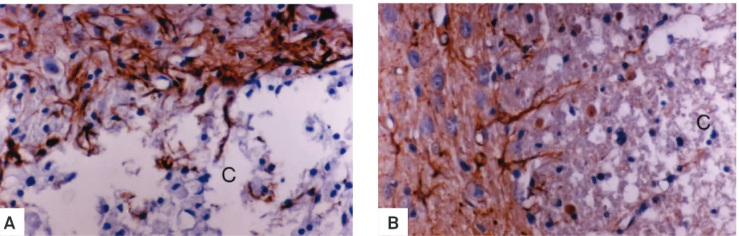
In rats from groups II and IV, there was no sign of astro-cyte loss and no leakage of ink from blood vessels in the injec-tion site. In groups III (Fig 1A) and I (Fig 1B), astrocyte disap-pearance began at 48 hours and some areas were still devoid of astrocytic processes 31 days after. In the EB-induced le-sion, leakage of carbon particles was seen from 48 hours to 15 days in group I (diabetic) and from 48 hours to 7 days in group III (non-diabetic). Tight junctions did not show any de-tectable ultrastructural change due to the lack of perivascu-lar astrocytes in groups injected with EB. Diabetic rats from group I presented delayed macrophagic activity (Fig 2B) and lesser remyelination (Fig 3B) in comparison to non-diabetic rats from group III (Fig 2A and 3A). Although oligodendro-cytes were the major remyelinating cells in the brainstem, Schwann cells invaded EB-induced lesions, irstly appearing at 11 days in non-diabetic rats and by 15 days in diabetic rats. Results indicate that short-term streptozotocin-induced di-abetes hindered BBB reconstruction and both oligodendro-cyte and Schwann cell remyelination in comparison to non-diabetic rats after local EB injection (Fig 4A and 4B).
DISCUSSION
A previous study6 had already demonstrated that normal Wistar rats presented leakage of carbon particles from 48 hours to 7 days after injection of this gliotoxic drug, as well as astrocyte disappearance for up to 31 days. he present re-sults in the non-diabetic group corroborate those indings. In streptozotocin-diabetic rats, however, the most important diference noted was that ink leakage persisted until the 15th day, suggesting that the diabetic state somehow delayed the functional repair of the barrier.
Fig 1. EB-induced lesions in groups III (A, non-diabetic) and I (B, diabetic). Note the absence of glial fibrillary acid protein (GFAP) - positive cells in the central areas (C) of both groups at 31 days post-injection. Increased astrocyte immunorreactivity to GFAP is seen at the Iesion boundaries. Greater amounts of cellular debris are found in the center of the lesions of the diabetic group. GFAP immuno-histochemical staining - Obj. 100x.
he BBB, a physical and metabolic barrier, is an impor-tant and complex structure designed to maintain a consimpor-tant neuronal extracellular environment by restraining the pen-etration of a wide range of hydrophilic molecules, proteins and cellular elements into the CNS7,8. It is composed of en-dothelial cells lining CNS microvessels that are interconnect-ed by tight junctions and are surroundinterconnect-ed by pericytes and astroglial processes. Besides the capillary endothelium and adjacent glia, this anatomical barrier may also include com-ponents of the extracellular matrix. he BBB involves several
mechanisms by which free exchange of non-lipid soluble mol-ecules between blood and cerebrospinal luid (CSF) space is restricted, such as active (energy-requiring) processes (e.g., amino-acid transporters) and passive mechanisms (e.g., en-dothelial tight junctions) that regulate CNS homeostasia, be-cause blood contains very high levels of potential neurotoxic substances and neural exposure to them would render inef-fective neuronal excitability9. Intravenous injection of an ex-ogenous contrast agent (that is normally excluded from the CNS space, but can enter with damage to the integrity of the
Fig 2. Central areas of ethidium bromide-induced lesions at 21 days in non-diabetic (A, group III) and diabetic (B, group I) rats containing macrophages (M) in phagocytic activity. (A) Myelin can be seen in different stages of breakdown, from the visualization of lamellae (L) to neutral fat droplets (n). v: blood vessel. Electron micrograph - 4.309x. (B) Diabetic rats show huge amounts of myelin-derived membranes in the extracellular space (asterisk) suggesting delayed phagocytic activity. Electron micrograph - 3.968x.
A
B
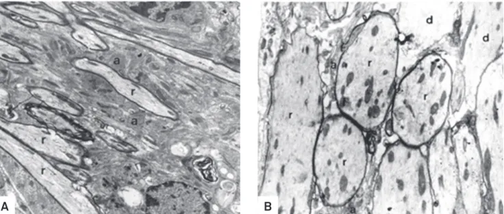
Fig 3. Oligodendrocyte-remyelinated areas in non-diabetic (A, group III) and diabetic (B, group I) rats at peripheral sites after ethidium bromide injection. (A) Axons in initial remyelination (r) among hypertrophic astrocytes (a) at 21 days. Electron micrograph - 6.064x. (B) Fewer axons (r) show thinner myelin sheaths even at 31 days. Many axons remain demyelinated (d). Electron micrograph - 7.152x.
BBB) allows magnetic resonance imaging (MRI) to visualize the breakdown of the BBB in patients with neurological dis-orders9, that can be even seen by light microscopy observa-tion in experimental condiobserva-tions using animal models6.
BBB breakdown is present in several common CNS disor-ders, such as stroke, trauma, brain tumors, multiple sclerosis, HIV-1 dementia, Alzheimer´s and Parkinson´s disease10. On the other hand, it is known that prolonged BBB disruption may lead to neurological deicits, as delayed cortical dysfunc-tion and epileptiform activity involving glutamatergic and GABAergic neurotransmission8.
Diabetes mellitus is a metabolic disorder associated with structural and functional alterations of various organ sys-tems. Although the peripheral neuropathy along with small and large blood vessel angiopathy can explain most of the diabetes-related organ failures, tissue injury is attributed mainly to chronic hyperglycemia11. In contrast to the high prevalence of renal and retinal disease in diabetic patients, the chronic diabetic complications of the CNS are hardly no-ticeable and the deleterious efects of persistent hyperglyce-mia on brain metabolism and cognitive function are often unrecognized. his glucotoxicity is not tissue speciic, even
though the severity of organ dysfunction in diabetes is highly variable11. Oxidative stress plays an important role in tissue damage caused by both insulin-induced hypoglycemia and streptozotocin-induced diabetes, which may be the result of deterioration in glucose homeostasia caused by these meta-bolic changes.
During severe energy deprivation following hypogly-cemia and diabetes, mitochondrial free radicals scavenger system is down regulated leading to reactive oxygen spe-cies generation and activation of processes conducting to DNA damage and neural cell death12. Neuroinlammation may expose endothelium to proinlammatory cytokines, such as IFN-gamma, TNF-alpha and IL-1beta, which in turn disorganize cell-cell junctions, afect leukocyte endothelial adhesion and migration, increase expression of class II ma-jor histocompatibility complex molecules and decrease the brain solute barrier13.
Exact mechanisms for breakdown of the BBB in patholog-ic conditions, including EB gliotoxpatholog-ic injection, are not com-pletely understood and may involve direct efects of these cy-tokines on endothelial regulation of BBB components, as well as indirect cytokine-dependent leukocyte mediated injury.
1. Graça DL, Bondan EF, Pereira LA, Fernandes CG, Maiorka PC. Behaviour of oligodendrocytes and Schwann cells in an experimental model of toxic demyelination of the central nervous system. Arq Neuropsiquiatr 2001;59:358-361.
2. Pereira LA, Dertkigill MS, Graça DL, Cruz-Höffling MA. Dynamics of remyelination in the brain of adult rats after exposure to ethidium bromide. J Submicrosc Cytol Pathol 1998;30:341-348.
3. Bondan EF, Lallo MA, Sinhorini IL, Pereira LAVD, Graça DL. The effect
of cyclophosphamide on the rat brainstem remyelination following local ethidium bromide injection in Wistar rats. J Submicrosc Cytol Pathol 2000;32:603-612.
4. Bondan EF, Lallo MA, Trigueiro AH, Ribeiro CP, Sinhorini IL, Graça DL. Delayed Schwann cell and oligodendrocyte remyelination after ethidium bromide injection in the brainstem of Wistar rats submitted to streptozotocin diabetogenic treatment. Braz J Med Biol Res 2006;39:637-646.
References
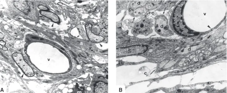
Fig 4. (A) Oligodendrocyte-remyelinated axons (arrowheads) near to a blood vessel (v) at 15 days after ethidium bromide injection. Note the presence of surrounding astrocyte processes (a). m: macrophage. Group III (non-diabetic) - Electron micrograph. 7.077x. (B) Demyelinated axons (d) at 31 days after EB injection in a perivascular area. Observe the presence of a tight junction (arrowhead) and Schwann cell (S) cytoplasm around the naked axons (arrows). v: blood vessel; c: collagen fibers. Group I (diabetic) - Electron micrograph. 10.864x.
5. Sanchez M, Bondan EF, Lallo MA, et al. [Immunohistochemical staining of the macrophagic and astrocytic response in the brainstem of Wistar rats submitted to the ethidium bromide gliotoxic model and treated with cyclophosphamide]. Arq Neuropsiquiatr 2006;64:787-793.
6. Bondan EF, Lallo MA, Dagli ML, Pereira LA, Graça DL. [Blood-brain barrier breakdown following gliotoxic drug injection in the brainstem of Wistar rats]. Arq Neuropsiquiatr 2002;60:582-589.
7. Ramos AT, Viott, AM, Machado GF. Astrócitos. In: Graça DL, Bondan EF, Pereira LAV, Maiorka PC (Eds.). Biologia da desmielinização e da remielinização: a base da esclerose múltipla. Santa Maria: Editora UFSM, 2011: 35-59.
8. Seiffert E, Dreier JP, Ivens S, et al. Lasting blood-brain barrier disruption induces epileptic focus in the rat somatosensory cortex. J Neurosci 2004;24:7829-7836.
9. Cifelli A, Arnold DL, Matthews PM. MRI visualization of multiple sclerosis. In: Lazzarini RA (Ed). Myelin biology and disorders. San Diego: Elsevier, 2004: 763-790.
10. Ballabh P, Braun A, Nedergaard M. The blood-brain barrier: an overview - structure, regulation, and clinical implications. Neurobiol Dis 2004;16:1-13.
11. Mooradian AD. Diabetic complications of the central nervous system. Endocr Rev 1988;9:346-356.
12. Singh P, Jain A, Kaur G. Impact of hypoglycemia and diabetes on CNS: correlation of mitochondrial oxidative stress with DNA damage. Mol Cell Biochem 2004;260:153-159.