Article
ISSN 0102-695X
doi: 10.1590/S0102-695X2011005000021
Received 19 Jun 2010 Accepted 27 Aug 2010 Available online 25 Feb 2011
and antioxidant effects of
Boehmeria nivea
root
extract in streptozotocin-induced diabetic rats
Shruti Sancheti,
1,§Sandesh Sancheti,
1,§Mayur Bafna,
1Hae-Ran
Kim,
1Young-Han You,
1Sung-Yum Seo
*,1 21Department of Biology, Kongju National University, Republic of Korea,
2Korean Collection of Herbal Extracts, Inc., Republic of Korea.
Abstract: The potential role of 80% methanolic extract of Boehmeria nivea (L.) Gaudich., Urticaceae, root in the treatment of diabetes, along with its antihyperlipidemic and antioxidant effects, was studied in streptozotocin-induced diabetic male Wistar rats. Preliminary screening of the extract revealed the presence of polyphenolics and
l avonoids. The animal study was conducted with variable doses of 125, 250 and 500 mg/ kg of extract for 21 days in diabetic rats. A signii cant effect was observed at a dose of
500 mg/kg, which was comparable to the standard drug, glibenclamide. Administration
of the extract at a 500 mg/kg dose resulted in a signii cant reduction of fasting blood
glucose, total cholesterol, triglycerides, blood urea, alanine aminotransferase, aspartate aminotransferase, urine sugar and urine ketone levels in diabetic rats in comparison
with the diabetic control group. Additionally, this dose signii cantly increased body
weight, hemoglobin, plasma total protein, high density lipoprotein cholesterol, liver glycogen content, superoxide dismutase, reduced glutathione and catalase levels in diabetic rats at the end of 21 days of treatment. Therefore, dietary supplementation with Boehmeria nivea root extract could be benei cial for correcting hyperglycemia,
hyperlipidemia and enhancing the antioxidant defense system.
Keywords:
antihyperglycemic antihyperlipidemic antioxidant
Boehmeria nivea root
diabetes streptozotocin
Introduction
Diabetes mellitus (DM) is a chronic life-threatening metabolic disorder (affecting carbohydrate, fat and protein metabolism) in which the level of glucose in the blood and/or urine is abnormally high, which is due to impaired carbohydrate utilization resulting from
an insufi ciency of the secretion or action of endogenous
insulin (Clark & Pazdernik, 2009; Maritim et al., 2003; Kumar & Murugesan, 2008). Along with hyperglycemia, hypertension, oxidative stress and dislipidemia (the traditional risk factors for onset and progression of chronic complications of diabetes), diabetes is associated with micro- and macro-vascular complications leading to cardiovascular disease, neuropathy, retinopathy and nephropathy, which are the major causes of morbidity and death (Maritim et al., 2003; Kumar & Murugesan, 2008; Kramer et al., 2009; Sharma et al., 2008).
Despite the considerable strides that have been made in the understanding and management of diabetes, the disease and disease-related complications are increasing unabated (Odetola et al., 2006). The worldwide prevalence of this major non-communicable disease has been projected to increase to approximately 366 million by the year 2030. Considering its increasing
prevalence, attendant complications and heavy economic and social burdens, DM is now considered as a public health nightmare (Wild et al., 2004; Adeneye & Adeyemi, 2009). In addition, current anti-diabetic medications
usually have adverse side effects, decreased efi cacy over
time, ineffectiveness against some long-term diabetic complications and low cost-effectiveness; therefore, the discovery and development of novel drugs for diabetes is rigorously needed (Hsu et al., 2009).
In recent years, there has been a renewed interest in the treatment of DM using herbal drugs, as they are a wonderful source of medicines that are frequently considered to be less toxic and have fewer side effects than synthetic ones. Furthermore, the World Health Organization has also recommended the investigation and evaluation of hypoglycemic agents from medicinal plants (Kumar & Murugesan, 2008; Udayakumar et al., 2009).
It is well known that the inhibition of intestinal
α-glucosidases limits postprandial glucose levels
α-glucosidase inhibitor (Sancheti et al., 2010).
Boehmeria nivea (L.) Gaudich., Urticaceae (common name: Ramie, China grass), is a perennial
herbaceous iber plant that is widely distributed in South
Korea, India, China and Japan. Its root is edible, having a sweet taste, and has been used traditionally as a diuretic, antipyretic, hepatoprotective, antioxidant and
anti-inlammatory agent (Sancheti et al., 2010; Lin et al., 1998;
Wu, 2005). Because no in vivo study has been carried out so far on the root extract of this plant with respect to
its anti-diabetic eficacy, the present study was designed
to study its antihyperglycemic, antihyperlipidemic and antioxidant effects in STZ-induced diabetic rats.
Materials and Methods
Animals
For this experiment, speciic-pathogen free
healthy male Albino Wistar rats weighing approximately 190-240 g were obtained from the Daehan Biolink Co., Chungcheongbuk-Do, Korea. Before commencement of the study, the animals were acclimatized for a period of one week to the laboratory conditions. They were kept in polycarbonate cages under controlled temperature (22±2
˚C) and 12 h light/12 h dark cycles. The animals were
allowed free access to laboratory chow diet and water ad libitum. For experimental purposes, the animals were kept fasting overnight, but were allowed free access to water. The protocol used in this study for the use of animals was approved by the University Animal Ethical Committee.
Preparation of Boehmeria nivea root extract (BNRE)
The dried and matured roots of B. nivea were obtained from the “Korean Collection of Herbal Extracts,”
a biotech company in Korea. The plant was identiied
by Professor Young-Han You, Department of Biology, Kongju national University, Kongju. A voucher specimen is available with the company (Korea Collection of Herbal Extracts, 2000) (voucher no.KCHE-0901BNR). The roots (6 kg, dry weight) were extensively extracted with 80% aqueous MeOH for three days. The extract was dried using a rotary vacuum evaporator below 40 °C. The vacuum dried crude extract was added to distilled water
and iltered. The iltrate was lyophilized (120 g) and used
for the experiments.
Preliminary phytochemical analysis
Total phenolic and total lavonoid contents
were determined by the methods reported earlier using calibration curves of gallic acid and quercetin, respectively (Zhang et al., 2006; Chang et al., 2002). All assays were performed in triplicate for three times and
results are expressed as mean±SD.
Dose ixation study
The dose ixation study was carried out as per
the method described by Pandikumar et al. (2009) with
slight modiications. The doses for the study were ixed
based on Irwin test for the extracts at 1, 2, 3, 4 and 5 g/ kg. The extracts were dissolved in distilled water. Non-diabetic, male rats weighing 190 to 240 g were used in this study. Three animals were used for each group. On the day preceding the experiment the animals were appropriately grouped and placed in the experiment room for acclimatization and were fasted for 12 h. Followed by the 12 h fasting, the animals were treated orally with the distilled water or the extract. At 0, 15, 30, 60, 120, 180 min and 24 h after treatment of the extracts, behavioral alterations were observed. The mortality caused by the extract within this period of time was also noted.
Induction of diabetes
A freshly prepared solution of streptozotocin (STZ) (55 mg/kg body weight) in citrate buffer (pH 4.5, 0.1 M) was injected into overnight-fasted rats (Saravanan et al., 2009). Three days after STZ induction,
the development of diabetes was conirmed by tail vein
blood glucose levels. Animals with blood glucose levels more than 250 mg/dL were included in the study.
Experimental design and treatment schedule
Thirty six rats were randomly divided into six equal groups and were treated daily for 21 days as follows:
(i) Group I (normal rats treated with distilled water). (ii) Group II (diabetic rats treated with distilled water). (iii) Group III (diabetic rats treated with BNRE at 125 mg/kg body weight).
(iv) Group IV (diabetic rats treated with BNRE at 250 mg/kg bw).
(v) Group V (diabetic rats treated with BNRE at 500 mg/ kg bw).
(vi) Group VI (diabetic rats treated with glibenclamide at 0.6 mg/kg bw).
Collection and processing of blood and tissue samples
On the 21st day (at the end of the study) after
an overnight fasting, the animals were sacriiced under
individually homogenized as per the method of Jung et
al. (2005) with minor modiications, to make 10% w/v
solutions in phosphate buffer (50 mM, pH 7.0) and stored
at -80 ˚C. The supernatants were used for the biochemical
analysis for the determination of the enzymes (SOD, GSH and CAT). The other half of the liver and muscles were used for the determination of glycogen content.
Blood glucose measurement
Blood was collected from the tip of the tail vein of the overnight fasted rats and the blood glucose was measured using an Accutrend Plus instrument (Roche Diagnostics, Germany) at regular time intervals after diabetes induction, i.e., 0th, 7th, 14th and 21st days of the
experiment. The fasting blood glucose (FBG) levels of the normal control group were also measured simultaneously. The results were expressed in terms of milligrams per deciliter (mg/dL) of blood.
Lipid proile measurement
Upon completion of the treatment, total cholesterol (TC), high-density lipoprotein cholesterol (HDL-C) and triglyceride (TG) levels in plasma were determined according to the instructions of the manufacturer (Asan Pharmaceutical, Seoul, Korea). The results were expressed in mg/dL.
Estimation of antioxidants in liver and kidney
SOD, GSH and CAT levels in normal, diabetic and treatment groups were determined as per the reported methods (Misra & Fridovich, 1972; Ellman, 1959; Aebi, 1984). All the parameters were expressed in terms of units/mg protein of the tissue.
Estimation of tissue glycogen
Glycogen content in liver and muscles were measured with the anthrone reagent using glucose as a standard (Morris, 1948; Maiti et al., 2004; Sadasivam & Manickam, 1996). The amount of tissue glycogen was expressed in micrograms of glucose per mg tissue.
Other biochemical parameters measurement
Hemoglobin (Hb), blood urea, aspartate aminotransferase (AST), alanine aminotransferase (ALT), plasma total protein (TP) and plasma albumin were estimated using commercially available standard assay kits (Asan Pharmaceutical, Seoul, Korea). Urine sugar and urine ketone were measured using Cybow urine reagent strips and a Cybow reader 720 (manufacturer: DFI Co., Ltd.).
Statistical analysis
Data were expressed as mean±S.D. for six animals in each group. Statistical analysis was done by one-way analysis of variance (ANOVA) followed by Tukey’s post-hoc test except for triglycerides data (Wilcoxon-Mann-Whitney Rank Sum Test was applied for TG data) using KaleidaGraph v. 4.0, Synergy Software. p<0.05 was considered to be statistically signiicant.
Results
Preliminary phytochemical analysis
The total polyphenolic content in BNRE was found to be 42±1.77 mg gallic acid equivalent/g of dry
weight of extract and the lavonoid content was estimated
as 12±0.92 mg quercetin equivalent/g of dry weight of extract.
Dose ixation study
BNRE showed no adverse effects in the dose range between 1 to 5 g/kg bw in normal male Wistar rats. There was no mortality report in the animals treated with the extracts at all the doses. Additionally, in previous studies, B. nivea root at 500 mg/kg dose was found as a potent hepatoprotective agent (Lin et al., 1998). Therefore, based on the results and literature search, 500 mg/kg bw dose (1/10th of the highest dose of 5 g/kg)
was selected as the highest dose for further biochemical studies. The animals were also treated with 125 and 250 mg/kg bw doses of BNRE for 21 days.
Effect on body weight, water and food intake
The body weights of all of the groups (control and treated) were estimated at the start and end of the
experiment. There was no signiicant intra-group variation
in the basal body weight on day 0 of the experiment. On the 21st day, the body weight in normal control rats was
signiicantly increased, whereas STZ-induced diabetic rats showed signiicant weight loss as compared to the initial
day 0 readings (Table 1). BNRE- and
glibenclamide-treated diabetic rats showed a recovery to inal body
weight that was close to that of normal control rats. The water and food intake in the diabetic control
rats were signiicantly increased as compared to the
normal control group. Twenty one days of BNRE and
glibenclamide treatment signiicantly reduced the water
and food intake in diabetic rats (Table 1).
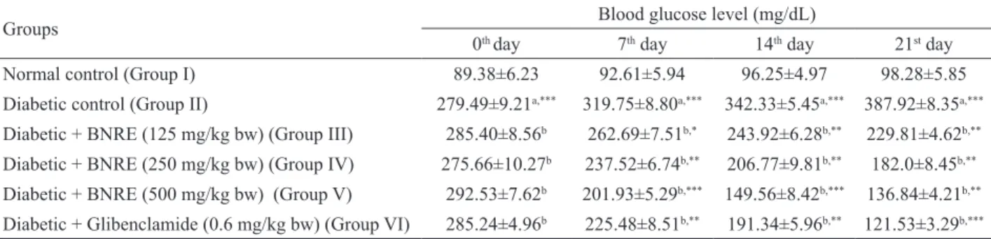
Effect on fasting blood glucose levels
blood glucose in diabetic control rats as compared to the normal control group. However, treatment with BNRE in
diabetic rats for 21 days resulted in a signiicant decrease
in fasting blood glucose levels, but to a varied extent, returning them to the control level at a 500 mg/kg dose (Table 2). Therefore, further studies were carried out only on group V (diabetic rats treated with 500 mg/kg dose of BNRE) along with groups I (normal control), II (diabetic control) and VI (diabetic rats treated with 0.6 mg/kg glibenclamide).
Plasma lipid proile
Plasma TC and TG levels were signiicantly
elevated in diabetic rats in comparison to normal control rats. Supplementation of BNRE for 21 days to the
diabetic rats resulted in a signiicant diminution of these
parameters, and the levels of these parameters returned toward the control level at a 500 mg/kg dose (Table 3).
HDL-C, a benevolent lipoprotein, was decreased in the diabetic groups with respect to the normal control group. After 21 days of treatment with the extract, there
was a signiicant elevation of this lipoprotein level in
group V, which was comparable to group VI (Table 3).
Estimation of antioxidants in liver and kidney
SOD, GSH and CAT levels were signiicantly
reduced in liver and kidney of diabetic rats as compared to that of normal rats (Table 4). Upon BNRE supplementation for 21 days at a dose of 500 mg/kg, the levels of all of these enzymes were corrected to a greater extent in diabetic rats
Table 1. Changes in body weight and food and water intake in STZ-induced diabetic animals before and after treatment with
Boehmeria nivea root extract (BNRE).
Groups Change in body weight (g) Food intake Water intake
Initial Final (g/rat per day) (mL/rat per day)
Normal control (Group I) 204.18±4.86 238.34±5.29 14.82±1.95 69.65±13.21
Diabetic control (Group II) 227.94±9.51 178.21±8.32a,** 32.91±3.72 a,** 172.97±27.78a,**
Diabetic + BNRE (125 mg/kg bw) (Group III) 210.45±7.12 195.43±8.52b 24.77±2.51b,* 114.09±6.42b,*
Diabetic + BNRE (250 mg/kg bw) (Group IV) 204.32±5.35 216.81±6.24b,** 19.72±2.19b,** 98.98±11.39b,**
Diabetic + BNRE (500 mg/kg bw) (Group V) 209.82±7.96 234.45±4.53b,*** 15.92±1.20b,** 85.21±8.06b,***
Diabetic + Glibenclamide (0.6 mg/kg bw) (Group VI) 218.57±6.41 236.61±3.92b,*** 16.64±2.78b,** 87.54±4.88b,**
Each value is a mean±S.D. for six rats in each group. Values are statistically signiicant at *p<0.05, ** p<0.01 and ***p<0.001. aDiabetic control was
compared with normal control. bTreated groups were compared with diabetic controls.
Groups Blood glucose level (mg/dL)
0th day 7th day 14th day 21st day
Normal control (Group I) 89.38±6.23 92.61±5.94 96.25±4.97 98.28±5.85
Diabetic control (Group II) 279.49±9.21a,*** 319.75±8.80a,*** 342.33±5.45a,*** 387.92±8.35a,***
Diabetic + BNRE (125 mg/kg bw) (Group III) 285.40±8.56b 262.69±7.51b,* 243.92±6.28b,** 229.81±4.62b,**
Diabetic + BNRE (250 mg/kg bw) (Group IV) 275.66±10.27b 237.52±6.74b,** 206.77±9.81b,** 182.0±8.45b,**
Diabetic + BNRE (500 mg/kg bw) (Group V) 292.53±7.62b 201.93±5.29b,*** 149.56±8.42b,*** 136.84±4.21b,**
Diabetic + Glibenclamide (0.6 mg/kg bw) (Group VI) 285.24±4.96b 225.48±8.51b,** 191.34±5.96b,** 121.53±3.29b,*** Table 2. Effect of 21 days of Boehmeria nivea root extract (BNRE) treatment on blood glucose levels in normal and diabetic rats.
Each value is a mean±S.D. for 6 rats in each group. Values are statistically signiicant at *p<0.05, **p<0.01 and ***p<0.001. aDiabetic control was
compared with normal control at the corresponding time-interval. bTreated groups were compared with diabetic controls.
Groups Total cholesterol
(mg/dL)
Triglycerides (mg/dL)
High density lipoprotein cholesterol (mg/dL)
Normal control (Group I) 74.12±3.38 98.43±4.17 42.86±2.51
Diabetic control (Group II) 122.96±4.36a,*** 146.62±3.48a,* 30.92±3.38a,***
Diabetic+BNRE (500 mg/kg bw) (Group V) 78.34±2.56b,*** 103.95±3.25b,* 39.34±1.12b,**
Diabetic+Glibenclamide (0.6 mg/kg bw) (Group VI) 77.92±1.80b,*** 101.27±2.13b,* 40.13±1.29b,**
Each value is a mean±S.D. for 6 rats in each group. Values are statistically signiicant at *p<0.05, **p<0.01 and ***p<0.001. aDiabetic control was
compared with normal control. bTreated groups were compared with diabetic controls.
and were comparable to that of group VI (diabetic rats treated with 0.6 mg/kg glibenclamide) (Table 4).
Glycogen level in tissues
The glycogen contents of the liver and muscle tissues in normal and diabetic controls, and diabetic rats supplemented with the extract are shown in Table 5. Hepatic and skeletal muscle glycogen contents were
signiicantly decreased in diabetic rats with respect to
the controls. However, treatment with BNRE at 500 mg/ kg dose led to an increase in liver and muscle glycogen content over the diabetic controls.
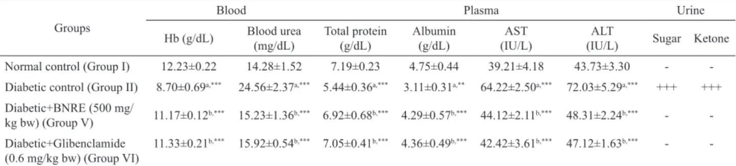
A signiicant reduction in the levels of Hb,
plasma TP and albumin was detected in diabetic rats as
compared to the normal control group. On the other hand,
the BNRE-treated rats signiicantly reversed these changes
to near normal levels (Table 6). Furthermore, the levels of blood urea, AST, ALT, urine sugar and urine ketone were
signiicantly increased in the diabetic control group, but
these values returned towards normal in BNRE-treated rats at 500 mg/kg dose, which was comparable to glibenclamide (0.6 mg/kg) (Table 6).
Discussion
In our previous study, the roots of B. nivea
exhibited potent α-glucosidase inhibition and possessed
a good antioxidant activity in vitro (Sancheti et al.,
2010). Based on these indings, the antidiabetic potential
Table 4. Effect of 21 days Boehmeria nivea root extract (BNRE) treatment on antioxidant proile of liver and kidney in normal and
diabetic rats.
Groups
Parameters (U/mg protein)
Superoxide dismutase Reduced glutathione Catalase
Liver Kidney Liver Kidney Liver Kidney
Normal control (Group I) 16.82±1.52 64.25±2.44 68.91±2.29 147.83±3.57 50.66±1.03 61.25±2.28 Diabetic control (Group II) 6.90±2.65a,*** 27.12±2.58a,*** 27.88±1.35a,** 48.09±2.81a,*** 30.51±2.36a,* 33.40±2.87a,**
Diabetic+BNRE (500 mg/kg bw) (Group
V) 14.99±2.05
b,** 62.11±2.96b,** 64.47±2.60b,** 143.55 ±1.26b,** 47.48±1.72b,** 60.82±3.05b,***
Diabetic+Glibenclamide (0.6 mg/kg bw)
(Group VI) 15.01±3.29
b,** 61.74±1.62b,** 65.05±1.56b,** 145.22±2.64b,*** 48.25±2.39b,** 60.25±2.14b,***
Each value is mean±S.E.M. for 6 rats in each group. Values are statistically signiicant at *p<0.05, **p<0.01 and ***p<0.001. aDiabetic control was
compared with normal control. bTreated groups were compared with diabetic controls.
Table 6. Effect of 21 days of Boehmeria nivea root extract (BNRE) treatment on blood Hb, blood urea, plasma proteins, AST, ALT, urine sugar and urine ketone in diabetic rats.
Groups
Blood Plasma Urine
Hb (g/dL) Blood urea (mg/dL)
Total protein (g/dL)
Albumin (g/dL)
AST (IU/L)
ALT
(IU/L) Sugar Ketone Normal control (Group I) 12.23±0.22 14.28±1.52 7.19±0.23 4.75±0.44 39.21±4.18 43.73±3.30 - -Diabetic control (Group II) 8.70±0.69a,*** 24.56±2.37a,*** 5.44±0.36a,*** 3.11±0.31a,** 64.22±2.50a,*** 72.03±5.29a,*** +++ +++
Diabetic+BNRE (500 mg/
kg bw) (Group V) 11.17±0.12
b,*** 15.23±1.36b,*** 6.92±0.68b,*** 4.29±0.57b,*** 44.12±2.11b,*** 48.31±2.24b,*** -
-Diabetic+Glibenclamide (0.6 mg/kg bw) (Group VI)
11.33±0.21b,*** 15.92±0.54b,*** 7.05±0.41b,*** 4.36±0.49b,*** 42.42±3.61b,*** 47.12±1.63b,*** -
-Each value is a mean±S.D. for six rats in each group. Values are statistically signiicant at *p<0.05, **p<0.01 and ***p<0.001. aDiabetic control was
compared with normal control. bTreated groups were compared with diabetic controls.
Table 5. Effect of 21 days of Boehmeria nivea root extract (BNRE) treatment on liver and muscle glycogen contents in diabetic rats.
Groups Glycogen content (µg of glucose/mg of tissue)
Liver Muscle
Normal control (Group I) 33.79±2.86 23.22±3.47
Diabetic control (Group II) 17.51±0.79a,*** 11.87±3.02a,***
Diabetic+BNRE (500 mg/kg bw) (Group V) 31.3±1.19b,*** 21.08±3.14b,**
of BNRE was scrutinized in animals. In the present study, the preliminary screening of BNRE revealed the
presence of total phenolics and lavonoids in considerable
amounts. Furthermore, it has been reported that, B. nivea contains rutin, maslinic acid, trans-p-hydroxycinamic acid and hederagenin (Xu et al., 2009). Interestingly, all of these compounds have been reported to possess strong antidiabetic potential (Kamalakkannan & Prince, 2006; Liu et al., 2007; Adisakwattana et al., 2008; Kim et al., 1998).
Diabetes is a chronic metabolic disorder affecting a major proportion of the population worldwide. A sustained reduction in hyperglycemia will decrease the risk of developing microvascular diseases and reduce complications (Kim et al., 2006). Based on the opinion of Ramkumar et al. (2009), the treatment of diabetes with medicines of plant origin that proves much safer than synthetic drugs is an integral part of many cultures throughout the world. Therefore, the aim of the present study was to investigate the antihyperglycemic, antihyperlipidemic and antioxidant potential of BNRE on STZ-induced diabetic rats. STZ-induced hyperglycemia has been described as a useful experimental model to study the activity of hypoglycemic agents (Szkudelski, 2001; Lenzen, 2008).
STZ-induced diabetes is characterized by a severe loss in body weight, polyphagia, polyuria and polydipsia (Babu et al., 2007). Rajkumar et al. (1997) have reported that increased catabolic reactions leading to muscle wasting might be the cause of the reduced weight in diabetic rats. Oral administration of the extract improved the body weight in diabetic rats (Table 1). The weight gain in diabetic rats might be due to the ability of the extract to reduce hyperglycemia (Genet et al., 1999). Additionally, BNRE gave rise to a decrease of daily food and water consumption (Table 1). These results indicated that BNRE may have a metabolic promotional effect on body tissue and might improve polyphagia and polydipsia.
Furthermore, prolonged administration of BNRE at
all doses for three weeks resulted in a signiicant diminution
of blood glucose levels as compared to the diabetic control rats (Table 2). The highest anti-hyperglycemic potency based on improving FBG levels was observed at a dose of 500 mg/kg, which was comparable to the standard drug, glibenclamide (0.6 mg/kg).
Abnormalities in the lipid proile are one
of the most common complications in DM, which is found in about 40% of diabetics. These patients have an increased risk of premature atherosclerosis, coronary
insuficiency and myocardial infarction (Ravi et al.,
2005). Taking this into consideration, the lipid profile of BNRE-administered diabetic rats was determined and compared with the diabetic control group. BNRE administration for 21 days significantly increased the level of cardioprotective HDL-C and decreased the
levels of TC and TG at a dose of 500 mg/kg bw of BNRE (Table 3). These results suggest beneficial effects of the natural extract in improving the imbalance in lipoprotein metabolism that are comparable to those of glibenclamide. The increased HDL-C levels in BNRE-administered diabetic rats indicates the possibility of increased transport of peripheral tissue cholesterol to the liver, decreasing the blood cholesterol level and thus acting as a protective factor. It also reduces the risk factor for atherosclerosis (Patel et al., 2009; Bopanna et al., 1997). The cholesterol lowering property of this extract may reduce the absorption of cholesterol from the intestine by binding with bile acids within the intestine and increasing bile acid excretion (Kritchevsky, 1978; Kelly & Tsai, 1978).
Hyperglycemia increases oxidative stress through overproduction of reactive oxygen species (ROS). The deleterious effects of superoxide anion and hydroxyl radical are counteracted by antioxidant enzymes, such as, SOD, GSH and CAT (Taleb-Senouci et al., 2009). The results showed the elevated levels of these enzymes in diabetic rats, which were diminished in the BNRE treated rats and were in compliance with that of the rats treated with glibenclamide (Table 4). These results indicated
the eficacy of the extract to reduce the oxidative stress
generated in the hyperglycemic model and this can be
positively correlated with the polyphenolic and lavonoid
contents in the extract (Aragão et al., 2010).
Glycogen is the primary intracellular storable form of glucose, and its levels in various tissues, especially
in liver and skeletal muscle, are a direct relection of insulin
activity, which regulates intracellular glycogen deposition by stimulating glycogen synthase and inhibiting glycogen phosphorylase. Because STZ causes selective destruction
of β-cells of islets of Langerhans, resulting in a marked
decrease in insulin levels, it is rational that glycogen levels
in tissues (skeletal muscle and liver) decrease as the inlux
of glucose in the liver is inhibited in the absence of insulin and recovers on insulin treatment (Vats et al., 2004; Golden et al., 1979; Weber et al., 1966). Our results showed that
BNRE supplementation to diabetic rats signiicantly
elevated both muscle and hepatic glycogen contents (Table 5).
Generally total Hb levels are far below the normal levels in diabetic subjects (Chandalia & Krishnaswamy, 2002; Gulfraz et al., 2008). This is due to the fact that Hb is extremely susceptible to damage by glucose through a process that leads to complete destruction of the essential heme group (Cussimanio et al., 2003). During oxidative stress, hydrogen peroxide (H2O2) and hydroperoxides are known to induce iron release from Hb, which promotes iron-mediated free radical reactions that could lead to
structural, conformational and functional modiications in erythrocytes (Kumar et al., 2009). A signiicant decrease
compared to the controls, which is in agreement with
previous reports. BNRE treatment signiicantly improved
these levels and was comparable to the standard drug, glibenclamide (Table 6).
Reductions in plasma TP and albumin were observed in diabetic rats. This might be due to microproteinuria and albuminurea, which are important clinical markers of diabetic nephropathy (Mauer et al., 1981), and/or due to increased protein catabolism (Almdal
& Vilstrup, 1988). A signiicant improvement in TP levels
upon BNRE treatment for 21 days (Table 6) indicated that it has favorable effect on reducing the severity of diabetes. Usually, elevated levels of urine sugar and urine ketone are associated with diabetes mellitus. Complete elimination of these within 21 days from the urine of diabetic rats through extract therapy (Table 6) is an additional advantage of this
treatment and indirectly conirmed the antidiabetic activity
of the extract.
The kidneys remove urea, uric acid, creatinine and ions as metabolic wastes to maintain the optimum chemical composition of body fluids. However, the concentrations of these metabolites increase in blood during renal diseases or renal damage associated with uncontrolled diabetes mellitus. Therefore, blood urea is considered as one of the significant markers of renal dysfunction (Almadal & Vilstrup, 1988). In the present study, there was an elevation in blood urea in the diabetic control rats, indicating renal damage, while a significant decrease in this parameter was observed in animals of the treated group (Table 6).
The administration of BNRE improved liver function by decreasing the plasma ALT and AST levels in diabetic rats (Table 6). Hepatospecific enzymes are activated when hepatocellular damage gives rise to abnormalities of liver function. AST and ALT activities in blood plasma are generally accepted as an index of liver damage, and ALT is used as a highly liver-specific enzyme. (El-Demerdash et al., 2005; Dhanasekaran et al., 2009; Kesari et al., 2007).
Based on the results, it can be concluded that the edible root of B. nivea has a significant antihyperglycemic, antihyperlipidemic and antioxidant effect in diabetic rats, which was comparable with the effect of glibenclamide. Therefore, this medicinal plant could be considered as a potential and alternative treatment for diabetes and needs further investigation.
Acknowledgement
This research was inancially supported by Seo
Chun Gun and the National Research Foundation of Korea.
References
Adisakwattana S, Moonsan P, Yibchok-Anun S 2008. Insulin-releasing properties of a series of cinnamic acid derivatives in vitro and in vivo. J Agric Food Chem 56: 7838-7844.
Adeneye AA, Adeyemi OO 2009. Further evaluation of antihyperglycaemic activity of Hunteria umbellata (K. Schum) Hallier f. seed extract in experimental diabetes. J Ethnopharmacol 126: 238-243.
Aebi H 1984. Catalase in vitro. Meth Enzymol 105: 121-126. Almdal TP, Vilstrup H 1988. Strict insulin therapy normalises
organ nitrogen contents and the capacity of urea nitrogen synthesis in experimental diabetes in rats. Diabetologia 31: 114-118.
Aragão DM, Guarize L, Lanini J, da Costa JC, Garcia RM, Scio E 2010. Hypoglycemic effects of Cecropia pachystachya in normal and alloxan-induced diabetic rats. J Ethnopharmacol 128: 629-633.
Babu PV, Sabitha KE, Srinivasan P, Shyamaladevi CS 2007. Green tea attenuates diabetes induced Maillard-type
luorescence and collagen cross-linking in the heart of
streptozotocin diabetic rats. Pharmacol Res 55: 433-440.
Bopanna KN, Kannan J, Gadgil S, Balaraman ER, Rathore SP 1997. Antidiabetic and antihyperglycaemic effects of neem seed kernel powder on alloxan diabetic rabbits.
Indian J Pharmacol 29: 162-167.
Chandalia HB, Krishnaswamy PR 2002. Glycated hemoglobin.
Curr Sci 83: 1522-1615.
Chang CC, Yang MH, Wen HM, Chern JC 2002. Estimation of
total lavonoid content in propolis by two complementary
colorimetric methods. J Food Drug Anal 10: 178-182. Clark DP, Pazdernik NJ 2009. Biotechnology: Applying the
Genetic Revolution. Elsevier Academic Press Inc., Amsterdam.
Cussimanio BL, Booth AA, Todd P, Hudson BG, Khalifah RG 2003. Unusual susceptibility of heme proteins to damage by glucose during non-enzymatic glycation. Biophys Chem 105: 743-755.
Dhanasekaran M, Baskar AA, Ignacimuthu S, Agastian P, Duraipandiyan V 2009. Chemopreventive potential of Epoxy clerodane diterpene from Tinospora cordifolia
against diethylnitrosamine-induced hepatocellular carcinoma. Invest New Drugs 27: 347-355.
El-Demerdash FM, Yousef I, El-Naga NIA 2005. Biochemical study on the hypoglycemic effects of onion and garlic in alloxan-induced diabetic rats. Food Chem Toxicol 43: 57-63.
Ellman GL 1959. Tissue sulphydryl groups. Arch Biochem Biophys 82: 70-77.
Gholamhoseinian A, Fallah H, Sharii far F 2009. Inhibitory
effect of methanol extract of Rosa damascena Mill.
lowers on alpha-glucosidase activity and postprandial
hyperglycemia in normal and diabetic rats. Phytomedicine 16: 935-941.
Golden S, Wals PA, Okakima F 1979. Glycogen synthesis by hepatocytes from diabetic rats. Biochem J 182: 727-734. Gulfraz M, Mehmood S, Ahmad A, Fatima N, Praveen Z,
Williamson EM 2008. Comparison of the antidiabetic activity of Berberis lyceum root extract and berberine in alloxan-induced diabetic rats. Phytother Res 22: 1208-1212.
Hsu YJ, Lee TH, Chang CL, Huang YT, Yang WC 2009. Anti-hyperglycemic effects and mechanism of Bidens pilosa
water extract. J Ethnopharmacol 122: 379-383.
Jung CH, Seog HM, Choi IW, Choi HD, Cho HY 2005. Effects of wild ginseng (Panax ginseng C.A. Meyer) leaves on lipid peroxidation levels and antioxidant enzyme activities in streptozotocin diabetic rats. J Ethnopharmacol 98: 245-250.
Kamalakkannan N, Prince PS 2006. Antihyperglycaemic and
antioxidant effect of rutin, a polyphenolic lavonoid, in
streptozotocin-induced diabetic wistar rats. Basic Clin Pharmacol Toxicol 98: 97-103.
Kelly JJ, Tsai AC 1978. Effect of pectin, gum Arabic and agar on cholesterol absorption, synthesis and turnover in rats. J Nutr 108: 630-639.
Kesari AN, Kesari S, Singh SK, Gupta RK, Watal G 2007. Studies on the glycemic and lipidemic effect of Murraya koenigii in experimental animals. J Ethnopharmacol 112: 305-311.
Kim DH, Yu KW, Bae EA, Park HJ, Choi JW 1998. Metabolism of kalopanaxsaponin B and H by human intestinal bacteria and antidiabetic activity of their metabolites. Biol Pharm Bull 21: 360-365.
Kim SH, Hyun SH, Choung SY 2006. Anti-diabetic effect of cinnamon extract on blood glucose in db/db mice. J Ethnopharmacol 104: 119-123.
Kramer CK, Leitao CB, Pinto LC, Boza J, Silveiro SP, Gross JL, Canani LH 2009. Risk factors for micro and macrovascular disease in black and white patients with type 2 diabetes mellitus. Rev Assoc Med Bras 55: 308-314.
Kritchevsky D 1978. Fiber, lipids and atherosclerosis. Am J Clin Nutr 31S: 65-74.
Kumar G, Banu S, Murugesan AG 2009. Inluence of Helicteres
isora administration for diabetes mellitus: Its effect on erythrocyte membrane and antioxidant status. Food Chem Toxicol 47: 1803-1809.
Kumar G, Murugesan AG 2008. Hypolipidaemic activity of
Helicteres isora L. bark extracts in streptozotocin induced diabetic rats. J Ethnopharmacol 116: 161-166.
Lenzen S 2008. The mechanisms of alloxan- and streptozotocin-induced diabetes. Diabetologia 51: 216-226.
Lin CC, Yen MH, Lo TS, Lin JM 1998. Evaluation of the
hepatoprotective and antioxidant activity of Boehmeria nivea var. nivea and B. nivea var. tenacissima. J Ethnopharmacol 60: 9-17.
Liu J, Sun H, Duan W, Mu D, Zhang L 2007. Maslinic acid reduces blood glucose in KK-Ay mice. Biol Pharm Bull 30: 2075-2078.
Maiti R, Jana D, Das UK, Ghosh D 2004. Antidiabetic effect of aqueous extract of seed of Tamarindus indica in streptozotocin-induced diabetic rats. J Ethnopharmacol 92: 85-91.
Maritim AC, Sanders RA, Watkins 3rd JB 2003. Diabetes, oxidative stress, and antioxidants: a review. J Biochem Mol Toxicol 17: 24-38.
Mauer SM, Steffes MW, Brown DM 1981. The kidney in diabetes. Am J Med 70: 63.
Misra HP, Fridovich IC 1972. The role of superoxide anion in the auto oxidation of epinephrine and a simple assay for super oxide dismutase. J Biol Chem 247: 3170-3175. Morris DL 1948. Quantitative determination of carbohydrates
with dreywood's anthrone reagent. Science 107: 254-255.
Pandikumar P, Prakash Babu N, Ignacimuthu S 2009. Hypoglycemic and antihyperglycemic effect of Begonia malabarica Lam. in normal and streptozotocin induced diabetic rats. J Ethnopharmacol 124: 111-115.
Patel SS, Shah RS, Goyal RK 2009. Antihyperglycemic, antihyperlipidemic and antioxidant effects of Dihar, a polyherbal ayurvedic formulation in streptozotocin induced diabetic rats. Indian J Exp Biol 47: 564-570. Odetola AA, Akinloye O, Egunjobi C, Adekunle WA, Ayoola
AO 2006. Possible antidiabetic and antihyperlipidaemic effect of fermented Parkia biglobosa (JACQ) extract in alloxan-induced diabetic rats. Clin Exp Pharmacol Physiol 33: 808-812.
Rajkumar L, Srinivasan N, Balasubramanian K, Govindarajulu P 1997. Increased degradation of dermal collagen in diabetic rats. Indian J Exp Biol 29: 1081-1083.
Ramkumar KM, Ponmanickam P, Velayuthaprabhu S, Archunan G, Rajaguru P 2009. Protective effect of Gymnema montanum against renal damage in experimental diabetic rats. Food Chem Toxicol 47: 2516-2521.
Ravi K, Rajasekaran S, Subramanian S 2005. Antihyperlipidemic effect of Eugenia jambolana seed kernel on streptozotocin-induced diabetes in rats.
Food Chem Toxicol 43: 1433-1439.
Sadasivam S, Manickam A 1996. Methods in Biochemistry. New Age International Private Limited, New Delhi.
Sancheti S, Sancheti S, Seo SY 2010. Evaluation of antiglycosidase and anticholinesterase activities of Boehmeria nivea. Pak J Pharm Sci 23: 236-240.
Saravanan G, Ponmurugan P, Senthilkumar GP, Rajarajan T 2009. Modulatory effect of S-allylcysteine on glucose metabolism in streptozotocin induced diabetic rats. J Funct Foods 1: 336-340.
hypolipidemic effects of lavonoid rich extract from
Eugenia jambolana seeds on streptozotocin induced diabetic rats. Food Chem Toxicol 46: 2376-2383. Szkudelski T 2001. The mechanism of alloxan and streptozotocin
action in B cells of the rat pancreas. Physiol Res 50: 537-546.
Taleb-Senouci D, Ghomari H, Krouf D, Bouderbala S, Prost J, Lacaille-Dubois MA, Bouchenak M 2009. Antioxidant effect of Ajuga iva aqueous extract in streptozotocin-induced diabetic rats. Phytomedicine 16: 623-631. Udayakumar R, Kasthurirengan S, Mariashibu TS, Rajesh M,
Anbazhagan VR, Kim SC, Ganapathi A, Choi CW 2009. Hypoglycaemic and hypolipidaemic effects of Withania somnifera root and leaf extracts on alloxan-induced diabetic rats. Int J Mol Sci 10: 2367-2382.
Vats V, Yadav SP, Grover JK 2004. Ethanolic extract of Ocimum sanctum leaves partially attenuates streptozotocin-induced alterations in glycogen content and carbohydrate metabolism in rats. J Ethnopharmacol 90: 155-160. Weber G, Lea MA, Fisher EA 1966. Regulatory pattern of
liver carbohydrate metabolizing enzymes; insulin as an inducer of key glycolytic enzymes. Enzymol Biol Clin (Basel) 7: 11-24.
Wild S, Roglic G, Green A, Sicree R, King H 2004. Global prevalence of diabetes: estimates for the year 2000 and projections for 2030. Diabetes Care 27: 1047-1053. Wu JN 2005. An Illustrated Chinese Materia Medica. Oxford
University Press, Inc., New York.
Xu Q, Chen G, Fan J, Zhang M, Li X, Yang S, Li X 2009. Chemical constituents of roots of Boehmeria nivea. Zhongguo Zhong Yao Za Zhi 34: 2610-2612.
Zhang Q, Zhang J, Shen J, Silva A, Dennis DA and Barrow CJ 2006. A simple 96-well microplate method for estimation of total polyphenols content in seaweeds. J Appl Phycol 18: 445-450.
*Correpondence
Sung-Yum Seo
Department of Biology, Kongju National University, Kongju 314-701, Republic of Korea