Carvedilol Attenuates Oxidative Stress in Chronic Chagasic
Cardiomyopathy
Patrícia Budni
1, Roberto Coury Pedrosa
2,3,4, Thais Regina Garlet
1, Eduardo Monguilhott Dalmarco
1, Juliana
Bastos Dalmarco
5, Manuel Rosa de Oliveira Lino
1, Edésio Luiz Simionato
5, Jorge Antônio Amara
6, Tânia Sílvia
Frode
1, Danilo Wilhelm Filho
1Universidade Federal de Santa Catarina1; Instituto do Coração Edson Saad2; Universidade Federal do Rio de Janeiro3; Hospital Universitário Clementino Fraga Filho, Rio de Janeiro, RJ4; Universidade Regional de Blumenau5; Hospital Universitário de Santa Catarina6, SC, Brazil
Abstract
Background: There is increasing evidence suggesting that Chagas disease involves oxidative damage and contributes to heart disease progression.
Objective: To evaluate the effect of carvedilol on oxidative stress markers in chronic Chagas disease.
Methods: The study population included 42 patients with Chagas cardiomyopathy and oxidative stress biomarkers were measured before and after a period of six months of treatment with carvedilol (37.5 mg/day). Patients were considered according to the Los Andes classification and the activity of superoxide dismutase, catalase, glutathione peroxidase, S-transferase and reductase, myeloperoxidase and adenosine deaminase; levels of reduced glutathione, thiobarbituric acid reactive species, carbonyl protein, vitamin E and nitric oxide were measured in blood.
Results: After treatment with carvedilol, all groups showed significant reductions in levels of carbonyl protein and reduced glutathione, whereas the levels of nitric oxide and adenosine activity increased significantly only in group IA. Moreover, most of the antioxidant enzymes showed decrease in activity in groups IA and IB.
Conclusion: The data suggest that treatment with carvedilol was effective in attenuating oxidative damage, an effect that may be particularly important in patients with chronic Chagas’ disease cardiomyopathy. (Arq Bras Cardiol 2012;98(3):218-224)
Keywords: Adrenergic beta-1 receptor antagonists; oxidative stress; chagas cardiomyopathy; chagas disease.
Mailling Address: Patricia Budni •
Universidade Federal de Santa Catarina, Centro de Ciências Biológicas – Cidade Universitária – Trindade – 80040-900 – Florianópolis, SC – Brazil E-mail: [email protected]
Manuscript received on 06/14/11, revised manuscript received on 08/15/11; accepted on 09/20/11.
intervention was effective to attenuate disease progression7,8.
Carvedilol acts as a beta-1-adrenergic blocker, a vasodilator and as an antioxidant and is considered a potent cardioprotective agent, as demonstrated in several experimental models of ischemia9. The protective action of carvedilol is superior to other
beta-blockers at equivalent doses10. The antioxidant effect has
been attributed largely to its carbazole structure and hydroxylated metabolites have higher antioxidant capacity when compared to that of carvedilol11.
The concept that carvedilol treatment can influence the course of CD has received more support in recent times; however, its antioxidant properties have been little explored in this disease, despite evidence of its antioxidant activity in experimental in vitro models12.
Carvedilol seems to act as an antioxidant, through electron donation directly to Reactive Oxygen Species (ROS) and Reactive Nitrogen Species (RNS)13.
Methods
Study design
This is a therapeutic intervention and prospective study, carried out with a sample that was part of an open cohort, consisting of patients followed at the Department of
Introduction
Chagas’ Disease (CD) is associated with the infection caused by the protozoan Trypanosoma cruzi (T. cruzi) and has a wide distribution in Central and Latin America. It is estimated that between eight and 15 million people are infected in the continent, and two to three million are Brazilians1,2.
The indeterminate phase represents the most common chronic form of the disease, with about 70% of cases, usually lasting 10 to 30 years, but in general, it may persist throughout life3. However, this form can be considered as belonging to the
category of patients with potential cardiac involvement4. Heart
involvement is the most common and serious consequence of chronic CD, with an estimated annual outpatient mortality of around 4%5.
Oxidative stress is common in inflammatory diseases, including CD, which is characterized by chronic inflammation6.
Cardiology, Hospital Universitário Clementino Fraga Filho, UFRJ HUCFF. Oxidative stress biomarkers were measured before and after six months of treatment with carvedilol (divided into three doses of 12.5 mg per day, including a final dose of 37.5 mg daily).
Patient selection
The study sample consisted of patients who were in the chronic phase of heart disease, defined according to electrocardiographic and echocardiographic assessments, who had spontaneously followed the flow chart of the ambulatory care unit of Chagas disease at HUCFF-UFRJ. The patients included in the study were 21 and 70 years, followed actively and regularly at the Chagas Disease outpatient clinic of the Service of Cardiology at HUCFF. They had been diagnosed with Chagas disease and had maintained dietary habits. Only chronic chagasic patients with no other associated diseases and who had been living far from endemic areas for over 20 years were included. The study protocol was approved by the Ethics Committee of HUCFF-UFRJ, according to the national and international guidelines for research involving human subjects (Resolution No. 1996 of the National Health Council), which regulate experiments involving human subjects (Protocol CEP N. 053/07). All patients received information about the study and signed the free and informed consent form.
The patients were followed by the same medical team. Medical visits were scheduled at the clinic regularly, with an mean interval of four months, and when necessary, patients underwent a biochemical evaluation, including thyroid function tests. Patients with hypertension, chronic obstructive pulmonary disease, cardiomyopathy of any origin other than Chagas’ disease, valvular heart disease, thyroid dysfunction, excessive tobacco/alcohol consumption, known immune dysfunction, abnormalities in serum electrolytes (potassium and calcium) or systemic disease were excluded.
All patients were clinically stable for at least three months at the time the blood sample was collected for this study. The diet of the patients was poor regarding the most important nutritional antioxidants and, therefore, the intake of vitamins C and E was negligible. Medications that could alter electrolyte balance were interrupted for 48 hours before the clinical and laboratory tests, and there were no clinical events during this period.
Patients were divided according to the modified Los Andes classification14 into four groups: 10 patients in group IA
(normal electrocardiogram and echocardiogram: no cardiac involvement); 20 patients in group IB (normal or borderline electrocardiogram and abnormal echocardiogram: mild cardiac involvement); eight patients in group II (abnormal electrocardiogram and echocardiogram, with no congestive heart failure: moderate cardiac involvement); and four patients in group III (abnormal electrocardiogram and echocardiogram with heart failure: severe cardiac involvement).
Serological diagnosis of Chagas’ disease
The serological diagnosis of Chagas’ disease was carried out in all patients through screening for anti-T. cruzi antibodies, using two methods. The dilution considered a positive
serological reaction was established by the reference center laboratory of Manguinhos-Fiocruz (RJ).
The seropositive patient was one with two positive serological tests in two different collections. In case of doubt between the methods, serum samples were retested by immunofluorescence and if any discrepancy remained, this method’s result was considered. Blood was collected by the same person, on the same day of the week in the morning and in fasting conditions.
Drugs and reagents
The drug carvedilol, (RS)-1-(9H-carbazole-4-iloxy)-3-[2-(2-ethoxyphenyl) ethylamine] propan-2-ol, was kindly provided by Pharmaceutical Laboratory of the State of São Paulo (Brazil), EMS Sigma Pharmaceuticals.
Reagents related to the analysis of oxidative stress biomarkers were purchased from Sigma Chemical Co. (St. Louis, USA).
Sample preparation
Blood samples were collected in tubes previously cooled to a temperature of 10°C, containing heparin as an anticoagulant or without heparin to obtain serum. Plasma, serum and acid extracts were stored in liquid nitrogen until the analysis of the parameters was carried out. Enzymatic evaluations were performed in hemolyzed samples, while GSH levels were obtained from acid extracts of whole blood. Thiobarbituric acid reactive species (TBARS) and protein carbonyl (PC) content were analyzed in plasma.
Antioxidant enzyme assays
The catalase (CAT) enzyme activity was determined by the speed of degradation of hydrogen peroxide (H2O2) (10 mM) at 240 nm14,15. Superoxide dismutase (SOD) activity was
determined at 480 nm according to the method of adrenalin autoxidation16. Glutathione reductase (GR) activity was
determined at 340 nm by measuring the rate of oxidation of NADPH17. Glutathione peroxidase (GPx) was determined
at 340 nm, by the reduction of tert-Butyl hydroperoxide (tBOOH) by oxidation of reduced glutathione (GSH)18.
Activity of Glutathione S-transferase (GST) was determined at 340 nm using 1-chloro-2 ,4-dinitrobenzene as substrate according to Habig et al19. All activities were conducted in
hemolysates and expressed as milliliter of whole blood.
Reduced glutathione (GSH) Assay
GSH is a tripeptide with important endogenous antioxidant activity that prevents oxidative damage to important cellular components caused by ROS and was measured in the acid extract to 412 nm, using the method described by Beutler et al20.
Protein carbonyl (PC)
Lipid peroxidation assessment (TBARS levels)
TBARS is a lipid peroxidation and oxidative stress index. It was determined spectrophotometrically at 535 nm in plasma using substances that react with thiobarbituric acid, according to the method described by Bird and Draper22.
Vitamin E Assessment
Vitamin E was determined in plasma and carried out by high performance liquid chromatography with UV detection at 292 nm23.
Inlammatory marker assay
Levels of Nitric Oxide (NO) were indirectly measured by the Griess method24. Myeloperoxidase (MPO) activity was
assessed according to the method developed by Rao et al25,26,
while the activity of adenosine deaminase (ADA) was assessed according to the method of Giusti and Galanti27. All these
parameters were measured in serum.
Statistical Analysis
Statistical comparisons of markers of oxidative stress and inflammatory responses within the different groups were performed according to Analysis of Variance - One way ANOVA complemented by Tukey-Kramer test. The data were analyzed by using generalized linear models for repeated measures. For all analysis the SPSS version 11.5.0 software were used, which was acquired under license from the Federal University of Santa Catarina on 09/06/2002. The minimum significance level adopted was 5%.
Results
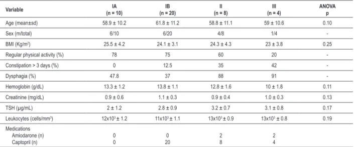
Table 1 contains the clinical observations regarding the analyzed groups. Of the 42 selected patients at the start of the
study, age ranged from 31 to 67 years, mean of 58.8 years, of which 17 were males and 25 were females. Body Mass Index (BMI) was 24.3 kg/m2.
Symptoms potentially related with esophagopathy occurred more frequently in groups II and III, with statistical significance (p < 0.001). As for symptoms potentially related to colopathy (constipation > 3 days), there was significant difference only in group IA (in which 100% of subjects showed no such symptoms) compared to groups II and III (Table 1).
Radiological evaluation showed that the cardiothoracic ratio increased with the degree of cardiac involvement (p = 0.0001). No x-rays of the esophagus and/or barium enema were performed. The echocardiogram showed an ejection fraction in patients from group III that was significantly lower than in other groups (p = 0.0001). The left ventricular end-diastolic diameter was corrected for body surface area and values > 32 mm/m2 were considered abnormal. Based on
this criterion, 12 patients had left ventricular dilatation. The right myocardial involvement expressed by the pulmonary hypertension was present in groups II and III (Table 2).
The response in the different groups was very similar after treatment with carvedilol. All groups showed significant decreases in the levels of GSH and PC, which were accompanied by a nonsignificant decrease in TBARS levels.
Vitamin E levels were also maintained, along with no changes in inflammatory markers, except NO and ADA levels, which showed a significant increase only in patients from group IA. Regarding the enzymatic profile, there was a generalized response of decreased activity in basically all groups. More specifically, there was a significant decrease in SOD activity in all studied groups, with significant decreases in groups IA and IB regarding the activities of GPx and GST. In turn, we observed the maintenance of GR activity and significant increases in CAT only in groups IA and IB. GSH
Table 1 - Biometric and demographic characteristics of the 42 chagasic patients
Variable (n = 10)IA (n = 20)IB (n = 8)II (n = 4)III ANOVAp
Age (mean±sd) 58.9 ± 10.2 61.8 ± 11.2 58.8 ± 11.1 59 ± 10.6 0.10
Sex (m/total) 6/10 6/20 4/8 1/4
-BMI (Kg/m2) 25.5 ± 4.2 24.1 ± 3.1 24.3 ± 4.3 23 ± 3.8 0.25
Regular physical activity (%) 78 75 60 20 -Constipation > 3 days (%) 0 12.5 35 42
-Dysphagia (%) 47.8 37 88 91
-Hemoglobin (g/dL) 13.3 ± 1.2 13.8 ± 1.1 12.8 ± 1.6 10 ± 1.8 0.11 Creatinine (mg/dL) 0.9 ± 0.6 1.1 ± 0.3 0.9 ± 0.4 1.0 ± 0.3 0.13 TSH (µg/mL) 2 ± 1.2 2.8 ± 0.9 3.2 ± 0.7 3.1 ± 0.8 0.17 Leukocytes (cells/mm3) 12x103 ± 1.2 11x103 ± 1.1 13x103 ± 0.9 13x103 ± 0.8 0.19
Medications Amiodarone (n) Captopril (n)
0 0
0 20
2 8
2 4
levels were significantly decreased in patients from IA group treated with carvedilol (0.18 ± 0.12 mmol mL-1) when
compared to untreated patients (0.31 ± 0.17 mmol mL-1, p < 0.05), while the levels of TBARS and vitamin E remained unchanged in both groups (Table 3).
However, levels of PC were significantly decreased in patients treated with carvedilol (0.04 ± 0.01 nmol mg-1)
when compared to untreated ones (0.15 ± 0.07 nmol mg-1,
p < 0.01). When compared within the same group IA, the activity of antioxidant enzymes such as SOD, GST and GPx showed significant decrease compared to those found in untreated patients, while CAT activity was increased and GR activity remained unchanged (Table 4). In relation to inflammatory markers after treatment with carvedilol, there was a significant increase in NO levels compared to untreated patients (10.93 ± 3.19 mM and 17.96 ± 3.2 mM, respectively; p < 0.05) and ADA activity (10.03 ± 1.28 Ul−1
and 17.17 ± 2.5 Ul−1, respectively; p < 0.05), while MPO
activity remained unchanged (Table 3).
Patients from Group IB treated with carvedilol showed a similar response when compared to patients in group IA. GSH levels (0.16 ± 0.13 mmol mL-1) were also significantly
decreased when compared to untreated patients (0.22 ± 0.17 mmol mL-1, p < 0.01), while TBARS and vitamin E levels
remained unchanged in both groups of patients (Table 3). Similarly as shown in the IA group of patients, PC values were also significantly reduced in patients treated with carvedilol (0.05 ± 0.02 nmol mg-1) compared to untreated
patients (0.16 ± 0.19 nmol mg-1, p <0.05). The profile of
antioxidant enzymes in this group was also similar to that found in group IA, while the inflammatory markers in this group remained unchanged (Tables 3 and 4).
In patients classified as II, the behavior of the analyzed parameters were also very similar to those in groups IA and IB (Table 3). GSH levels were significantly lower in patients treated with carvedilol compared to untreated ones (0.18 ± 0.11 and 0.29 ± 0.10 mmol mL-1, respectively, p < 0.05),
whereas there were no significant differences in levels of
Table 2 – Data concerning radiology, electrocardiography and echocardiogram results of chagasic patients
Variable IA
(n = 10)
IB (n = 20)
II (n = 8)
III (n = 4)
ANOVA p
Chest x-ray
- Cardiothoracic index (m±dp) -Compatible with CHF (%)
0.45 ± 0.02 0
0.45 ± 0.02 0
0.48 ± 0.04 20
0.52 ± 0.02 100
0.0001*
0.001♣
Electrocardiogram
RBBB (%) RBBB+LAHB (%) Inactive area (%)
0 0 0
55.7 68.9 2
62.3 72.1 55
97.8 99.1 89.3
0.001♥ 0.003♥ 0.001♥
Echocardiogram
LVEF (%) LVEDDi (mm/m2)
Pulmonary hypertension (%)
65.2 28.3 ± 1.8
0
61.6 29.2 ± 1.2
0
42.4 32.3 ± 4.8
25
37.6 35.9 ± 1.9
44
0.0001·● 0.001·● 0.001·● LAHB – left anterior hemiblock; LVEF – left ventricular ejection fraction; LVEDDi - Left Ventricular End-Diastolic Diameter Index; CHF - Congestive heart failure; RBBB – right bundle-branch block; Tukey (IA ¹ III)*·; (IA ¹ III)♣; (IB ¹ III)♥; (IA ¹ III) ●.
Table 3 – Comparison within the same group of levels of GSH, TBARS and CP, Vitamin E and levels of •NO, MPO and ADA in the blood of
chagasic patients at two different moments of treatment
Group IA (n = 10) Group IB (n = 20) Group II (n = 8) Group III (n = 4)
Before treatment
After 6 months of
treatment
Before treatment
After 6 months of treatment
Before treatment
After 6 months of treatment
Before treatment
After 6 months of treatment
GSH 0.31 ± 0.17 0.18 ± 0.12* 0.22 ± 0.17 0.16 ± 0.13** 0.29 ± 0.10 0.18 ± 0.11* 0.38 ± 0.15 0.16 ± 0.09* TBARS 13.11 ± 9.98 9.5 ± 4.28 10.02 ± 6.18 7.71 ± 1.17 11.34 ± 4.60 8.33 ± 1.55 15.19 ± 5.04 9.50 ± 2.22 CP 0.15 ± 0.07 0.04 ± 0.01** 0.16 ± 0.19 0.05 ± 0.02* 0.17 ± 0.07 0.05 ± 0.01*** 0.15 ± 0.06 0.05 ± 0.01* Vit. E 17.36 ± 8.11 12.44 ± 2.85 17.12 ± 8.93 15.70 ± 4.68 19.64 ± 9.25 12.24 ± 2.18 11.72 ± 3.40 11.18 ± 3.83
•NO 10.93 ± 3.19 17.96 ± 3.2 * 11.18 ± 1.38 16.07 ± 1.50 15.49 ± 3.42 18.86 ± 2.60 13.17 ± 4.62 12.10 ± 1.00 MPO 417.3 ± 40.1 544.2 ± 70.1 430.97 ± 31.53 420.7 ± 27.9 409.54 ± 80.95 352.13 ± 50.36 440.92 ± 68.15 395.08 ± 60.10 ADA 10.03 ± 1.28 17.17 ± 2.5 * 10.67 ± 1.08 15.36 ± 2.29 14.02 ± 2.27 12.90 ± 2.14 10.63 ± 3.52 9.05 ± 4.06
ADA - adenosine deaminase (UI-1); GSH - reduced glutathione (μmol mL−1); MPO - myeloperoxidase (mU mL−1);•NO - nitric oxide (mM ); CP - carbonyl protein
(nmol mg−1); TBARS - thiobarbituric acid reactive species (nmol mL−1); Vit - Vitamin E (μmol mL−1); Values represent mean ± SD, *p < 0,05; **p < 0.01;***p < 0.001
TBARS and vitamin E. PC values were also significantly decreased after treatment with carvedilol (0.05 ± 0.01 nmol mg-1) compared to untreated patients (0.17 ± 0.07 nmol
mg-1, p <0.001). After treatment with carvedilol, the values
of inflammatory markers remained unchanged (Table 3); the activities of SOD and GST enzymes decreased significantly, while the activities of the CAT, GPx and GR enzymes showed no significant differences when compared to those in untreated patients (Table 4).
As observed in the other groups, the group of patients classified as group III showed a decrease in GSH levels in patients treated with carvedilol (0.16 ± 0.09 mmol mL-1, p
< 0.05) when compared to untreated patients (0.38 ± 0.15 mmol mL-1) (Table 3). Also similar to values found in groups
IA, IB and II, there were no significant differences in the levels of TBARS and vitamin E, while the protein damage marker once more showed decreased values in patients treated with carvedilol (0.05 ± 0.01 nmol mg-1) when compared to
untreated patients (0.15 ± 0.06 nmol mg-1, p < 0.05) (Table 3).
The activities of CAT, GR, GST and GPx were not significantly different in relation to the treatment with carvedilol, but SOD activity was decreased among patients treated with carvedilol (69.20 ± 6.54 ml-1), when compared to untreated patients
(145.44 ± 44.12 mL USOD-1, p < 0.05). Similar to the results
obtained in groups IB and II, inflammatory markers remained unchanged after treatment with carvedilol (Table 3).
Discussion
In the present study, untreated patients had higher levels of biomarkers of damage to lipids (TBARS) and proteins (PC), with generally higher activity of antioxidant enzymes, which seem to represent the maintenance of the oxidative stress profile related to Chagas’ disease cardiac involvement.
Apparently, the group III, considered the most severe stage of heart failure and, thus, that in which the number of participating individuals was lower due to the high mortality rate in this phase, showed a higher oxidative stress in relation to groups characterized by lower severity, especially considering the groups IA, IB and to a lesser extent, also group
II. In a similar study by Keith et al. involving cardiac patients with varying degrees of involvement, markers of oxidative damage such as lipid peroxides and malondialdehyde, as well as antioxidant defenses such as levels of glutathione, vitamins E and C, and GPx activity also showed a direct correlation between these markers and disease severity28. This association has been
demonstrated by our laboratory in previous studies, as well as other related studies29.
According to the data shown in this study, it appears that patients with lower cardiac involvement and therefore with a higher antioxidant capacity (groups IA and IB) seem to have a greater capacity to neutralize the oxidative damage detected in their blood, a response that seems to be enhanced by treatment with carvedilol. A similar response has been detected by our laboratory in chagasic patients after six months of supplementation of vitamins E and C. In a study with mice infected with T. cruzi, the results were similar, where the activities of antioxidant enzymes CAT, GPx and GR, as well as GSH levels were increased in response to infection, while oxidative insult occurred in the myocardium of these animals30.
Conversely to what was found in most of the other antioxidant enzymes, an increase in CAT activity was detected in the blood of patients belonging to groups IA and IB. Chow et al. showed that rats with vitamin E deficiency had decreased CAT activity, while the activities of SOD and GPX were not changed, which are in accordance with the present study, suggesting that vitamin E in the diet provides protection against the CAT inactivation under such experimental conditions31. Considering that the beta-oxidation
of lipids in peroxisomes increases the production of H2O232, and
that vitamin E protects against CAT inactivation33, treatment with
carvedilol could be responsible for the increased activity of this enzyme in the blood of these patients.
Carvedilol protects against overproduction of ROS at concentrations of 0.1 to 100 mM, which are consistent with the dose (37.5 mg / day) used in this study34. Early studies with
this compound indicated that carvedilol is much more potent in inhibiting the production of hydroxyl radical (•OH) than other
beta-adrenergic antagonists35.
Studies have shown that hydroxylated metabolites are also potent antioxidants and may contribute to the overall
Table 4 – Comparison of antioxidant enzyme activity within the same group in the blood of chagasic patients at two different moments
of treatment
Group IA (n = 10) Group IB (n = 20) Group II (n = 8) Group III (n = 4)
Before treatment
After 6 months of treatment
Before treatment
After 6 months of treatment
Before treatment
After 6 months of treatment
Before treatment
After 6 months of treatment
SOD 144.99 ± 29.00 64.04 ± 6.05 *** 171.52 ± 41.16 66.37 ± 8.56 *** 141.26 ± 46.62 70.9 ± 11.2 ** 145.4 ± 44.1 69.20 ± 6.54 * CAT 8.87 ± 2.55 13.27 ± 3.88 * 9.21 ± 2.01 11.62 ± 4.10 * 8.43 ± 3.14 9.50 ± 4.06 7.54 ± 3.93 12.51 ± 6.43 GR 5.02 ± 0.71 4.78 ± 1.26 4.94 ± 1.43 4.86 ± 1.70 4.76 ± 1.16 4.00 ± 1.09 4.69 ± 0.81 4.73 ± 1.52 GST 30.61 ± 2.58 24.07 ± 3.68* 35.41 ± 9.42 24.58 ± 9.64 *** 34.10 ± 5.64 20.81 ± 2.9 *** 26.66 ± 7.51 23.28 ± 5.33 GPX 2.35 ± 0.22 1.48 ± 0.54 *** 2.32 ± 0.35 1.49 ± 0.39 *** 2.75 ± 0.73 2.36 ± 0.35 2.48 ± 0.17 2.14 ± 0.40
CAT - catalase (mmol min−1 mL−1); GPx - glutathione peroxidase (μmol min−1 mL−1); GR - glutathione reductase (μmol min−1 mL−1); GST - glutathione S-transferase
(μmol min−1 mL−1); SOD - superoxide dismutase (USOD mL−1); Values represent mean ± standard deviation *p < 0,05; **p < 0,01;***p < 0,001 represent signiicant
antioxidant action of this drug and may increase its power by 5-10 fold36,37. Several carvedilol metabolites found in human
plasma showed an even stronger antioxidant activity (50-80 times higher than carvedilol) to inhibit oxidation of LDL by macrophages38. Thus, it is possible that the in vivo antioxidant
activity of carvedilol is also attributed to its metabolites39.
Its antioxidant potential can also be linked to its ability to bind to Fe (III) and Cu (II), preventing the oxidation of lipids and proteins mediated by these transition metals, which could explain the decrease in biomarkers of lipid (TBARS) and protein (PC) damage in the present study, after treatment with carvedilol. When analyzing the marker of damage to proteins, carvedilol consistently reduced the formation of PC when compared to patients before treatment.
Except for the group classified as IA, with lower cardiac involvement, all inflammatory markers remained unchanged in the other groups. It is known that NO, in combination with the O●–
2 leads to the formation of peroxynitrite, a potent biological
oxidant that eliminates T. cruzi in a dose-dependent manner, and that MPO is an enzyme that participates in immune defense reactions through the formation of hypochlorous acid as a defense mechanism against pathogens40. That could explain the
increase found in the levels of NO and ADA in the group that has a better immune response against infection by the parasite. Furthermore, the cardioprotection of carvedilol may be related
not only to its antioxidant capacity, but also in preventing the infiltration of inflammatory cells in the ischemic myocardium, as well as inhibiting the vascular smooth muscle remodeling by decreasing the migration and proliferation of vascular smooth muscle cells, as shown in an animal model.
Conclusion
These data suggest that treatment with carvedilol was effective in attenuating the oxidative damage caused by Chagas’ disease itself, an effect that may be particularly important for patients with chronic Chagas’ disease and cardiomyopathy.
Potential Conflict of Interest
No potential conflict of interest relevant to this article was reported.
Sources of Funding
There were no external funding sources for this study.
Study Association
This article is part of the thesis of doctoral submitted by Patrícia Budni, from Universidade Federal de Santa Catarina.
References
1. Dias JCP. Doença de Chagas, ambiente, participação e estado. Cad Saúde Pública. 2001;17(supl):S165-9.
2. Duarte JD, Magalhães LP, Santana OO, Silva LB, Simões M, Azevedo DO, et al. Prevalence and prognostic value of ventricular dyssynchrony in chagas cardiomyopathy. Arq Bras Cardiol. 2011;96(4):300-6.
3. Dias JC. The indeterminate form of human chronic Chagas’ disease: a clinical epidemiological review. Rev Soc Bras Med Trop. 1989;22(3):147-56.
4. Pinto Dias JC. The treatment of Chagas’ disease (South American trypanosomiasis). Ann Intern Med. 2006;144(10):772-4.
5. Organización Panamericana de la Salud (OPAS). Estimación cuantitativa de la enfermedad de Chagas en las Américas. Montevideo (Uruguay): PAHO Publishing; 2006. p. 1-28.
6. Vilas-Boas F, Feitosa GS, Soares MB, Pinho-Filho JA, Mota AC, Almeida AJ, et al. Bone marrow cell transplantation in Chagas’ disease heart failure: report of the first human experience. Arq Bras Cardiol. 2011;96(4):325-31.
7 . de Oliveira TB, Pedrosa RC, Wilhelm- Filho D. Oxidative stress in chronic cardiopathy associated with Chagas disease. Int J Cardiol. 2007;116(3):357-63.
8. Maçao LB, Wilhelm-Filho D, Pedrosa RC, Pereira A, Backes P, Torres MA, et al. Antioxidant therapy attenuates oxidative stress in chronic cardiopathy associated with Chagas’ disease. Int J Cardiol. 2007;123(1):43-9.
9. Feuerstein GZ, Ruffolo RR Jr. Carvedilol, a novel multiple action antihypertensive agent with antioxidant activity and the potential for myocardial and vascular protection. Eur Heart J. 1995;16(Suppl F):38-42.
10. Packer M, Bristow MR, Cohn JN, Colucci WS, Fowler MB, Gilbert EM, et al. The effect of carvedilol on morbidity and mortality in patients with chronic heart failure. U.S. Carvedilol Heart Failure Study Group. N Engl J Med. 1996;334(21):1349-55.
11. Yue TL, McKenna PJ, Lysko PG, Ruffolo RR Jr, Feuerstein GZ. Carvedilol, a new antihypertensive, prevents oxidation of human low density lipoprotein by macrophages and copper. Atherosclerosis. 1992;97(2-3):209-16.
12. Donetti E, Soma MR, Barberi L, Paoletti R, Fumagalli R, Roma P, et al. Dual effects of the antioxidant agents probucol and carvedilol on proliferative and fatty lesions in hypercholesterolemic rabbits. Atherosclerosis. 1998;141(1):45-51.
13. Ferrari R, Agnoletti L, Ceconi C, Curello S, Nesta F, Manfredini R. Endothelial dysfunction in congestive heart failure: effects of carvedilol. Arq Bras Cardiol. 1999;4(1):53-63.
14. Bern C, Montgomery SP, Herwaldt BL, Rassi A Jr, Marin-Neto JA, Dantas RO, et al. Evaluation and treatment of Chagas disease in the United States: a systematic review. JAMA. 2007;298(18):2171-81.
15. Aebi H. Catalase in vitro. Methods Enzymol. 1984;105:121-6.
16. Misra HP, Fridovich I. The role of superoxide anion in the autoxidation of epinephrine and a simple assay for superoxide dismutase. J Biol Chem. 1972;247(10):3170-5.
17. Calberg I, Mannervik B. Glutathione reductase. Methods Enzymol. 1985;113:484-90.
18. Flohé L, Gunzler WA. Assays of glutathione peroxidase. Methods Enzymol. 1984;105:114-21.
19. Habig WH, Pabst MJ, Jacoby WB. Glutathione S-transferases: the first enzymatic step in mercapturic acid formation. J Biol Chem. 1976;249(22):7130-9.
21. Levine RL, Garland D, Oliver CN, Amici A, Climent I, Lenz AG, et al. Determination of carbonyl content in oxidatively modified proteins. Methods Enzymol. 1990;186:464-78.
22. Bird RP, Draper AH. Comparative studies on different methods of malondyhaldehyde determination. Methods Enzymol. 1984;90:299-305.
23. Nicoletti G, Crescibene L, Scornaienchi M, Bastone L, Bagalà A, Napoli ID, et al. Plasma levels of vitamin E in Parkinson’s disease. Arch Gerontol Geriatr. 2001;33(1):7-12.
24. Fröde-Saleh TS, Calixto JB, Medeiros YS. Analysis of the inflammatory response induced by substance P in the mouse pleural cavity. Peptides. 1999;20(2):259-65.
25. Rao TS, Currie JL, Shaffer AL, Isakson PC. Comparative evaluation of arachidonic acid (AA)- and tetradecanoylphorbol acetate (TPA)-induced dermal inflammation. Inflammation. 1993;17(6):723-41.
26. Fröde TS, Medeiros YS. Myeloperoxidase and adenosine deaminase levels in the pleural fluid leakage induced by carrageenan in the mouse model of pleurisy. Mediators Inflamm. 2001;10(4):223-7.
27. Giusti G, Galanti B. Colorimetric method: adenosine deaminase: In: Bergmeyer HU. (ed.). Methods enzymatic analysis. 3rd ed. Weinheim: Verlag
Chemie; 1984. p. 315-23.
28. Keith M, Geranmayegan A, Sole MJ, Kurian R, Robinson A, Omran AS, et al. Increased oxidative stress in patients with congestive heart failure. J Am Coll Cardiol. 1998;31(6):1352-6.
29. Péres-Fuentes R, Guegan JF, Barnabé C, López-Colombo A, Salgado-Rosas H, Torres-Rasgado E, et al. Severity of chronic Chagas disease is associated with cytokine/antioxidant imbalance in chronically infected individuals. Int J Parasitol. 2003;33(3):293-9.
30. Wen JJ, Vyatkina G, Garg N.Oxidative damage during chagasic cardiomyopathy development: role of mitochondrial oxidant release and inefficient antioxidant defense. Free Radic Biol Med. 2004;37(11):1821-33.
31. Chow CK. Glucose and dietary vitamin E protection against catalase inactivation in 651 the red cells of rats. Int J Vitam Nutr Res. 1980;50(4):364-9.
32. Chow CK. Oxidative damage in the red cells of vitamin E-deficient rats. Free Radic Res Commun. 1992;16(4):247-58.
33. Tardif JC, Gregoire J, Schwartz L, Title L, Laramée L, Reeves F, et al. Effects of AGI-1067 and probucol after percutaneous coronary interventions. Circulation. 2003;107(4):552-8.
34. Ruffolo RR Jr, Feuerstein GZ. Pharmacology of carvedilol: rationale for use in hypertension, coronary artery disease, and congestive heart failure. Cardiovasc Drugs Ther. 1997;11(Suppl 1):247-56.
35. Feuerstein GZ, Ruffolo RR Jr. Carvedilol, a novel vasodilating beta-blocker with the potential for cardiovascular organ protection. Eur Heart J. 1996;17(Suppl B):24-9.
36. Yue TL, McKenna PJ, Lysko PG, Gu JL, Lysko KA, Ruffolo RR Jr, et al. SB 211475, a metabolite of carvedilol, a novel antihypertensive agent, is a potent antioxidant. Eur J Pharmacol. 1994;251(2-3):237-43.
37. Yue TL, Wang X, Gu JL, Ruffolo RR Jr, Feuerstein GZ. Carvedilol, a new vasodilating beta-adrenoceptor blocker, inhibits oxidation of low-density lipoproteins by vascular smooth muscle cells and prevents leukocyte adhesion to smooth muscle cells. J Pharmacol Exp Ther. 1995;273(3):1442-9.
38. Lopez BL, Christopher TA, Yue TL, Ruffolo R, Feuerstein GZ, Ma XL. Carvedilol, a new beta-adrenoreceptor blocker antihypertensive drug, protects against free-radical-induced endothelial dysfunction. Pharmacology. 1995;51(3):165-73.
39. Denicola A, Rubbo H, Rodriguez D, Radi R. Peroxynitrite-mediated c y t o t o x i c i t y t o Tr y p a n o s s o m a c r u z i . A r c h B i o c h e m B i o p h y s . 1993;304(1):279-86.