Brazilian Journal of Microbiology (2010) 41: 133-145 ISSN 1517-8382
MODE OF ACTION AND IN VITRO SUSCEPTIBILITY OF MASTITIS PATHOGENS TO MACEDOCIN ST91KM AND PREPARATION OF A TEAT SEAL CONTAINING THE BACTERIOCIN
Renee Pieterse, Svetoslav D. Todorov*, Leon M.T. Dicks
Department of Microbiology, University of Stellenbosch, 7600 Stellenbosch, South Africa
Submitted: November 26, 2008; Returned to authors for corrections: April 20, 2009; Approved: July 22, 2009.
ABSTRACT
Mastitis is considered to be the most economically costly disease affecting the dairy industry. Regular
dosage of animals with antibiotics, including use of prophylactic concentrations, may select for resistant
strains. The purpose of this study was to determine the mode of action of a new bacteriocin (macedocin
ST91KM), to evaluate the antimicrobial resistance of mastitis pathogens to antibiotics commonly used in
treatment remedies, and to introduce the possible use of an alternative antimicrobial agent. The bacteriocin
macedocin ST91KM, produced by Streptococcus gallolyticus subsp. macedonicus ST91KM, is bactericidal
to Streptococcus agalactiae, Streptococcus dysgalactiae, Streptococcus uberis and Staphylococcus aureus
associated with mastitis infections, including strains resistant to methicillin and oxacillin. Sensitive cells
were deformed and secreted nucleotides, K+ and -galactosidase when exposed to macedocin ST91KM.
Adsorption of the peptide to target cells decreased in the presence of solvents, suggesting that receptors on
the cell surfaces have lipid moieties. No adsorption was recorded in the presence of MgCl2, KI and
Na2CO3, suggesting that ionic strength plays an important role. A teat seal preparation containing
macedocin ST91KM effectively released the peptide and inhibited the growth of S. agalactiae. Macedocin
ST91KM could form the basis for alternative dry cow therapy to prevent mastitis infections in dairy cows
as it is effective against pathogens that display resistance to conventional antibiotic therapy.
Key words: Macedocin ST91KM; activity against mastitis pathogens; teat seal preparation; milk
INTRODUCTION
Mastitis concerns cows is primarily caused by infection
with Staphylococcus aureus, Streptococcus agalactiae,
Streptococcus uberis, Streptococcus dysgalactiae and
Escherichia coli (25). After entering the mammary gland
through the teat canal, the bacteria multiply rapidly, leading to
inflammation and tissue damage (9). Antibiotics are routinely
administered at drying-off to treat sub clinical cases of mastitis
and prevent further infection (4,37). Although fairly successful,
this practice may lead to the development of antibiotic resistant
strains, as reported for coagulase-negative staphylococci, S.
aureus and streptococci (6,8,20,28). If any of these strains are
present in a dairy product, it may lead to transfer of antibiotic
resistance genes to normal intestinal microorganisms
(14,15,16,17).
Pieterse, R. et al.
Bacteriocins are ribosomally synthesized peptides, usually
active against bacteria of the same or closely related species
(12). These peptides produced by lactic acid bacteria are
generally considered safe and may present a cost-effective
alternative to treat mastitis caused by S. aureus, S. agalactiae
and S. dysgalactiae. In a study which involved the
incorporation of the bacteriocin lacticin 3147 into a teat seal
(1280AU/mL), 99.9 % of S. aureus cells were killed (30).
Peptide AS-48, produced by Enterococcus faecalis FAIRE 92,
inhibited the growth of a S. aureus strain isolated from mastitic
milk (5). Treatment of S. aureus-infected udders with
liposome-encapsulated peptide AS-48 (6400AU/mL) resulted
in an 85% reduction in somatic cell counts and a 99%
reduction in viable cell numbers of S. aureus (5).
Streptococcus gallolyticus subsp. macedonicus ST91KM,
isolated from Bulgarian goat yoghurt, produces the bacteriocin
macedocin ST91KM (26). The peptide has a narrow spectrum
of antibacterial activity and inhibits the growth of
Lactobacillus sakei, S. agalactiae, S. dysgalactiae,
Streptococcus uberis, S. aureus and Staphylococcus
epidermidis. The peptide is approximately 2.0–2.5 kDa in size
and remained stable after incubation for 2h at pH2.0 to 10.0.
No decrease in activity was recorded after treatment at 100°C
for 100min, but the peptide was inactivated at 121°C for
20min. No change in activity was recorded after treatment
with SDS, urea, Tween 20, Tween 80 and EDTA. Treatment
with pronase, pepsin and trypsin inactivated the bacteriocin.
As far as we could determine, only one other bacteriocin
has been described for S. macedonicus, i.e. the lantibiotic
macedocin produced by S. macedonicus ACA-DC 198 (7).
In this study, parameters affecting the adsorption and some
aspects of the mode of action of macedocin ST91KM to target
organisms were evaluated. Minimum inhibitory concentration
(MIC) of macedocin ST91KM was determined and compared
to that of -lactam penicillins and erythromycin. Macedocin
ST91KM was incorporated in a teat seal preparation and its
activity against S. agalactiae studied in vitro.
MATERIALS AND METHODS
Growth conditions and preparation of macedocin ST91KM
Streptococcus gallolyticus subsp. macedonicus ST91KM
was grown in MRS broth (Biolab, Biolab Diagnostics,
Midrand, SA) at 30°C. The target strains used in the study
were grown either in MRS broth or BHI broth (Merck,
Darmstadt, Germany), at temperatures indicated in the
respective culture collection catalogues. S. agalactiae
RPSAG2, isolated from a clinical mastitis case, was obtained
from the Western Cape Provincial Veterinary Laboratory
(Stellenbosch, South Africa) and was cultured in BHI broth at
37°C. All strains were stored at -80°C in MRS or BHI
supplemented with 40% (v/v) glycerol.
Macedocin ST91KM was prepared as follows: Strain
ST91KM was inoculated (2%, v/v) into 100mL MRS and
incubated for 17h at 30°C. The cells were harvested (1000xg,
10min, 4°C), the pH of the cell-free culture supernatant
adjusted to 6.0 with 1M NaOH, and then heated for 10min at
80°C to inactivate proteolytic enzymes. The supernatant was
filter-sterilised (0.20µm pore size membrane, Minisart®,
Sartorius, Aubagne, France) and the activity of macedocin
ST91KM determined by using the agar-spot method (36).
Active samples of macedocin ST91KM were stored at 4ºC.
Antimicrobial activity assays
Overnight cultures of the target strains (OD600nm=0.1–0.2),
listed in Table 1, were inoculated (0.1%, v/v) into 10mL MRS
or BHI soft agar (0.7%, w/v, agar), poured into sterile Petri
dishes and allowed to solidify. A dilution series of macedocin
ST91KM was prepared and 10µL spotted onto the surface of
the solid media. Antimicrobial activity was expressed in
arbitrary units (AU)/mL, calculated as follows: abx100, where
“a” represents the dilution factor 2 and “b” the last dilution that
produces an inhibition zone of at least 2 mm in diameter.
Macedocin ST91KM against mastitis pathogens
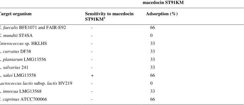
Table 1. Activity spectrum of macedocin ST91KM and adsorption to target cells
macedocin ST91KM
Target organism Sensitivity to macedocin
ST91KMa
Adsorption (%)
E. faecalis BFE1071 and FAIR-E92 - 66
E. mundtii ST4SA - 0
Enterococcus sp. HKLHS - 33
L. curvatus DF38 - 33
L. plantarum LMG13556 - 33
L. salvarius 241 - 33
L. sakei LMG13558 + 66
Lactococcus lactis subsp. lactis HV219 - 0
L. innocua LMG13568 - 33
S. caprinus ATCC700066 - 66
a - = not inhibited, and + = inhibited by macedocin ST91KM
Effect of macedocin ST91KM on cell growth and membrane permeability
S. agalactiae RPSAG2 was grown in 50mL BHI broth
(Merck) for 5h at 37ºC to an optical density of 0.17. Twenty
mL macedocin ST91KM was added to the culture, representing
a final macedocin ST91KM concentration of 230AU/mL.
Optical density readings were recorded every hour for 10h.
The control was S. agalactiae RPSAG2 cultured in the same
way, but to which 20mL sterile distilled water was added. The
experiment was repeated in triplicate.
In a separate experiment, an overnight culture of L. sakei
LMG13558 (OD600nm=2.2) was harvested (8000xg, 15min,
25°C), the cells washed twice with 2.0mL 5mM phosphate
buffer (pH 6.5), and then incubated in the presence of
macedocin ST91KM (0.1:10 ratio). After 1h at 37°C, the cells
were harvested (10000xg, 15min, 4°C) and the supernatant
filtered through a 0.20 µm pore size filter membrane
(Ministart®, Sartorius). The presence of nuclear material was
recorded by absorbance readings at 260nm (SmartSpec™ Plus
Spectrophotometer, Bio-Rad Laboratories, Hecules, CA USA).
The presence of K+ in the filtrate was determined by atomic
absorption spectrometry (Avanta , GBC Scientific
Equipment, Victoria, Australia). Controls were cells prepared
the same way, but not treated with macedocin ST91KM.
In another experiment, extracellular levels of
-galactosidase activity were determined according to the
methods of Nagy et al. (22) and Hsu et al. (10). Cells of L.
sakei LMG13558 were harvested (10000xg, 15min, 25°C) after
11h incubation at 30°C, washed twice with 0.03M sodium
phosphate buffer (pH 6.8) and then re-suspended in 2.0mL of
the same buffer. The cell suspension was treated with 2mL
macedocin ST91KM (800AU/mL) for 5min at 25°C, followed
by the addition of 0.2mL 0.1M ONPG (O-nitrophenyl-
-D-galactopyranoside) in 0.03M sodium phosphate buffer (pH
6.8). After 10min incubation at 37°C for 10min, 2.0mL 0.1M
sodium carbonate was added to stop the reaction of
-galactosidase. The cells were harvested (10000xg, 15min,
25°C) and absorbance readings of the cell-free culture
supernatant recorded at 420nm, using a spectrophotometer
(SmartSpec™ Plus Spectrophotometer). Cells of L. sakei
LMG 13558 treated with sterile water instead of macedocin
ST91KM served as control. All experiments were performed
Pieterse, R. et al.
Adsorption of macedocin ST91KM to target cells
Adsorption to target cells (Table 1) was tested according
to Yildrim et al. (38). The bacterial strains were grown
overnight in MRS or BHI broth at 37°C to OD600nm=0.1-0.2
and the cells harvested (10000xg, 15min, 4°C). Cells were
washed twice in 5mM phosphate buffer (pH 6.5) and
re-suspended in the same buffer to the original volume. Each cell
suspension (0.7mL) was mixed with an equal volume of
macedocin ST91KM and incubated at 37°C for 1h. The cells
were harvested (10000xg for 15min) and activity of unbound
macedocin ST91KM in the cell-free supernatant determined
using the agar-spot method as described before. Adsorption of
macedocin ST91KM to the target cells was calculated
according to the following formula: % Adsorption = 100 –
[(macedocin ST91KM activity after treatment/original
macedocin ST91KM activity) x 100]. The experiment was
performed in triplicate.
Effect of pH and temperature on adsorption of macedocin ST91KM to L. sakei
Cells from an overnight culture of L. sakei LMG 13558
(OD600nm = 2.2) were harvested and re-suspended in original
volume of physiological water and the pH corrected to values
between 2.0 and 10.0 with sterile 1M NaOH or 1M HCl (36).
Macedocin ST91KM was added to the cell suspension and
incubated at 4, 10, 25, 30, 37, 45 and 60°C for 1h. The cells
were harvested (10000xg, 15min, 25°C) and the pH of the
supernatants adjusted to 6.0 with sterile 1M NaOH.
Macedocin ST91KM activity was determined as described
before. All experiments were performed in triplicate.
Effect of surfactants, inorganic salts and organic compounds on the adsorption of macedocin ST91KM to L. sakei
Cell suspensions of L. sakei LMG 13558 were prepared as
described before and were treated with 1% (w/v) NaCl,
K2HPO4, KH2PO4, MgCl2, KCl, KI, Tris, (NH4)3C6H5O7,
CH3COONa, Na2CO3, EDTA (C10H16O8N2), SDS and 1% (v/v)
Triton X-100, Triton X-114, -mercaptoethanol, and 80% (v/v)
ethanol, methanol and chloroform, respectively (36). The pH
of each suspension was adjusted to 6.5 with 1M NaOH or 1M
HCl. Macedocin ST91KM was added to the treated cells, as
before, and incubated at 37°C for 1h. The cells were harvested
(10000xg, 15min, 25°C) and activity of the cell-free
supernatant determined as before. The experiment was
performed in triplicate.
Effect of macedocin ST91KM on cell morphology
Strain ST91KM was cultured in MRS broth for 15h at
30°C. The cells were harvested (8000xg, 10min, 4°C) and the
bacteriocin precipitated from the cell-free culture supernatant
with 60% saturated ammonium sulphate (31). The precipitate
was suspended in 25mM ammonium acetate buffer (pH 6.5) to
one-tenth of the original volume and loaded onto a SepPak C18
cartridge (Water Millipore, MA, USA). Peptides were eluted
with 20%, 40%, 60% and 80% (v/v) isopropanol, respectively,
in 25mM ammonium acetate buffer (pH 6.5) according
Todorov et al. (35). Fractions of 1mL were dried under
vacuum at 50°C and stored at -20°C until required.
Cells from an overnight culture (OD600nm=0.16) of S.
agalactiae RPSAG2 were harvested (8000xg, 10min, 4°C) and
washed five times with sterile distilled water. The dried
bacteriocin fractions of macedocin ST91KM were reconstituted
and concentrated with milliQ water, filter-sterilised (0.20µ m
pore size membrane filter, Minisart®, Sartorius Aubagne,
France) and incubated with the prepared cells overnight at 8°C
(approximately 2000AU/mL). The cells were then washed five
times with sterile distilled water and re-suspended in 1mL
sterile distilled water. Untreated cells of S. agalactiae
RPSAG2 served as control. Images of cell surface areas of
treated and untreated cells were visualised by atomic force
microscopy (Multimode AFM from Veeco, Santa Barbara
USA). The cell suspension was applied onto freshly cleaved
mica surface and allowed to dry for 5min before subjected to
AFM. All images were obtained in air and with tapping mode.
A silicone non-contact cantilever from Nansensors (Neuchatel,
Switzerland) with a resonance frequency of 160 kHz and a
spring constant of approximately 50N/m was used. Height and
size information was acquired by using imaging software from
Macedocin ST91KM against mastitis pathogens
Antimicrobial agents
Peptide ST91KM, eluted from a SepPak C18 cartridge
with 40% (v/v) isopropanol, as desribed before, were used for
antimicrobial activity tests. The antibiotics used in this study
were selected based on their approval and frequent use in the
treatment of mastitis in South Africa. The antimicrobial agents
selected included -lactam penicillins: Penicillin G (PEN)
(USB, Amersham Life Science), ampicillin (AMP) (Roche
Diagnostics, Mannhein, Germany), oxacillin (OX)
(Sigma-Aldrich, St. Louis, USA), methicillin (MET) (Sigma-(Sigma-Aldrich,
St. Louis, USA) and a macrolide antibiotic, erythromycin (EM)
(Sigma-Aldrich, St. Louis, USA). The antimicrobial agents
were diluted in sterile deionised water to prepare stock
solutions, each containing 1280µg/mL of the active antibiotic.
The final concentration range used for antimicrobial activity
tests for each antibiotic was 32.0, 16.0, 8.0, 4.0, 2.0, 1.0, 0.5,
0.25 and 0.12µg/mL.
Antimicrobial activity tests
Minimum inhibitory concentration (MIC) tests were
performed using the microdilution broth method, with
U-bottomed 96-well microtitre plates, recommended by the
Clinical and Laboratory Standards Institute (CLSI) (23). The
antimicrobial agents are listed in Table 4. Serial two-fold
dilutions of each antimicrobial agent were prepared in
cation-adjusted Mueller-Hinton broth (CAMHB) (Oxoid) and 50µL
pipetted into the wells of sterile microtitre plates. A positive
control well contained 50µL CAMHB, without an
antimicrobial agent. Inoculum for each test organism (listed in
Table 4) was prepared as follows: Three to five colonies were
selected from a 24-h-old agar plate culture and suspended in
2mL sterile 0.85% saline. The turbidity was adjusted to 0.5
according to the MacFarland standard. Ten µL of this
suspension was transferred to 10mL CAMHB to obtain a
bacterial suspension of approximately 5x10⁵CFU/mL. From
this suspension, 50µL was pipetted into each well. The
microtitre plates were sealed and incubated for 20h at 37°C.
The MIC was defined as the lowest concentration of the
antimicrobial agent at which the bacterial growth was
completed inhibited as detected by the unaided eye. MIC
breakpoints were used to determine resistance and
susceptibility for each antibiotic tested. The exact
concentration of peptide ST91KM in the bacteriocin
preparations was unknown, therefore the antimicrobial activity
of the peptide was expressed as arbitrary units per millilitre
(AU/mL), calculated as follows: abx20, where “a” is equal to 2,
and “b” the lowest dilution of the antimicrobial agent that
prevented visible growth of the microorganisms tested.
Activity was expressed per millilitre by multiplication with 20.
The protein concentration, expressed as mg/mL, in the
bacteriocin preparations was assayed using the Bradford
method. The final minimum inhibitory concentration of
macedocin ST91KM was expressed as AU/mL protein.
Teat seal formulation with macedocin ST91KM
Macedocin ST91KM, precipitated from culture
supernatant with ammonium sulphate, as described before, was
suspended in 25mM ammonium acetate buffer (pH 6.5) to
one-tenth of its original volume. The teat seal consisted of 65%
(w/w) bismuth subnitrate in a paraffin base, similar in
composition to that of commercially available intramammary
teat sealers such as Orbeseal (Pfizer Animal Health). A second
formulation containing 1% (w/w) Tween 80 in the teat seal
paste was also prepared. Macedocin ST91KM (100µL) was
added to 1g of the teat seal paste with or without 1% (w/w)
Tween 80 and mixed to form an emulsion. The teat seal
formulations were kept in 5mL sterile syringes at 4°C.
In vitro effect of macedocin ST91KM combined with the teat seal against S. agalactiae
The bacteriocin activity of macedocin ST91KM was
determined using the agar well diffusion method. An overnight
culture of S. agalactiae RPSAG2 was diluted by inoculating
10µL in 10mL sterile physiological saline. One mL was
inoculated into 9mL semi-solid (0.7%, w/v) BHI agar (BHI
Pieterse, R. et al.
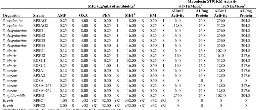
Table 4. Minimum inhibitory concentration (MIC, expressed as µg/mL) of selected antibiotics and activity of macedocin ST91KM (expressed as AU/mg protein) against mastitis-causing pathogens
Macedocin ST91KM Activity
MIC (µg/mL) of antibioticsa ST91KMppt.c ST91KMcond
Organism Strain AMP OXA PEN METb EM
AU/mL Activity
AU/mg Protein
AU/mL Activity
AU/mg Protein
S. agalactiae RPSAG1 0.25 S 4.00 R 0.50 I 8.00 R 0.50 I 640 76.8 2560 204.8
S. agalactiae RPSAG2 0.25 S 4.00 R 0.25 I 16.00 R 0.25 S 1280 76.8 5120 204.8
S. dysgalactiae RPSD1 0.25 S 8.00 R 0.25 I 8.00 R 0.25 S 640 76.8 2560 204.8
S. dysgalactiae RPSD2 0.25 S 4.00 R 0.25 I 16.00 R 0.25 S 640 76.8 2560 204.8
S. dysgalactiae RPSD3 0.25 S 4.00 R 0.25 I 8.00 R 0.25 S 640 76.8 2560 204.8
S. dysgalactiae RPSD4 0.25 S 4.00 R 0.50 I 16.00 R 0.50 I 640 76.8 2560 204.8
S. uberis RPSU1 0.12 S 8.00 R 0.25 I 16.00 R 0.25 S 640 76.8 10240 204.8
S. uberis RPSU2 0.25 S 8.00 R 0.25 I 16.00 R 0.25 S 160 75.2 640 217.6
S. uberis EDSU1 0.12 S 8.00 R 0.25 I 32.00 R 0.25 S 640 76.8 5120 204.8
S. uberis EDSU2 0.25 S 8.00 R 1.00 I 16.00 R 0.50 I 160 75.2 1280 217.6
S. aureus RPSA1 0.12 S 8.00 R 0.50 R 16.00 R 0.50 S 640 76.8 1280 217.6
S. aureus RPSA2 0.25 S 8.00 R 0.50 R 16.00 R 0.50 S 640 76.8 1280 217.6
S. aureus EDSA 0.25 S 4.00 R 0.50 R 16.00 R 0.50 S 0 0 0 0
S. aureus EDSA0267 0.25 S 8.00 R 8.00 R 16.00 R 0.25 S 640 76.8 1280 217.6
S. aureus EDSA0269 0.12 S 8.00 R 0.50 R 16.00 R 0.50 S 640 76.8 1280 217.6
S. epidermidis RPSE1 0.25 S 16.00 R 0.50 R 16.00 R 0.25 S 2560 76.8 10240 204.8
E. coli RPEC1 1.00 S >32 (R) 32.00 (R) >32.00 (R) >32 (R) 0 0 0 0
E. coli RPEC2 2.00 S >32 (R) 32.00 (R) >32.00 (R) >32 (R) 0 0 0 0
aampicillin (AMP), oxacillin (OX), penicillin G (PEN), methicillin (MET), erythromycin (EM);
sensitive (S), intermediate (I), resistant (R);
b MET interpretative data based on OX
No veterinary-specific interpretative criteria available for E.coli for OX, PEN, MET and EM, breakpoint interpretation is thus given in parentheses.
c
macedocin ST91KM precipitated in 60% saturated ammonium sulphate,
d macedocin ST91KM precipitated in 60% saturated ammonium sulphate, eluted with 40% (v/v) isopropanol, in 25mM ammonium acetate buffer (pH 6.5). Fractions of 1mL were dried under vacuum at 50°C,
Macedocin ST91KM against mastitis pathogens
into the agar. Teat seal preparations combined with bacteriocin
ST91KM, with and without 1% (w/w) Tween 80, were
carefully dispensed into the wells to ensure that the seal was in
contact with the wall of the wells. The agar plates were
incubated at 37°C overnight for 16h and the zones of inhibition
measured.
RESULTS
Addition of macedocin ST91KM (229AU/mL) to S.
agalactiae RPSAG2 after 5h of growth, decreased the cell
density from OD600nm=0.16 to 0.10 within 1h and remained at
this level for a further 4h (Fig. 1). Optical density readings of
the control culture (no bacteriocin added) increased to
OD600nm=1.21 over 10h (Fig. 1).
Optical density readings measured at 260nm and 420nm
are presented in Table 2. Potassium levels of cells treated with
macedocin ST91KM was 534mg K+/L compared to 509mg
K+/L recorded for untreated cells.
0,0 0,2 0,4 0,6 0,8 1,0 1,2 1,4
0 1 2 3 4 5 6 7 8 9 10
O . D . (6
0 0 n m )
Time (h)
Control (no bacteriocin added) Macedocin ST91K M
bacteriocin
Figure 1. The effect of macedocin ST91KM on S. agalactiae RPSAG2. The arrow indicates the point at which macedocin ST91KM was added.
Table 2. Effect of macedocin ST91KM on the permeability of L. sakei LMG13558 cells
nucleotides (O.D. 260nm)
-galactosidase (O.D. 420nm)
Potassium ions (mg/L)
Treated cells 2.979 1.265 534
Untreated cells 0.624 0.116 509
Macedocin ST91KM, no
cells 0.581 0.631 8
Macedocin ST91KM adsorbed to both sensitive and
non-sensitive cells (Table 1). Sixty-six percent of the peptide
adsorbed to L. sakei LMG13558. However, 66% of the peptide
also adsorbed to non-sensitive strains of Enterococcus faecalis
(BFE1071 and FAIR-E92) and Streptococcus caprinus
(ATCC700066). Limited, or very little adsorption (up to 33%)
of macedocin ST91KM has been recorded for the other
resistant strains (Table 1).
Treatment of cells with macedocin ST91KM at 10–60°C
and at pH8 and 10 lead to a significant increase in adsorption
(Table 3). Adsorption of the peptide to target cells decreased
from 66 to 33% below 37°C and was completely inhibited at
Pieterse, R. et al.
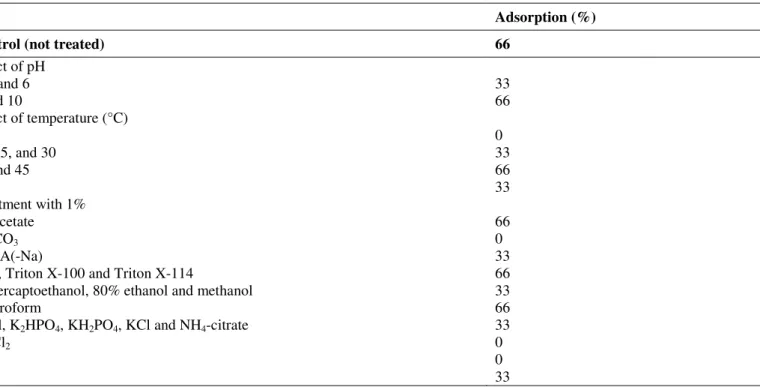
Table 3. Effect of pH, temperature, surfactants and salts on the adsorption of macedocin ST91KM to L. sakei LMG 13558
Adsorption (%)
Control (not treated) 66
Effect of pH
2, 4 and 6 33
8 and 10 66
Effect of temperature (°C)
4 0
10, 25, and 30 33
37 and 45 66
60 33
Treatment with 1%
Na-acetate 66
Na2CO3 0
EDTA(-Na) 33
SDS, Triton X-100 and Triton X-114 66
-mercaptoethanol, 80% ethanol and methanol 33
Chloroform 66
NaCl, K2HPO4, KH2PO4, KCl and NH4-citrate 33
MgCl2 0
KI 0
Tris 33
Addition of inorganic and organic salts reduced adsorption
of the bacteriocin. MgCl2, KI and Na2CO3 salts completely
prevented adsorption of the peptide to target cells (Table 3).
SDS, Triton X-100 and Triton X-114 did not affect adsorption
of macedocin ST91KM to target cells, while
-mercaptoethanol, 80% ethanol and methanol reduced
adsorption to 33%. Chloroform had no effect on adsorption.
Cells of S. agalactiae RPSAG2 treated with partially
purified macedocin ST91KM (approximately 2000AU/mL)
were deformed and had irregular surfaces compared to cells
that have not been treated (Fig. 2).
(A) Untreated cells (B) Cells treated with macedocin ST91KM
Figure 2. Changes in cell morphology of S. agalactiae RPSAG2 treated with macedocin ST91KM. A: before treatment, B: after
Macedocin ST91KM against mastitis pathogens
Susceptibility of pathogens to antibiotics is shown in
Table 4. Data were interpreted based on veterinary-specific
criteria, except for oxacillin against streptococci, which was
interpreted based on data obtained from trials on humans (24).
All isolates were resistant to methicillin and oxacillin, but
susceptible to ampicillin (Table 4). Streptococci showed
intermediate resistance to penicillin G. Three of the ten
streptococci tested had intermediate MICs against
erythromycin. S. agalactiae RPSAG1, S. dysgalactiae RPSD4
and S. uberis EDSU2 displayed the highest MICs against
penicillin G. Strains of S. aureus and coagulase-negative
staphylococci (CNS), S. epidermidis, were resistant to
penicillin G. The staphylococci were all susceptible to
erythromycin. The only Gram-negative mastitis pathogens
tested was E. coli. Both isolates were resistant to oxacillin,
methicillin and erythromycin. The strains of E. coli tested
were susceptible to ampicillin.
Antimicrobial susceptibility to the macedocin ST91KM is
shown in Table 4. Two preparations of macedocin ST91KM
were used: ST91KM precipitate (ST91KMppt) and ST91KM
concentrate (ST91KMcon). The minimum concentration of
macedocin ST91KM required to completely inhibit visible
growth of the sensitive strains was 74.8AU/mg protein for
ST91KMppt and 216AU/mg protein for ST91KMcon. The
purification and concentration of macedocin ST91KM in
ST91KMcon resulted in an average increase of 63.5% in the
AU/mg protein. Only Gram-positive isolates (93.3%) were
sensitive to the bacteriocin. Of the four S. aureus strains tested,
three were sensitive to macedocin ST91KM.
S. agalactiae RPSAG2 and S. epidermidis RPSE1 were
the most sensitive, as growth was completely inhibited after a
1:64 (1280AU/mL) and 1:128 (2560AU/mL) dilution,
respectively. Most other sensitive strains were inhibited at a
dilution of 1:32 (640AU/mL). Similar results were obtained
with the ST91KM concentrate when tested against S.
agalactiae RPSAG2 and S. epidermidis RPSE1. The ST91KM
concentrate inhibited the growth of these pathogens at dilutions
of 1:512 (10240AU/mL) and 1:256 (5120AU/mL),
respectively.
The activity of the bacteriocin added to the teat seal
formulation was 160AU/mL macedocin ST91KM ppt per
gram. Clear zones of inhibition against the test organism S.
agalactiae (RPSAG2) were observed (data not shown). No
inhibitory effect was seen in wells that contained the teat seal
only. Macedocin ST91KM was released from the teat seal
preparation without Tween 80 (1%, w/w) showing an
inhibition zone of 14 mm, slightly smaller than the inhibition
zone without the addition of Tween 80.
DISCUSSION
A decrease in cell growth, from 106CFU/mL to
105CFU/mL after 1 h of addition of macedocin ST91KM (Fig.
1) suggests that its mode of activity is bactericidal. A
bactericidal mode of activity has been demonstrated for
lantibiotics lacticin 3147 (19) and macedocin ACA-DC 198
(7). Addition of 300AU/mL lacticin 3147 to cells of
Lactococcus lactis subsp. cremoris HP resulted in a 30%
decline in viable cell numbers within 15min. An increase in
potency of lacticin 3147 to 1200AU/mL resulted in complete
repression of growth (19). A similar mode of action was
demonstrated for macedocin ACA-DC 198 against L. sakei
LMG13558. Treatment of the cells with macedocin
ACA-DC198 (400AU/mL) lowered the cell numbers from 108 to
107CFU/mL after 1h. Treatment of the target cells with higher
levels of macedocin ACA-DC198 (800AU/mL) did not result
in a more rapid reduction of viable cell numbers (7).
Optical density readings measured at 260nm and 420nm
increased after addition of macedocin ST91KM (Table 2),
suggesting that nuclear material and -galactosidase leaked
from damaged cells. Potassium levels were only slightly
higher at 534 mg K+/L, compared to untreated cells (509 mg
K+/L). This was attributed to leakage from the cells of L. sakei
(LMG13558). Similar results have been reported for buchnerin
LB, a bacteriocin produced by Lactobacillus buchneri (38).
However, a much weaker efflux of K+ was reported for
macedocin ST91KM.
The mode of action of macedocin ST91KM did not only
Pieterse, R. et al.
revealed similar levels of adsorption of the peptide (Table 1).
Yildirim et al. (38) reported a high percentage adsorption of
buchericin LB to resistant strains of L. lactis (94%),
Pediococcus cerevisiae (100%) and S. aureus (80%). Manca
de Nadra et al. (18), on the other hand, reported weak
adsorption of pediocin N5p to resistant strains (13 to 20%) and
higher adsorption to sensitive strains (30–100%). This
indicated that the bactericidal action of pediocin N5p is
dependent on specific receptors on sensitive strains.
SDS, Triton X-100 and Triton X-114 did not affect
adsorption of macedocin ST91KM to target cells, while the
solvents, -mercaptoethanol, 80% ethanol and methanol
reduced adsorption to 33%. This suggests that the binding sites
on target cells may have lipid moieties.
Deformation of target cells treated with macedocin
ST91KM suggests that the peptide forms pores in the cell
membrane. This is in line with the mode of action of other
bacteriocins, such as lacticin 3147 (19).
All isolates were resistant to methicillin and oxacillin, but
susceptible to ampicillin (Table 4). Streptococci showed
intermediate resistance to penicillin G. This is in agreement
with findings reported by Rossitto et al. (29) for isolates of S.
dysgalactiae and S. uberis, where 1.3% and 50.4% of isolates
showed intermediate resistance to penicillin, respectively.
However, Guérin-Faublée et al. (8) reported that only S. uberis
(14% of isolates tested) had an intermediate MIC for penicillin
G. Other strains of S. agalactiae and S. dysgalactiae were
susceptible to penicillin G (8). Three of the ten streptococci
tested had intermediate MICs against erythromycin.
Interestingly, these strains (S. agalactiae RPSAG1, S.
dysgalactiae RPSD4 and S. uberis EDSU2) also displayed the
highest MICs against penicillin G.
Strains of S. aureus and coagulase-negative staphylococci,
Staphylococcus epidermidis, were resistant to penicillin G.
This is in agreement with findings reported by Gentilini et al.
(6) and Moroni et al. (20). Oxacillin is recommended as the
antimicrobial agent to evaluate susceptibility to methicillin and
cloxacillin (23). The MIC data suggests that all the isolates
tested are also resistant to cloxacillin, even though the
antibiotic was not tested. Most antimicrobial products
currently used to treat and prevent intramammary infections in
cows contain -lactam penicillins, i.e. penicillin, cloxacillin
and ampicillin (11). According to the antimicrobial
susceptibility patterns of the isolates tested, ampicillin would
be the drug of choice to treat both negative and
Gram-positive bacterial infections. However, the Clinical and
Laboratory Standards Institute state that methicillin
(oxacillin)resistant staphylococci should be considered (oxacillin)resistant to all
-lactams (including ampicillin), regardless of in vitro results
(23). One CNS, S. epidermidis was susceptible to ampicillin.
CNS strains are often referred to as minor pathogens (27).
However, in a study by Rajala-Scultz et al. (28), 77% of all
isolates associated with bovine mastitis were CNS and 77.3%
of these strains were resistant to ampicillin.
Both strains of E. coli were resistant to oxacillin,
methicillin and erythromycin. This is not unusual, as
Gram-negative strains are not usually susceptible to these antibiotics
(23). E. coli strains tested were susceptible to ampicillin. Some
strains of E. coli that have been isolated from mastitis
infections are resistant to ampicillin (13,34).
The bactericidal action of some bacteriocins such as
lantibiotics is to form pores in the membrane of sensitive
bacterial cells in a specific target-mediated manner, binding to
membrane-bound cell wall receptors (12). It is thus possible
that the insensitive strain of S. aureus does not have the
specific cell wall receptors required for binding to macedocin
ST91KM. Variations in the sensitivity of different strains of S.
uberis could be attributed to slight differences in the initial
inoculum size.
Bacteriocins that have been considered as antimicrobial
agents against mastitis pathogens include the lantibiotics
lacticticin 3147 and nisin (30,33). Lacticin 3147 and nisin are
both broad-spectrum lantibiotics, exhibiting antibacterial
activity against Gram-positive mastitis pathogens. Steptococci
are more sensitive to these lantibitics than S. aureus as was the
case for macedocin (3,30). The potency of lacticin 3147
against S. dysgalactiae reported by Ryan et al. (30) would
Macedocin ST91KM against mastitis pathogens
the macedocin ST91KM concentrate at 2560AU/mL. Further
purification of macedocin ST91KM would be necessary to
improve potency.
Given that most of the mastitis pathogens tested showed
resistance to antibiotics commonly used in dry cow and
lactation therapeutics ( -lactam penicillins), this macedocin
could be a possible candidate for an alternate antimicrobial
agent. Methicillin-resistance in S. aureus (MRSA) is a
widespread problem (32) and the antimicrobial susceptibility
data would suggest that the S. aureus pathogens isolated from
bovine milk samples are methicillin-resistant. The risk of
contaminated food products containing MRSA entering the
food chain could increase the risk of the transfer of resistance
genes to human S. aureus isolates (15). The development of
bacteriocins as antimicrobial agents against pathogens,
especially those resistant to therapeutic antibiotics, should be
considered. A comparative study evaluating the effectiveness
of the bacteriocins nisin and mutacin B-Ny266 and the
antibiotics oxacillin and vancomycin have shown that these
bacteriocins are at least as effective against oxacillin-resistant
S. aureus strains (21).
Dry cow therapy is recommended practice for all cows at
the end of lactation. Dry cow therapy would involve the
administration of antibiotics in a prophylactic manner to reduce
the likelihood of new infections developing during this period.
Inert teat sealants have been used in combination with
antibiotics such as cloxacillin (2), the lacticin 3147 (30) and
alone (1) for dry cow therapy.
Given that macedocin ST91KM was effective at inhibiting
various mastitis pathogens, it was incorporated into a teat seal
formulation (65%, w/w, bismuth subnitrate in paraffin base) to
determine if macedocin ST91KM was effectively released to
inhibit target organisms. Clear zones of inhibition against the
test organism S. agalactiae (RPSAG2) were observed in the
teat seal formulation containing macedocin ST91KM. The
activity of the bacteriocin added to the teat seal formulation
was 160AU/mL macedocin ST91KM ppt per gram. No
inhibitory effect was seen in wells that contained the teat seal
only. Ryan et al. (30) performed a similar evaluation
incorporating lacticin 3147 into a teat seal, but found that
unless Tween 80 (2% w/w) was added; no inhibitory effect was
seen, indicating that this bacteriocin was hydrophobic.
Macedocin ST91KM was however released from the teat seal
preparation without Tween 80 (1%, w/w) showing an
inhibition zone of 14mm, slightly smaller than the inhibition
zone without the addition of Tween 80.
In conclusion, the action of macedocin ST91KM resulted
in efflux of cellular components in sensitive strains. The
peptide adsorbed to both sensitive and non-sensitive cells,
indicating that activity is not species-specific but rather
dependent on specific cell-surface receptors. Binding sites for
the peptide could be lipid in nature, as the addition of solvents
reduced adsorption. Salts prevented the adsorption of
macedocin ST91KM to target cells, possibly due to
competitive ion adsorption on the cell surface. Optimal
adsorption of macedocin ST91KM was recorded at
physiological pH and temperature, suggesting that the peptide
could be included in a teat seal. Macedocin ST91KM is also
heat stable remaining active at 100°C (26). This may be an
important consideration to ensure that macedocin ST91KM
would remain active after storage at a variety of storage
condition in a final teat seal product. Macedocin ST91KM
could potentially be used as an antimicrobial agent against
pathogens associated with mastitis due the rapid bacteriocidal
mode of action against the mastitis pathogen S. agalactiae
RPSAG2. In vivo studies would however be necessary to fully
evaluate the effectiveness of this bacteriocin and its possible
synergetic effect with currently applied antibiotics.
ACKNOWLEDGEMENTS
This work was supported by a grant from the National
Research Foundation (NRF) of South Africa. Dr. Svetoslav D.
Todorov received a post-doctoral grant from the Claude Leon
Foundation, Cape Town, South Africa. Dr. Martina Meinken
from Central Analytical Facilities, AFM unit, University of
Pieterse, R. et al.
REFERENCES
1. Berry, E.A.; Hillerton, J.E. (2002). The effect of an intramammry teat seal on new intramammry infections. J. Dairy Sci., 85: 2512-2520. 2. Bradley, A.; Newton, H.; Benchaoui, H.; Tilt, N.; Cracknell, V.; Rowan,
T. (2005). Orbeseal® and Orbenin® for the treatment of intra-mammary
infections at drying off and prevention of new infections during the dry period and early lactation in dairy cows. pp. 339-344 in H. Hogeveen (Ed.): Mastitis in Dairy Production Current Knowledge and Future Solutions. Wageningen Academic Publishers, The Netherlands. 3. Broadbent, J.R.; Chou, Y.C.; Grilles, K.; Kondo, J.K. (1989). Nisin
inhibits several gram-positive, mastitis-causing pathogens. J. Dairy Sci., 72: 3342-3345.
4. Crist, W.L.; Harmon, R.J.; O’Leary, J.; McAllister, A.J. (1997). Mastitis
and its control. Available from:
http://www.ca.uky.edu/agc/pubs/asc/asc140/asc140.pdf. [Accessed 11 June 2007].
5. Davidse, E.; Balla, E.; Holzapfel, W.H.; Muller, C.J.C.; Cloete, S.W.P.; Dicks, L.M.T. (2004). Peptide AS-48 (Enterococcus faecalis) for the prevention and treatment of mastitis in dairy cows. Online J. Veterinary Res., 8: 22-32.
6. Gentilini, E.; Denamiel, G.; Bentancor, A.; Rebuelto Fermepin, M.R.; De Torres, R.A. (2002). Antimicrobial susceptibility of coagulase-negative staphylococci isolated from bovine mastits in Argentina. J. Dairy Sci., 85: 1913-1917.
7. Georgalaki, M.D.; Van den Bergh, E.; Kritikos, D.; Devreese, B.; Van Beeuman, J.; Kalantzopoulos, G.; De Vuyst, L.; Tsakalidou, E. (2002). Macedocin, A food-grade lantibiotic produced by Streptococcus macedonicus ACA-DC 198. Appl. Environ. Microbiol., 68: 5891-5903. 8. Guérin-Faublée, V.; Tardy, F.; Bouveron, C.; Carret, G. (2002).
Antimicrobial susceptibility of Streptococcus species isolated from clinical mastitis in dairy cows. Int. J. Antimicrob. Agents, 19: 219-226. 9. Hébert, A.; Sayasith, K.; Sénéchal, S.; Dubreuil, P.; Lagacé, J. (2000).
Demonstration of intracellular Staphylococcus aureus in bovine mastitis alveolar cells and macrophages isolated from naturally infected cow milk. FEMS Microbiol. Letts., 193: 57-62.
10. Hsu, C.A.; Yu, R.C.; Chou, C.C. (2005) Production of -galatosidase by bifidobacteria as influenced by various culture conditions. Int. J. Food Microbiol., 104:197-200.
11. IVS Desk Reference (2005). Cape Town: CTP Book Publishers. 12. Jack, R.W.; Tagg, J.R.; Ray, B. (1995). Bacteriocins of gram-positive
bacteria. Microbiol. Revs., 59: 171-200.
13. Kaspar, H. (2006). Results of the antimicrobial agent susceptibility study raised in a representative, cross-sectional monitoring study on a national basis. Int. J. Med. Microbiol., 296: 69-79.
14. Khan, S.A.; Nawaz, M.S.; Khan, A.A.; Steele, R.S.; Cerniglia, C.E. (2000). Characterization of erythromycin-resistant methylase genes from
multiple antibiotic resistant Staphylococcus spp isolated from milk samples of lactating cows. Am. J. Vet. Res., 61: 1128-1132.
15. Lee, J.H. (2003). Methicillin (Oxacillin)-resistant Staphylococcus aureus strains isolated from major food animals and their potential transmission to humans. Appl. Environ. Microbiol., 69: 6489-6494.
16. Loch, I.M.; Geln, K.; Zadoks, R.N. (2005). Macrolide and lincosamide resistance genes of environmental streptococci from bovine milk. Vet. Microbiol., 111: 133-138.
17. Lüthje, P.; Schwarz, S. (2006). Antimicrobial resistance of coagulase-negative staphylococci from bovine subclincial mastitis with particular reference to macrolide-lincosamide resistance phenotypes and genotypes. J. Antimicrob. Agents. Chemother., 57: 966-969.
18. Manca de Nadra, M.C.; Sandino de Lamellas, D.; Strasser de Saad, A.M. (1998). Pediocin N5p from Pediococcus pentosaceus, adsorption on bacterial strains. Int. J. Food Microbiol., 39: 79-85.
19. McAuliffe, O.; Ryan, M.P.; Ross, R.P.; Hill, C.; Breuwer, P.; Abee, T. (1998). Lacticin 3147, a broad-spectrum bacteriocin which selectively dissipates the membrane potential. Appl. Environ. Microbiol., 64: 439-445.
20. Moroni, P.; Vellere, F.; Antonini, M.; Pisoni, G.; Ruffo, G.; Carli, S. (2004). Antibiotic susceptibility of coagulase-negative staphylococci isolated from goats’ milk. Int. J. Antimicrob. Agents, 23: 637-640. 21. Mota-Meira, M.; LaPointe, G.; Lacroix, C.; Lavoie, M.C. (2000). MICs
of mutacin B-Ny266, nisin A, vancomycin, and oxacillin against bacterial pathogens. Antimicrob. Agents Chemother., 44: 24-29.
22. Nagy, Z.; Kiss, T.; Szentirmai, A.; Biro, S. (2001). -Galactosidase of Penicillium chrysogenum: production, purification and characterisation of the enzyme. Prot. Express. Purific., 21: 24-29.
23. National Committee for Clinical Laboratory Standards Institute. (2002). Performance Standards for Antimicrobial Disk and Dilution
Susceptibility Tests for Bacteria Isolated from Animals; Approved
Standard. 2nd ed. NCCLS document M31-A2, Wayne, Pennsylvania, USA.
24. National Committee for Clinical Laboratory Standards Institute. (2004). Performance Standards for Antimicrobial Disk and Dilution
Susceptibility Tests for Bacteria Isolated from Animals, Informational
Supplement. NCCLS document M31-S1, Wayne, Pennsylvania, USA. 25. Philot, W.N.; Nickerson, S.C. (1999). Mastitis: Counter Attack.
Westfalia Surge LLC: Illinois, USA.
26. Pieterse, R.; Todorov, S.D.; Dicks, L.M.T. (2008). Bacteriocin ST91KM, produced by Streptococcusgallolyticus subsp. macedonicus ST91KM, is a narrow-spectrum peptide active against bacteria associated with mastitis in dairy cattle. Can. J. Microbiol., 54: 525-531.
27. Quinn, P.J.; Carter, M.E.; Markey, B.; Carter, G.R. (1999). Clinical Veterinary Microbiology, Harcourt Publishers Ltd.: London.
Macedocin ST91KM against mastitis pathogens
29. Rossitto, P.V.; Ruiz, L.; Kikuchi, Y.; Glenn, K.; Luiz, K.; Watts, J.L.; Cullor, J.S. (2002). Antibiotic susceptibility patterns for environmental streptococci isolated from bovine mastitis in central California dairies. J. Dairy Sci., 85: 132-138.
30. Ryan, M.P.; Meaney, W.J.; Ross, R.P.; Hill C. (1998). Evaluation of lacticin 3147 and a teat seal containing this bacteriocin for inhibition of mastitis pathogens. Appl. Environ. Microbiol., 64: 2287-2290.
31. Sambrook, J.E.; Eritsch, F.; Mariatis, J. (1989). Molecular Cloning: A Laboratory Manual. 2nd ed. New York: Cold Spring Harbour Laboratory Press, Cold Spring Harbour.
32. Scott, G. (2005) Antibiotic resistance. Medicin., 33:47-51.
33. Sears, P.M.; Smith, B.S.; Stewart, W.K.; Gonzalez, R.N.; Rubino, S.D.; Gusik, S.A.; Kuliusek, E.S.; Projam, S.J.; Blakburn, P. (1992). Evaluation of a nisin-based germicidal formulation on teat skin of live cows. J. Dairy Sci., 75: 3185-3190.
34. Srinivasan, V.; Gillespie, B.E.; Lewis, M.J.; Nguyen, L.T.; Headrick, S.I.; Schukken, Y.H.; Oliver, S.P. (2007). Phenotypic and genotypic
antimicrobial resistance patterns of Escherichia coli isolated from dairy cows with mastitis. Vet. Microbiol., 124; 319-328.
35. Todorov, S.; Vaz-Velho, M.; Gibbs, P. (2004). Comparation of two methods for purification of plantaricin ST31, a bacteriocin produced from Lactobacillus plantarum ST31. Braz. J. Microbiol., 35: 157-160. 36. Todorov, S.D. (2008). Bacteriocin production by Lactobacillus
plantarum AMA-K isolated from Amasi, a Zimbabwean fermented milk product and study of adsorption of bacteriocin AMA-K to Listeria spp. Braz. J. Microbiol., 38: 178-187.
37. Twomey, D.P.; Wheelcock, A.I; Flynn, J.; Meaney, W.J.; Hill, C.; Ross, R.P. (2000). Protection against Staphylococcus aureus mastitis in dairy cows using a bismuth-based teat seal containing the bacteriocin, lacticin 3147. J. Dairy Sci., 83: 1981-1988.