The Computational Role of Short-Term Plasticity and the
Balance of Excitation and Inhibition in Neural Microcircuits
– Experimental and Theoretical Analysis –
Tiago Jorge de Pinho Carvalho
Oeiras, February 2009
Dissertation presented to obtain the Doutoramento (Ph.D.) degree in Biology at the
Instituto de Tecnologia Química e Biológica
ISBN: 978-989-20-1525-5
Com o apoio financeiro da FCT e do FSE no âmbito do Quadro Comunitário
President of the Jury:
- Professor Miguel Nuno Sepúlveda Gouveia Teixeira, Instituto de
Tecnologia Química e Biológica, Universidade Nova de Lisboa.
Members of the Jury:
- Professor Dean Buonomano, University of Califórnia, Los Angeles, USA;
- Professor Alex Reyes, New York University, USA;
- Dr. Rui Costa, National Institutes of Health, USA;
- Professor Ana Maria Ferreira de Sousa Sebastião, Faculdade de
Medicina, Universidade de Lisboa;
- Dr. Sukalyan Chatterjee, Centro de Neurociências e Biologia Celular,
Universidade de Coimbra;
- Professor Jorge Carneiro, Instituto de Tecnologia Química e Biológica,
Universidade Nova de Lisboa.
TABLE OF CONTENTS
TABLE OF CONTENTS... i
ACKNOWLEDGEMENTS... v
SUMMARY (English) ... vii
SUMÁRIO (Português) ... ix
CHAPTER 1 Introduction ... 1
Basic General Concepts ...5
Balance of Excitation and Inhibition in Neural Microcircuits ...9
EPSP-Spike potentiation ... 11
Gain Modulation... 13
Scope of Chapter 2 ... 15
Short-Term Synaptic Plasticity (STP)...17
Mechanisms of STP... 18
Computational Models Incorporating STP ... 19
Scope of Chapter 3 ... 20
Final Introductory Remarks ...21
CHAPTER 2 Differential effects of excitatory and inhibitory
plasticity on synaptically-driven neuronal Input-Output functions
... 23
Abstract ...27
Introduction ...29
Results ...33
Discussion...51
Supplemental Figures ... 61
Supplemental Information Regarding the Mechanisms Underlying Gain Control ... 67
Supplemental Methods... 75
Acknowledgments ... 81
CHAPTER 3 Short-term plasticity and metaplasticity of short-term
plasticity enhance the discrimination of spatiotemporal stimuli in
simulated neural networks ...83
Abstract ... 87
Introduction... 89
Methodology ... 91
Results ... 99
Discussion ... 115
Acknowledgements ... 119
CHAPTER 4 Final Discussion ...121
APPENDIX A Spike-Timing-Dependent Plasticity (STDP) ...127
APPENDIX B Neural Dynamics in Organotypic Cortical Networks
...135
Summary ... 137
Methods... 139
Results ... 143
Discussion ... 155
| Acknowledgements
ACKNOWLEDGEMENTS
First and foremost, I would like to thank my close family for their
unconditional support in these difficult times. In particular, I thank my mom,
who has revealed herself as a strong and truly extraordinary human being that
constantly leaves me full of admiration for her tenderness, tolerance and wise
decisions. To my brother, whom I do not think is aware of how much I love and
care about him. To my dad, while I am sure that he is very proud and would
like to enjoy this moment with me, I just hope he will one day re-connect with
the simple people that truly care about him.
I am happy for finally having the chance to properly acknowledge
Professor Arnaldo Videira for the opportunities and trust he gave me, and
Margarida Duarte for her patience and dedication in the beginning of my
scientific career. I feel extraordinarily lucky to have had such a supportive start,
from which everything else developed.
Naturally, there are people that had a more direct role in contributing to
the success of this Ph.D., as supervisors but above all as mentors. To
Sukalyan I am in debt for his support and for his clarity and rationality of
thoughts, especially in times where everything seemed so cloudy and
complicated. Sukalyan’s lessons extend well beyond science and I was
fortunate enough to have benefited from his “human-being lessons” as well. To
Dean, well... these were years of intense scientific learning! I knew nothing
about neuroscience and Dean was audacious enough to accept me in his lab.
We shared long hours trying to figure out “how the brain works”, during which
my imagination often drifted to implausible scenarios. But as a true mentor
teaching his apprentice, Dean kept patiently pulling me back down to earth to
steer my focus to the biologically meaningful explanations. Dean is an
about the mysteries of the brain. Let there be no doubts that the final outcome
of this thesis project would not be the same if it were not for Dean’s feedback,
support and motivation, both inside the lab as well as outside of it. Of course
these acknowledgements extend naturally to everyone else in the lab, whom I
had the pleasure to work with.
To my eternal roommate Cleide, for her friendship and for making my
life in Los Angeles so pleasant and colorful.
And finally, for everything that it encompasses, my touched thank you
to Lotta who has shared with me the vast majority of the intense up and down
moments, and helped me deal with some of the burden of this intense Ph.D.
trip. The most important memories and lessons I learned from these last four
and a half years extend well outside the scope of this thesis and I was very
fortunate to have Lotta sharing her feelings with me while being my safe
harbor when work in the lab was not going as well (as it always happens with
any serious research – otherwise where are the problems to be solved?). My
mood and motivation would not have endured such a journey if it was not for
such special companionship and heart felt love. Love you!
Needless to say, the extraordinary financial support from FCT provides
| Summary
SUMMARY (English)
The computations performed by the brain ultimately rely on the
functional connectivity between neurons embedded in complex networks. It is
well known that the neuronal connections, the synapses, are plastic, i.e. the
contribution of each presynaptic neuron to the firing of a postsynaptic neuron
can be independently adjusted. The modulation of effective synaptic strength
can occur on time scales that range from tens or hundreds of milliseconds, to
tens of minutes or hours, to days, and may involve pre- and/or post-synaptic
modifications. The collection of these mechanisms is generally believed to
underlie learning and memory and, hence, it is fundamental to understand their
consequences in the behavior of neurons.
Virtually all neurons in the brain receive inputs from excitatory and
inhibitory synapses, and their response to a brief stimulus is determined by the
magnitude and net balance of fast excitatory and inhibitory currents. While it is
well established that both synapse types are plastic, the computational benefit
of their concerted regulation is not understood. Computer simulations showed
that excitatory plasticity primarily controls the threshold of the neuronal
input-output function, while balanced changes in excitation and inhibition modify the
gain, independently of the threshold. These theoretical results generated
testable predictions that were confirmed experimentally in rat hippocampal
slices with a collection of electrophysiological techniques. These data support
the existence of two types of functional synaptic plasticity: threshold and gain
plasticity.
In addition to these long-term changes in synaptic strength it is known
that the effective synaptic efficacy between two neurons can dynamically
decrease or increase in a use-dependent manner, on a time scale on the order
known as short-term synaptic plasticity (STP), enhances the ability of small
networks of neurons to discriminate time-varying stimuli, such as Poisson
spike patterns. Additionally, it is proposed that STP may be governed by
specific learning rules in which synapses ‘learn’ when to be strong – i.e., whether to express short-term synaptic depression or facilitation. Computer
simulations show how the discrimination of spatiotemporal patterns greatly
benefits from metaplasticity of short-term plasticity.
Together, the work presented here contributes to bridging the gap
between the understanding of synaptic and cellular properties and neural
systems, by studying network dynamics and plasticity at the level of isolated
| Sumário
SUMÁRIO (Português)
As computações efectuadas pelo cérebro dependem, em última
análise, das conexões entre neurónios embebidos em redes complexas. É
sabido que estas conexões, as sinapses, são plásticas, i.e. a contribuição de
cada neurónio pré-sináptico para a activação do neurónio pós-sináptico pode
ser ajustada independentemente. O controlo do peso efectivo de cada sinapse
pode ocorrer a diferentes níveis – em escalas de dezenas ou centenas de
milisegundos, até minutos, horas ou dias – e pode envolver modificações pré-
e/ou pós-sinápticas. Está estabelecido que os fenómenos de memória e
aprendizagem dependem do conjunto destes mecanismos e, portanto, é
fundamental conhecer e perceber as consequências de todas estas
modificações sinápticas no comportamento dos neurónios.
Praticamente todos os neurónios no cérebro recebem informação de
sinapses excitatórias e inibitórias, e, portanto, a resposta dos neurónios a
estímulos transientes é determinada pela magnitude e pelo balanço entre as
correntes excitatórias e inhibitórias rápidas. Enquanto que está estabelecido
que ambas as sinapses são plásticas, não é conhecido qual o benefício
computacional da sua regulação concertada. Simulações computacionais
mostraram que a plasticidade excitatória controla principalmente o limiar da
função de entrada-saída (I/O) do neurónio, enquanto que modificações
paralelas da excitação e inibição modificam o ganho, independentemente do
limiar. Estes resultados teóricos geraram hipóteses testáveis e que foram
confirmadas experimentalmente em fatias de hipocampo de rato, através de
um conjunto de técnicas electrofisiológicas diferentes. Estes dados sugerem a
existência de dois tipos de plasticidade sináptica funcional: plasticidade do
Para além das modificações de longo termo da força sináptica é
sabido que a eficácia efectiva entre dois neurónios pode decrescer ou
aumentar dinâmicamente dependendo da actividade, numa escala da ordem
das dezenas às centenas de milisegundos. Aqui mostra-se que este
fenómeno, denominado de plasticidade sináptica de curto termo, aumenta a
capacidade de pequenas redes de neurónios de discriminarem estímulus
temporais, tais como padrões de Poisson. Adicionalmente, é proposto que a
plasticidade de curto termo pode ser governada por regras específicas em que
as sinapses “aprendem” quando ser fortes – i.e., quando expressar depressão
ou facilitação de curta duração. Simulações computacionais mostram que a
discriminação de padrões espacio-temporais beneficia desta metaplasticidade
da plasticidade de curta duração.
No seu conjunto o trabalho aqui apresentado contribui para preencher
o vazio entre o conhecimento das propriedades sinápticas e celulares e o
comportamento de redes neurais complexas, ao estudar plasticidade sináptica
| Chapter 1
CHAPTER 1
Introduction | Chapter 1
The brain is the center of the nervous systems of all vertebrate animals
(Kandel and Schwartz, 1985). The human brain weights approximately 1,400
grams (Byrne, 2003) and is the most complex biological structure known
(Shepherd, 1994), with its 100 billion neurons, each of which connecting to as
many as 1,000 - 10,000 other cells (Kandel and Schwartz, 1985). It is now
generally accepted that human behavior, consciousness, feelings and emotion
are intrinsically dependent and related to brain activity (Damásio, 1994).
Nevertheless, what constitutes the human essence is still a point of scientific
and philosophical debate (Czerner, 2001; Stevens, 2005).
From a scientific point of view, it is safe to say that animal behavior (in
which humans are included) is limited by the biological substrate of the
nervous system, namely the number and types of cells and their connectivity
patterns. Through this highly complex structure flow patterns of neuronal
activity that on one hand carry representations of the sensorial world and, on
the other hand, determine and execute the behavioral interaction with the
external world.
One of the fundamental goals of neuroscientists is to understand how
the brain transmits and processes information, i.e. to ‘crack the neural code’.
To achieve this goal there is a need to understand how and when ‘simple’
cellular phenomena become meaningful neural computations. This thesis is positioned somewhere in between the details of molecular biology or
biochemistry and the large-scale neuronal patterns observable with fMRI or
EEG, and it attempts to describe how certain subcellular events directly
Basic General Concepts | Introduction | Chapter 1
Basic General Concepts
There are two main categories of cells in the brain: neurons and glial
cells. While each of the following properties may not be exclusive of each
category, in general, neurons differ from glial cells by their ability to generate
propagated action potentials (also called spikes) and in their ability to
communicate with other neurons through specialized subcellular structures
called the synapses. Neurons are polarized cells in the sense that their citoarchitecture is divided into specialized compartments with different
biological functions: the dendrites and the axon (usually one). Neurons receive
information from certain locations in the dendritic plasma membrane that
contain neurotransmitter receptors; and release neurotransmitter at specialized
locations in the axon (the presynaptic terminal) (Byrne, 2003).
The work presented here focuses exclusively on neurons as they may
be the main contributors to information propagation in the brain. There are
many different cellular types of neurons, but they can be broadly classified into
two general groups according to the neurotransmitter they release: excitatory
and inhibitory neurons (Miles, 2000).
Due to the action of constantly active ionic pumps (Na+-K+-ATPases),
the intracellular side of neurons is negatively charged with relation to the
extracellular milieu, which generates a difference in potential (Wright, 2004)
(reference values may be around -60 to -70 mV). Thus, if positive ions enter
the cell the membrane potential will become less negative and the neuron
depolarizes. Conversely, if negative ions enter the neuron its potential will
become more negative and the cell hyperpolarizes (Brock et al., 1952a, b).
When excitatory neurons become active they generate an action
potential that travels down the axon, causing the release of glutamate from the
-amino-3-hydroxyl-5-methyl-4-isoxazole-propionate) channels on the postsynaptic side resulting in the influx of Na+, causing the postsynaptic neuron to depolarize
slightly and transiently, i.e., producing an excitatory postsynaptic potential
(EPSP) (Brock et al., 1952b). If there are sufficient (~10-30) presynaptic
terminals releasing glutamate in close temporal proximity (~5-20 ms) the
summed depolarization in the postsynaptic neuron might reach threshold (~
-40 mV) and triggers the generation of an action potential in the postsynaptic
neuron. This flow of neuronal activity throughout networks of neurons is related
to information transmission in the brain.
Roughly 20% of the neurons in the brain are inhibitory (Beaulieu et al.,
1992) and the mechanisms of synaptic transmission and action potential
generation are similar to described above. However, when inhibitory neurons
elicit an action potential it is generally the neurotransmitter GABA
(Gamma-aminobutyric acid) that is released instead of glutamate. GABA binds to
GABAA receptors on the surface of the postsynaptic neuron, causing an inflow
of Cl- (an inhibitory postsynaptic potential – IPSP) (Brock et al., 1952b),
causing the neuron to hyperpolarize..
Each cortical neuron receives inputs from many excitatory and
inhibitory synapses, and during active cortical processing neurons receive a
barrage of excitatory and inhibitory inputs (Hirsch et al., 1998; Destexhe and
Pare, 1999; Steriade, 2001). In simple terms, it is the compound interaction
between the depolarizing and hyperpolarizing effects of EPSPs and IPSPs that
will determine whether or not the postsynaptic neuron will elicit an action
potential (Pouille and Scanziani, 2001; Wehr and Zador, 2003; Wilent and
Contreras, 2005).
Another prominent feature of neurons is synaptic plasticity, which means that the ability of synapses to depolarize (or hyperpolarize) a neuron
Basic General Concepts | Introduction | Chapter 1
induce changes in the strength of synapses in a way determined by the
patterns of pre- and/or postsynaptic activity (Malenka and Bear, 2004;
Caporale and Dan, 2008).
There are many forms of synaptic plasticity, but here the focus will be
on short-term forms of synaptic plasticity (STP), which last tens to hundreds of
milliseconds, and long-term forms of synaptic plasticity (long-term potentiation,
LTP; or long-term depression, LTD), which persist for tens of minutes or
longer. Collectively, the multiple forms of synaptic plasticity are responsible for
the dynamics of neural circuit function and are thought to play important roles
Balance of Excitation and Inhibition | Introduction | Chapter 1
Balance of Excitation and Inhibition in Neural
Microcircuits
A virtually ubiquitous network motif in the brain is the disynaptic
microcircuit, in which an input generates an EPSP via a direct excitatory
synapse followed by a fast IPSP, via an indirect feed-forward inhibitory
synapse (Buzsaki and Eidelberg, 1981; Buzsaki, 1984; Ferster, 1986; Pouille
and Scanziani, 2001; Wehr and Zador, 2003; Wilent and Contreras, 2005;
Kapfer et al., 2007).
Both the excitatory (Bliss and Lomo, 1973; Dudek and Bear, 1992;
Malenka and Bear, 2004) and inhibitory (Komatsu, 1994; Xie et al., 1995;
McLean et al., 1996; Lu et al., 2000; Gaiarsa et al., 2002; Chevaleyre and
Castillo, 2003) synapses are known to undergo long-term plasticity, suggesting
that the tuning of each plays a critical role in controlling the response of the
post-synaptic neuron. Previous experimental evidence indicates that excitation
and inhibition (Ex-Inh) are balanced or co-tuned (Galarreta and Hestrin, 1998;
Wehr and Zador, 2003, Gabernet and Scanziani), suggesting that this balance
is important for cortical function and that there are mechanisms in place to
actively maintain it.
Wehr and Zador (Wehr and Zador, 2003) recorded intracellularly from
neurons in primary auditory cortex of anaesthetized rats and analyzed the
response of the neuron to brief auditory tones (25-70 ms duration). By
recording the total synaptic current evoked by the auditory stimulation, while
voltage-clamping the neuron at different holding potentials, they were able to
decompose the synaptic response into excitatory and inhibitory conductances.
Their main observation was that excitatory and inhibitory conductances were
the stimuli that elicited maximal excitatory synaptic drive would also evoke
maximal inhibitory synaptic drive.
More recently, Froemke et al. (Froemke et al., 2007) also recorded the
response evoked by brief auditory tones (50 ms duration), from neurons in the
auditory cortex of anaesthetized rats. After determining the excitatory and
inhibitory synaptic conductances at different sound frequencies, they paired
nucleus basalis stimulation (the main source of cortical acetylcholine) with
tones at a frequency different than the preferred one. They observed that this
protocol rapidly potentiated tone-evoked EPSCs and depressed IPSCs at the
paired sound frequency. Interestingly however, they observed that tens of
minutes after the pairing there was a gradual increase in the IPSCs elicited by
the paired frequency, thus re-balancing again EPSCs and IPSCs.
The functional significance of this balance has not been fully elucidated
yet, but a number of non-mutually exclusive hypothesis have been put forth
regarding the function of balanced Ex-Inh, including:
• keeping excitation in check and preventing epileptic-like activity
(Freund and Buzsaki, 1996; Galarreta and Hestrin, 1998);
• controlling the period over which effective EPSP summation can occur
– the temporal integration window (Wehr and Zador, 2003; Gabernet et
al., 2005; Wilent and Contreras, 2005);
• balanced Ex-Inh is critical for computations such as temporal
Balance of Excitation and Inhibition | Introduction | Chapter 1
EPSP-Spike potentiation
Since the discovery of potentiating and depressing forms of long-term
synaptic plasticity (Bliss and Lomo, 1973; Dudek and Bear, 1992), there has
been extensive molecular and physiological characterization of these
phenomena. Physiological studies classically focus on the enhancement or
depression of excitatory subthreshold responses; specifically, by looking at the
slope of EPSPs before and after the plasticity inducing protocol. The initial
slope of the EPSP is a measure of the synaptic excitatory drive onto the cell
(Wigstrom and Gustafsson, 1985; McCormick et al., 1993) and thus it is a valid
measure to assess the changes induced by the plasticity protocols.
Aside from the debate regarding the fine details of long-term synaptic
plasticity, it is important to realize that changes in EPSP strength are only functionally significant when they translate into changes in the firing patterns of the postsynaptic neuron. Accordingly, early in the development of the field there was an interest in understanding the relationship between evoked
extracellular EPSPs (field EPSPs or fEPSPs – a measure of neuronal input),
and the simultaneously recorded extracellular population spike amplitude (Pop.
Spike – the output of the neurons). After the induction of LTP there is an increase in the slope of fEPSPs, but it was not known how that would relate
with the Pop. Spike amplitude.
Andersen et al. (Andersen et al., 1980) observed that tetanization of
the Schaffer collaterals in acute hippocampal slices induced a long lasting left-shift of this E-S function (fEPSP slope vs. Pop. Spike amplitude). Functionally, this would mean that after the LTP induction protocol a fEPSP of the same
slope could drive more neurons to fire action potentials, and hence the term
E-S potentiation.
Kairiss et al. (Kairiss et al., 1987) confirmed these results in the
the E-S relationship it was also observed a decrease in the E-S slope. The slope of these types of Input/Output (I/O) functions is related to the sensitivity
or gain of the neuronal response: a smaller gain means that a given change in the fEPSP is accompanied by a smaller change of the population spike. Kairiss
et al. suggested that the LTP inducing protocol may also be inducing a parallel
increase in the strength of inhibitory synapses, which could be the cause of the
slope change of the E-S curve.
A commonality in these earlier studies is that the protocol used to
induce synaptic plasticity (extracellular tetanic stimulation) was likely to induce
plasticity at excitatory synapses, but may also induce plasticity of
excitatory-to-inhibitory and excitatory-to-inhibitory-to-excitatory synapses as well.
More recently, Marder and Buonomano (Marder and Buonomano,
2004) re-examined the issue of E-S potentiation with the goal of determining
the mechanisms underlying the observed left-shift of the E-S function. Marder
and Buonomano performed single-cell intracellular recordings and plotted the EPSP slope versus the action potential probability for a series of increasing
stimulation intensities. Intracellular I/O functions can be defined by two
parameters: the gain (similar to extracellular I/O functions) and the threshold
(the EPSP slope that elicits action potentials with 50% probability), which is a
measure of the position of the curve on the x-axis. To induce LTP Marder and
Buonomano used an associative pairing protocol, which was shown to elicit
plasticity at the excitatory synapses with no changes in IPSPs or intrinsic
excitability. As expected, LTP caused an increase in the EPSP slope, but
when the stimulation intensity is reduced until the EPSP slope is back to the
original levels there is also a reduction in the activation of feed-forward
inhibitory neurons. Thus, after LTP the same EPSP slope is accompanied by less inhibition and thus the neuron has an increased probability of firing an
Balance of Excitation and Inhibition | Introduction | Chapter 1
Gain Modulation
Finally, another line of research that has received considerable
attention, and that relates to the work presented here, concerns the gain
modulation of the neuronal Input/Output function. Above, it was discussed that
increases in excitatory synaptic strength could induce leftward shifts of the I/O function (E-S potentiation). Given that, during active cortical processing,
neurons are being constantly bombarded with excitatory and inhibitory inputs
from other neurons, we will consider in this section the impact of that
background synaptic activity on the neuronal I/O characteristics. Gain
modulation consists in changing the sensitivity of neurons without changing
their selectivity or, in other words, changing the slope of the relationship
between input ‘intensity’ and spike output (Cardin et al., 2008).
Chance et al. (Chance et al., 2002) analyzed the mechanisms
underlying gain modulation by recordings from layer 5 pyramidal neurons, in
rat brain slices, in dynamic clamp mode (Robinson and Kawai, 1993; Sharp et
al., 1993) with two whole-cell somatic recording electrodes. Briefly, using this
configuration Chance et al. were able to measure the voltage through one
electrode and simultaneously inject current through the other electrode,
directly into the soma of the neuron. To simulate background synaptic noise
they modeled independent excitatory and inhibitory conductances from many
‘synapses’, and the computer would calculate in a continuous fashion how
much current should be injected through the electrode to mimic that synaptic
activity.
In their definition of the neuronal I/O function Chance et al. considered
the output firing rate of the neuron versus an injected step of constant depolarizing current (2-3 sec.). After determining a ‘basal’ I/O response they
the excitatory and inhibitory ‘synaptic’ conductances had a divisive gain
modulation effect (the slope of the curve decreases). To determine the
mechanistic causes underlying this gain modulation they analyzed separately
the shunting and the membrane potential (Vm) fluctuation contributions,
caused by increased background activity. They concluded that increased
shunting shifts additively the curve to the right, while increased Vm fluctuations
facilitate responses at all depolarizing levels, but more so for smaller driving
currents. The combination of these two effects results in divisive gain
modulation.
The work of Chance et al. was followed up by Shu et al. (Shu et al.,
2003), where they used UP states in cortical slices to emulate physiologically
increases in background synaptic activity, essentially confirming the previous
results.
Recently, Cardin et al. (Cardin et al., 2008) attempted to further
elucidate the cellular mechanisms of gain modulation in vivo. They performed
intracellular recordings in primary visual cortex of anesthetized cats and
analyzed the effects on the neuronal I/O function of two different visual stimuli
(drifting grating and broadband stimuli), which elicit background synaptic inputs
with different characteristics. The neuronal I/O function was again defined as
the mean firing rate caused by constant depolarizing current pulses (100 ms)
of different intensities.
Increasing the contrast of drifting grating stimuli caused non-linear
increases in the Vm standard deviation, membrane depolarization and
decrease in the input resistance of neurons. Accordingly with previous results
(Chance et al., 2002; Shu et al., 2003), drifting grating visual stimuli of
increasing contrast caused a decrease in the slope of the I/O function (gain
modulation). On the other hand, broadband stimuli of increasing contrasts did
Balance of Excitation and Inhibition | Introduction | Chapter 1
recorded neurons, but still caused a linear increase in the Vm standard
deviation. Curiously, this visually evoked increase in Vm fluctuations alone
failed to affect neuronal gain, raising the hypothesis that the mechanisms of
gain modulation in vivo may involve other properties besides input noise
caused by the input.
Scope of Chapter 2
All together, these brief descriptions of some previous work give an
idea of the state of the art in the fields that relate to the work presented in
Chapter 2.
The gain modulation observed in the studies just mentioned (Chance et
al., 2002; Shu et al., 2003; Cardin et al., 2008) relied on background synaptic
input, which may be caused by changes in the state of the animal and thus
allowing for rapid and online modulation of neural computations. However,
during longer-term processes, such as learning and memory, it should also be
possible to modulate the gain of neurons in a more permanent manner. Here,
it will be considered whether synaptic plasticity could mediate such an effect.
Accordingly, we analyzed the changes induced by excitatory and
inhibitory synaptic plasticity in synaptically determined I/O functions of single neurons. In the context of the E-S potentiation field, we contribute by analyzing
not only the LTP-induced left shifts of I/O functions, but by also analyzing the
changes in gain, thus completing the possible modifications that may occur to
the I/O function. In addition, we also determine the contribution of inhibitory
synaptic plasticity in the modulation of single-neuron I/O functions.
These results establish a framework to understand the effects of
excitatory and inhibitory plasticity on both the gain and threshold shifts of the
because they may provide answers to intriguing questions such as: why are
there plasticity protocols that potentiate both EPSPs and IPSPs onto the same
postsynaptic neuron? (Brown et al., 1990; Komatsu, 1994; Xie et al., 1995;
Shew et al., 2000, Froemke Merzenich). What is the computational benefit of
this apparently self-defeating form of plasticity? From a computational
perspective, what is the functional difference between potentiating excitatory
and depressing inhibitory inputs?
One concept that is fundamental throughout this thesis is that, whether
one is considering small or large networks of neurons, the computations that
can be performed are ultimately dependent on the conversion of input to
output by each individual neuron that makes up the network.
Here, we propose that one of the functions of the feed-forward
neuronal circuit architecture, along with the presence of plasticity at both the
excitatory and inhibitory branches, is to allow neurons to control independently
the two features that ultimately determine their role in a local computation: the
Short-Term Synaptic Plasticity | Introduction | Chapter 1
Short-Term Synaptic Plasticity (STP)
In the second chapter of this thesis we focused on how long-term
synaptic plasticity affects the output of neurons, with regards to brief and transient synaptically activated inputs (I/O function). The changes in gain and threshold that are presented are possible because synapses are plastic and
their strength can increase or decrease in an activity dependent manner
(Malenka and Bear, 2004; Caporale and Dan, 2008). In this framework
synapses are simple communication channels, that increase or decrease how
‘loud’ they transmit, and the computations and integration of information are
performed by the neuron, which is often considered the computational unit of
the brain (Abbott and Regehr, 2004).
However, another noteworthy property of synapses is their ability to
keep track of their history of prior activity through a phenomenon called
Short-Term synaptic Plasticity (STP), in which the effective synaptic strength between two neurons can also dynamically decrease or increase but in a use-dependent manner, on a time scale on the order of tens to hundreds of milliseconds.
During active cortical processing, neurons (and thus synapses) are
active at rates between 5-20 Hz, or even higher (Abbott et al., 1997; Steriade
et al., 2001). Thus, due to the ubiquitous presence of STP, the effective
synaptic strength between two neurons in the brain will change dynamically
depending on the frequency of firing of the presynaptic neuron and on the
specific characteristics of each synapse (Markram and Tsodyks, 1996; Abbott
et al., 1997; Reyes et al., 1998).
While STP was discovered long time ago (in the neuromuscular
junction, Eccles et al., 1941; Eccles and Mac, 1949), a quick search in
forms of synaptic plasticity, even though its computational effects in the brain
may be no less important.
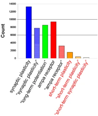
Figure 1 – Number of Pubmed results for the respective search term. Searching with
quotes forces phrase searching.
Mechanisms of STP
The mechanisms underlying short-term synaptic plasticity have not
been completely unraveled yet, but there is a growing consensus that synaptic
depression due to long trains of stimulating pulses may be attributed to
depletion of the readily releasable pool of synaptic vesicles (Rosenmund and
Stevens, 1996; Schneggenburger et al., 2002; Zucker and Regehr, 2002).
Short-Term Synaptic Plasticity | Introduction | Chapter 1
(Sullivan, 2007). Synapses that have a low probability of vesicle release are
likely to show some degree of short-term facilitation (Dobrunz and Stevens,
1997). In these cases, trains of action potentials may cause accumulation of
residual calcium, which binds to sensor proteins that trigger transmitter
release, thus enhancing synaptic transmission (Katz and Miledi, 1968; Zucker
and Regehr, 2002).
A recent alternative is that, in some systems, depression and
facilitation may be caused by common mechanisms, involving Ca2+-dependent
regulation of Ca2+ sensor proteins that regulate the presynaptic calcium
channels responsible for triggering transmitter release (Mochida et al., 2008).
Computational Models Incorporating STP
Buonomano and Merzenich (Buonomano and Merzenich, 1995)
showed through computer simulations that STP (specifically, paired-pulse
facilitation, PPF), together with slow IPSPs, could allow networks of neurons to
discriminate time-varying stimuli such as brief-pulse intervals, input rates,
simple temporal patterns or even phonemes. In their paper, the input layer
feeds to a model of LIV, which in turn feeds a model of LII/III. Importantly, each
brief, tap-like, stimulus activates the same set of input neurons to make sure that the network is not using spatial information, i.e., which input neurons get activated. Equally important is the fact that there was no learning or changes in
the synaptic weights but, due to the time-dependent properties of PPF and
slow IPSPs, the second stimulus of a pair coming at either 100 or 200 ms after the first will elicit different patterns of activation in each layer. A set of output
neurons was connected to LII/III and a simple supervised learning rule was
used in those synapses, so that the output neurons could learn the different
patterns elicited by each of the different stimuli, and in this manner become
Other studies have proposed a computational role for STP including
temporal processing (Buonomano, 2000), working memory (Mongillo et al.,
2008) and gain control (Abbott et al., 1997; Chance et al., 1998; Galarreta and
Hestrin, 1998). However, most of these studies have not considered synapse
specific STP, nor ‘plasticity’ of STP itself.
Scope of Chapter 3
It is well established that each of the multiple spikes in a train do not
contribute equally to the depolarization of the post synaptic cell, due to STP
(Markram and Tsodyks, 1996; Reyes et al., 1998). Given that neurons in the
cortex are active at high firing rates, it is likely that STP plays a role in shaping
information processing and transmission. In Chapter 3, we consider the
computational potential of STP for the discrimination of spatiotemporal spike
patterns in simple feed-forward neural networks.
In addition, multiple forms of STP have been observed in the brain, in a
synapse specific manner (Markram et al., 1998; Reyes et al., 1998; Rozov et
al., 2001; Zucker and Regehr, 2002). We propose that this diversity may be
the result of ‘adaptative’ (or plastic) mechanisms that determine how much and
for how long a synapse should depress or facilitate, depending on the
computation at hand. This would imply that STP is much more than a general
mechanism to regulate gain control or other general properties but takes an
active role in performing computations, in a synapse specific manner. Indeed,
we propose that STP itself may be plastic and propose a learning rule for this
Introduction | Chapter 1
Final Introductory Remarks
Throughout this work, we considered two well known properties of
biological neural networks, namely long-term (Chapter 2) and short-term
(Chapter 3) forms of synaptic plasticity. We show, using a combination of
experiments and computer simulations, how the dynamics of synaptic plasticity
| Chapter 2
CHAPTER 2
Differential effects of excitatory and inhibitory
plasticity on synaptically-driven neuronal
| Chapter 2
Differential effects of excitatory and inhibitory
plasticity on synaptically-driven neuronal
Input-Output functions
Tiago P. Carvalho1,2 & Dean V. Buonomano2,3,4
1
Gulbenkian Ph.D. Program in Biomedicine, P-2781-901 Oeiras, Portugal.
2
Departments of Neurobiologyand 3Psychology, and 4Brain Research Institute, University
of California, Los Angeles, CA 90095, USA.
| Chapter 2
Abstract
Ultimately, whether or not a neuron produces a spike determines its
contribution to local computations. In response to brief stimuli the probability a
neuron will fire can be described by its input-output function, which depends on
the net balance and timing of excitatory and inhibitory currents. While
excitatory and inhibitory synapses are plastic, most studies examine plasticity
of subthreshold events. Thus, the effects of concerted regulation of excitatory
and inhibitory synaptic strength on neuronal input-output functions are not well
understood. Here, theoretical analyses reveal that excitatory synaptic strength
controls the threshold of the neuronal input-output function, while inhibitory
plasticity alters the threshold and gain. Experimentally, changes in the balance
of excitation and inhibition in CA1 pyramidal neurons also altered their
input-output function as predicted by the model. These results support the existence
of two functional modes of plasticity that can be used to optimize information
Introduction | Chapter 2
Introduction
A large number of studies have characterized the mechanisms and
learning rules underlying synaptic plasticity, and it is generally accepted that
changes in synaptic strength contribute to learning and memory (Martin et al.,
2000; Malenka and Bear, 2004). However, since alterations in behavior must
ultimately be caused by changes in neuronal firing, it is not synaptic plasticity
per se, but how synaptic plasticity modifies the output of neurons, that underlies learning. Thus, to understand the relationship between synaptic
plasticity and learning it is important to elucidate how synaptic plasticity alters
the input-output characteristics of neurons.
We use the term neuronal Input-Output (I/O) function to refer to the relationship between the excitatory input to a neuron and the probability it will
generate an action potential (Fig. 1B,C) (Daoudal and Debanne, 2003; Staff
and Spruston, 2003; Marder and Buonomano, 2004; Campanac and Debanne,
2008). A neuron's I/O curve, generally represented as a sigmoidal function, is
characterized by two components: the threshold and the gain. Here we define
the I/O threshold as the EPSP slope that elicits a spike 50% of the time (this
usage is similar to that in the artificial neural network literature in which
threshold refers to the midpoint of the activation function, Rumelhart et al.,
1986). The gain refers to the rate of change or sensitivity of the I/O function
(Fig. 1C). The I/O threshold and gain of a neuron are directly related to its
computational role, as both of these features can be used to quantify the ability
of neurons to discriminate sensory stimuli (Mountcastle and Powell, 1959;
Maffei and Fiorentini, 1973; Dean et al., 2005) and optimize the encoding of
sensory information (Laughlin, 1981). Indeed, at the psychophysical level
similar measures are used to quantify behavioral performance, where the
threshold and gain are related to the point of subjective equality and just
Previous studies have established that LTP alters the threshold of the
I/O function – a phenomenon referred to as EPSP-spike (E-S) potentiation
(Andersen et al., 1980). Specifically, an EPSP of the same strength (as
measured by the slope), that was not effective in eliciting spikes, can fire the
cell after the induction of LTP. While the mechanisms underlying the
LTP-induced shift in the I/O function continue to be debated (Daoudal and
Debanne, 2003; Frick et al., 2004; Marder and Buonomano, 2004; Campanac
and Debanne, 2008), the balance of excitation and inhibition is known to be an
important contributing factor. For example, one reason that an EPSP of a
given size can elicit a spike after LTP, but not before, is due to an increase in
the excitation/inhibition ratio. After LTP, a smaller stimulation intensity is
required to elicit the same size EPSP and consequently fewer inhibitory
neurons will be recruited and those that are will have a longer latency, which
facilitates the generation of the action potential (Marder and Buonomano,
2004). However, in contrast to the threshold, previous studies have not
examined how excitatory plasticity influences the gain of neuronal I/O
functions. Additionally, to date no general framework exists as to how
excitatory and inhibitory synaptic plasticity interact to control the I/O function of
a neuron.
To understand how synaptic plasticity alters the behavior of neurons it
is necessary to characterize the I/O function in response to synaptically
evoked activity. It is important to note that the issue of long-term changes in
I/O functions produced by synaptic plasticity is distinct from the rapid ‘online’
changes in gain of the firing rate curve – such as the modulation produced by
the position of the eyes (Trotter and Celebrini, 1999) or attention (McAdams
and Reid, 2005) – that are critical for many sensory and motor computations
(Salinas and Thier, 2000). It has been shown that the gain modulation of the
firing rate curves is dependent on background synaptic activity (Chance et al.,
2002; Murphy and Miller, 2003; Prescott and De Koninck, 2003; Cardin et al.,
Introduction | Chapter 2
injected depolarizing current steps, and address how firing rate is modulated
on a rapid time scale for online computations. The distinct question addressed
here pertains to the probability a neuron will spike in response to a brief
stimulus depending on the strength of the active excitatory and inhibitory synapses. The focus on the early response to stimuli is important, particularly
in sensory systems, because it is the transient response that is critical to many
sensory computations (Durstewitz and Deco, 2008; Rabinovich et al., 2008)
and brief sensory stimuli often elicit only one or a few spikes (DeWeese et al.,
2003; Wang et al., 2005). Indeed, in many cases steady-state responses are
unlikely to contribute to computations (Rolls and Tovee, 1994; Thorpe et al.,
1996; Hung et al., 2005; Rabinovich et al., 2008).
While it is established that both EPSPs (Bliss and Lomo, 1973; Dudek
and Bear, 1992) and IPSPs (Komatsu, 1994; McLean et al., 1996; Lu et al.,
2000; Gaiarsa et al., 2002; Chevaleyre and Castillo, 2003) undergo LTP and
LTD, the trade-off between different types of synaptic plasticity and the
computation being performed is not understood. For example, from a
computational perspective, what is the functional difference between
potentiating excitatory inputs and depressing inhibitory ones? What is the
computational benefit of potentiating both EPSPs and IPSPs onto the same
postsynaptic neuron (Kairiss et al., 1987; Komatsu, 1994; Xie et al., 1995;
Shew et al., 2000; Lamsa et al., 2005; Froemke et al., 2007), which
superficially seems self-defeating?
To address these questions we first developed a computational model
which shows that the threshold and gain of neuronal I/O functions can be
independently controlled by change in excitatory and/or inhibitory synaptic
strength. We next examined experimentally the prediction of the model by
determining the I/O function of neurons in response to manipulation of
excitatory and inhibitory synaptic strengths. Our findings indicate that
while keeping the gain constant. On the other hand, balanced changes in
synaptic excitation and inhibition can adjust the gain of the neuron's I/O
function while maintaining a constant threshold. This study establishes a
framework for understanding the potential function and trade-off between
invoking excitatory and inhibitory plasticity in isolation or in parallel, and
proposes that I/O function plasticity could be used to optimize the encoding of
Results | Chapter 2
Results
Theoretical analysis of the effects of excitatory and inhibitory plasticity on neuronal I/O functions
To examine the effects of changing excitatory and inhibitory synaptic
strengths on the neuronal I/O function, we simulated a feed-forward disynaptic
circuit (Fig. 1A) and examined the response of a single postsynaptic excitatory
neuron (Ex) to increasing input intensity, which we represented as an increase
in the number of active excitatory and inhibitory synapses (Fig. 1B; see
Methods). In accordance with real neurons, the likelihood of eliciting an action
potential is probabilistic as a result of an incorporated “noise” current –
representing background synaptic activity and other stochastic processes. The
estimation of the spike probability across increasing intensities was fit with a
sigmoid function and, as observed experimentally, high intensities led not only
to an increased probability of firing but also to a decrease in the spike latency
Results | Chapter 2
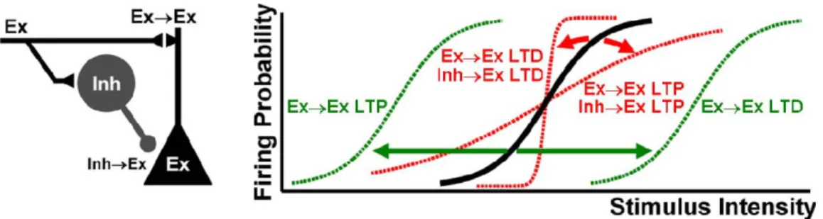
Fig. 1: Excitatory and inhibitory synaptic strengths control the gain and threshold of the neuronal input-output function.
A) Topology of the simulated feed-forward inhibitory circuit.
B) Sample voltage responses of the Ex unit at different input intensities (see text), for a particular combination of Ex→Ex and Inh→Ex synaptic weights (number 2 in panel D). Voltage traces were colored gray after the peak to ease the visualization of overlapping lines.
C) I/O function of the Ex unit in panel B, obtained by plotting the action potential probability versus the EPSP slope of the voltage traces (in bins, see text and Experimental Procedures).
D) Parameter scan of the excitatory and inhibitory synapse space. At each coordinate an I/O function was determined for the corresponding Ex→Ex and Inh→Ex synaptic weights. The numbers in the foreground depict the individual I/O functions plotted in panel E. Top: the gain (inverse) of each I/O function is plotted in color (range: [0.09 1.10] ms/mV). Hot colors depict an I/O function with a shallow slope, while cold colors depict an I/O function with a very sharp slope. Black depicts coordinates in which the inhibitory synapses were so strong that the Ex unit never fired. In gray the Ex unit fired occasionally, but not yielding enough points to be fitted with a sigmoid. Bottom: as above, but plotting the threshold of the same I/O curves (range: [10 20] mV/ms). Hot colors depict I/O functions with high threshold while cold colors depict I/O functions with low threshold. The dashed arrow highlights that a single I/O function is defined by two properties (gain and threshold).
E) Sample individual I/O functions. The gain and threshold of these sigmoids are highlighted in the corresponding plots in panel D by the corresponding numbers.
To understand how different excitatory and inhibitory synaptic weights,
corresponding to LTD or LTP of EPSPs and/or IPSPs, modify the I/O function
of a neuron, we parametrically varied the strength of Ex→Ex and Inh→Ex synapses. For each pair of synaptic weights, we plotted the threshold and gain
of the corresponding I/O function, hence describing the behavior of the neuron
across synapse space (Fig. 1D). These results show that, for fixed levels of
inhibitory synaptic strength, modifying the strength of a neuron's excitatory
synapses shifts the threshold to the left or right, but has little effect on the gain
of the I/O function (Fig. 1E, top). The horizontal shift in the threshold indicates
that some of the previously subthreshold EPSPs are now suprathreshold. This
is because, as excitatory synapses get stronger, it is possible to elicit the same
Buonomano, 2004). This scenario is equivalent to LTP of the Ex→Ex synapses in the absence of other forms of plasticity. In contrast, inhibitory
plasticity alone altered both the threshold and gain of the I/O function (Fig. 1E,
bottom). Interestingly, regulating the excitatory and inhibitory synaptic weights
in a balanced manner allowed neurons to change the gain of their I/O function while maintaining the same threshold, essentially establishing an
‘iso-threshold’ band along the diagonal of the excitatory and inhibitory synapse
space (Fig. 1E, middle). In contrast to the previously observed shifts in the
threshold, the change in the gain as a function of excitatory and inhibitory
synaptic strength has not been previously described experimentally or theoretically.
These theoretical results suggest that one reason excitatory and
inhibitory synapses are plastic is to allow for the independent control of the gain and threshold of neuronal I/O functions. That is, if the gain has to be
changed while maintaining the threshold, parallel excitatory and inhibitory
plasticity should be engaged, whereas if the threshold should be changed
while maintaining the gain, only excitatory plasticity should be induced.
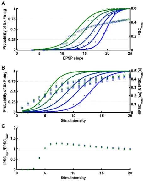
Synaptic inhibition increases the threshold and gain of I/O functions in CA1 pyramidal neurons
To test the above predictions, we performed experiments in which we
analyzed the I/O function of CA1 pyramidal neurons in hippocampal slices in
response to manipulations of the strength of the excitatory or inhibitory
synapses. Like most neurons, CA1 pyramidal cells receive robust feed-forward
excitation and inhibition; however, in contrast to the majority of cortical areas,
the CA1 subfield has little recurrent connectivity, thus providing a reasonable
approximation to the simulated disynaptic circuit used above. Effective
Results | Chapter 2
and directly through the induction of single-cell LTP. Given the difficulty in
inducing plasticity exclusively at Inh→Ex synapses, uncertainties regarding the protocols that induce inhibitory plasticity, and the variability of results (Xie et
al., 1995; Lu et al., 2000; Shew et al., 2000; Gaiarsa et al., 2002; Chevaleyre
and Castillo, 2003), we limited our manipulations of inhibitory strength to
pharmacological means to alter Inh→Ex transmission independently of the Ex→Ex and Ex→Inh strengths.
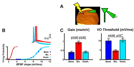
While recording in whole-cell configuration, we first examined the
effects of low concentrations (2-3 μM) of the GABAA antagonist bicuculline on
the neuronal I/O function. As already reported (Abraham et al., 1987; Marder
and Buonomano, 2003), there was a robust leftward shift of the threshold (Fig.
2B,C, dark blue vs. red, 9.9±1.1 vs. 4.6±0.6 mV/ms, p<0.001). Here we show
that in agreement with the above simulations (Fig. 1E, bottom), there was
also an increase in the gain of the I/O function (0.40±0.06 vs. 0.94±0.08
ms/mV, p<0.001). Upon washout of the drug, the threshold and gain of the I/O
function returned to baseline (Fig. 2B,C, light blue, gain: 0.42±0.05 ms/mV,
threshold: 10.0±1.0 mV/ms). The same results were also observed using
Fig. 2: Decrease in inhibitory strength decreases the threshold but increases the gain of neuronal I/O functions.
A) Schematic placement of the stimulating and whole-cell recording electrodes.
B) Example of a bicuculline experiment. Dark blue: I/O function of an intracellularly recorded CA1 pyramidal neuron, in standard ACSF. Red: I/O function of the same neuron in the presence of 3 μM bicuculline. Light blue: I/O function after 10 min. washout of bicuculline. Inset: Sample voltage traces for each of the conditions.
C) Average gain and threshold for the manipulations described in panel B (n=8).
Fig. 3: Dissociation of changes in gain and threshold.
A) Bicuculline followed by hyperpolarization experiment. Dark blue: I/O function of an intracellularly recorded CA1 pyramidal neuron, in whole-cell mode in standard ACSF.
Results | Chapter 2
B) Average gain and threshold for the manipulations described in panel A (n=12). Notice that the hyperpolarization, associated with the increase in stimulation intensity necessary to make the neuron fire, increases the I/O threshold in a statistically significant manner, without inducing significant changes in the gain.
Experimental dissociation of shifts in threshold and changes in gain
In the above experiments it could be argued that an increase in gain is
inextricably linked to the leftward shift in threshold. To establish that it is
possible to dissociate changes in threshold and gain, we tonically
hyperpolarized the cells (mean: 9.7±2.2 mV; range: 5-13 mV) after collecting
the baseline and bicuculline I/O curves (Fig. 3A,B). Tonic hyperpolarization
will alter all synaptic driving forces, however, under reduced inhibition (due to
bicuculline) its primary functional effect is a decrease in excitation (i.e., even
though EPSP amplitude may be larger, a neuron that was firing will cease to
do so because the peak EPSP is farther from action potential threshold). Thus,
hyperpolarization together with the necessary increase in stimulation intensity
to make the neuron fire shifts the I/O curve rightwards, towards values closer
to baseline but, interestingly, does not affect the gain (Fig. 3A,B, red vs.
orange, gain: 0.96±0.09 vs. 0.90±0.08 ms/mV, p>0.50, threshold: 4.4±0.4 vs.
7.2±0.5 mV/ms, p<10-5). These results show that changes in threshold and
gain can be dissociated and, indirectly, support the proposal that parallel
changes in excitation and inhibition may serve to maintain a constant threshold
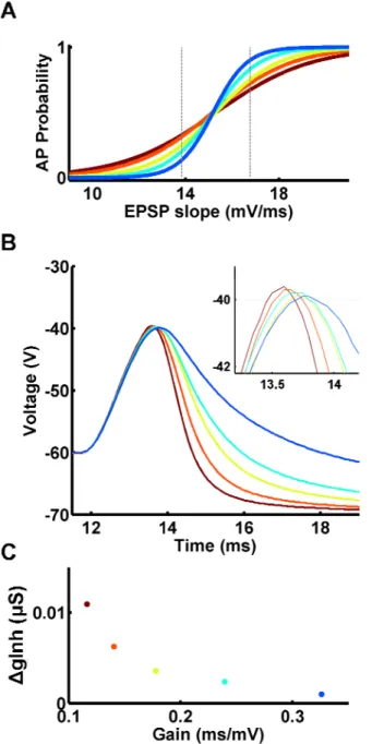
LTP alters the threshold while maintaining the gain of I/O functions
Early studies on LTP established that it produces a leftward shift of the
I/O curve (Bliss and Lomo, 1973; Andersen et al., 1980; Bliss et al., 1983). The
mechanisms underlying the leftward shift remain incompletely understood, in
part because some of the induction protocols used (e.g., presynaptic high
frequency stimulation) may induce plasticity at other synapses (Ex→Inh and/or Inh→Ex) (Kairiss et al., 1987; Komatsu, 1994; Xie et al., 1995; Shew et al., 2000) as well as changes in intrinsic excitability or dendritic integration
(Chavez-Noriega et al., 1990; Daoudal and Debanne, 2003; Xu et al., 2005;
Campanac and Debanne, 2008). Nevertheless, it has been shown that
single-cell associative pairing protocols can also induce left shifts in the I/O function
(Marder and Buonomano, 2004), which is consistent with our theoretical
framework. However, the effect of LTP of excitatory synapses on the gain of
the neuronal I/O function has not been addressed.
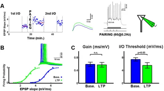
To examine this issue we performed intracellular experiments with high
resistance micropipettes (70-90 MΩ) to prevent washout of LTP (Lamsa et al., 2005). LTP was induced in single neurons with a pairing protocol that has
previously been shown not to induce changes in inhibition or intrinsic
excitability (Barrionuevo and Brown, 1983; Gustafsson et al., 1987; Marder
and Buonomano, 2004). Specifically, pairing intracellular depolarization (100
ms) with a train of 4 presynaptic stimuli (40Hz; 60 pairings at 0.2Hz) resulted in
a 79±17% increase in the EPSP slope (we only included experiments with LTP
> 10% in this analysis). The induction of LTP caused a left shift (7.4±0.5 vs.
5.6±0.8 mV/ms, p<0.05) and, in agreement with the theoretical predictions, did
not induce any change in the gain (0.59±0.07 vs. 0.57±0.07 ms/mV, p>0.80) of
Results | Chapter 2
Fig. 4: Potentiation of the excitatory strength decreases the threshold without changing the gain of neuronal I/O functions.
A) EPSP slopes recorded with a sharp microelectrode during the course of an associative LTP experiment. Voltage traces on the middle represent average sample PSPs from 5 min. after the 1st I/O and 5 min. before the 2nd I/O. The voltage trace on the right shows a sample of the pairing depolarization.
B) I/O functions before and after the associative LTP pairing protocol. The threshold of the I/O function decreases (left shift), but the gain is left unchanged. Inset: Sample voltage traces for each of the conditions
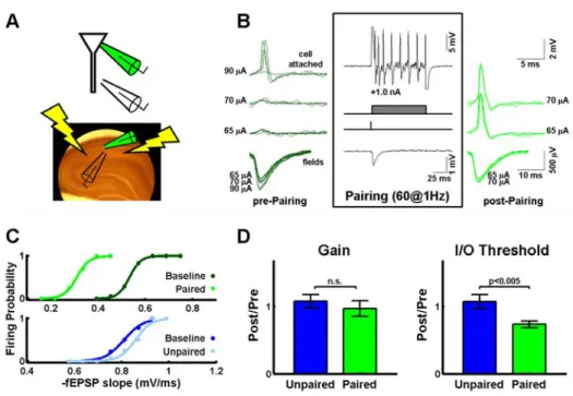
Fig. 5: LTP induced threshold left shifts with constant gain are not due to global changes in excitability.
A) Schematic placement of the stimulating and cell-attached and field recording electrodes.
B) Example of the potentiation protocol. Left: Sample voltage traces recorded from the cell-attached (top) and field (bottom) electrodes at 3 different intensities. There are four traces per intensity. Middle: Associative pairing protocol. Presynaptic stimulation was paired with 100 ms postsynaptic depolarization 60 times at 1Hz. Right: Voltage traces for the same intensities as before (the highest intensity was no longer used to optimize the estimation of the I/O functions). Notice the increased action potential probability. C) Sample I/O functions before and after the associative pairing protocol illustrated in panel B. Top: Paired pathway. The threshold of the I/O function decreases (left shift), but the gain is left unchanged. Bottom: Control pathway. The I/O function is unchanged supporting the existence of no global changes in excitability.
Results | Chapter 2
As mentioned above the mechanisms underlying the left shift in the I/O
function (E-S potentiation) remain controversial and other groups have
suggested that it could be due to changes in intrinsic excitability (Sourdet et
al., 2003; Frick et al., 2004; Losonczy et al., 2008). A further complicating set
of issues is that intracellular techniques can alter the neuronal I/O function as a
result of washout (Kato et al., 1993; Staff and Spruston, 2003; Lamsa et al.,
2005; Xu et al., 2005), changes in cell input resistance, or changes in the
balance of excitation and inhibition (Zhang et al., 1991; Staley and Smith,
2001). To avoid any potential methodological artifacts and determine if global
changes in intrinsic excitability could have influenced the above results we
performed experiments in tight-seal cell-attached configuration – which does
not rupture the cellular membrane – and included a second unpaired control
pathway in the LTP experiments. Given that the cell-attached technique does
not allow recording subthreshold responses, we estimated the average input to
the neuron by recording the field EPSP from an electrode placed in stratum
radiatum in a line perpendicular with the cell body layer (Fig. 5A) (Andersen et
al., 1980; Zalutsky and Nicoll, 1990). The high resistance cell-attached
configuration does not rupture the membrane (seal > 1 GΩ), but still allows the injection of positive current through the electrode and the recording of the
spikes (Perkins, 2006; Houweling and Brecht, 2008) (Fig. 5B, middle). By
pairing this depolarization (100 ms) with single presynaptic stimuli (60 pairings
at 1Hz), we consistently observed leftward shifts in the I/O functions (11/13
experiments) and, in agreement with the previous results, no change in gain
(Fig. 5C,D; threshold: 74±5% p<0.001, gain: 97±12% p>0.80). Importantly, the
unpaired control pathway onto the same cell showed no horizontal shift or
change in gain (threshold: 108±10% p>0.70; gain: 108±10% p>0.70). There
was a significant difference in the threshold between the paired and unpaired
pathways (p<0.005), but no difference in the gain (p>0.50, Fig. 5D). These
results establish that the pairing-LTP induced left shift is not a result of general
in Figure 4, the fact that there was no change in the gain of the I/O function is
consistent with the prediction made in Figure 1. However, it should be
stressed that the interpretation of I/O function in these cell-attached
experiments is constrained by the fact that the extracellular fEPSP was used to
construct the I/O function.
Together these results demonstrate that LTP produces a leftward shift
in the absence of a change in gain, and that this effect is not likely to be a
result of any cell-wide form of intrinsic plasticity. In contrast, a decrease in
inhibition is accompanied by a change in gain, in addition to the change in
threshold.
Mechanisms of the changes in gain and threshold induced by synaptic plasticity
The simulations and experiments above indicate that increasing
excitatory (E-LTP) or decreasing inhibitory synaptic strength (I-LTD) both
produce left shifts in the threshold of the I/O function; however, the latter also
induces an increase in the gain (the potential computational relevance of these
forms of plasticity is addressed in the Discussion). Next, we used the
computational model to understand the origin of the change in gain associated
with changes in synaptic inhibitory strength. It is important to point out that
excitatory and inhibitory synaptic plasticity produce fundamentally different
changes in the post-synaptic potential (PSP) waveform: excitatory plasticity
changes the slope and peak of the PSP, while changes in inhibition alter the
peak and width of the PSP (Fig. S1, Buonomano and Merzenich, 1998a;
Pouille and Scanziani, 2001). As a consequence of the inherent asymmetry
between excitatory and inhibitory plasticity, imposed primarily by the delay of
inhibition in relation to excitation, small changes in excitation are proportionally
Results | Chapter 2
On the other hand, the fact that inhibitory plasticity determines the width of the
PSP is an important factor in determining the gain of the I/O function because
the wider the PSP the longer it borders action potential threshold − hence, subsequent small increases in the PSP slope will result in sharp increases in
spike probability and the I/O gain (Fig. S2).
There are a number of interrelated properties that jointly contribute to
determining the I/O gain, and whether or not it changes after synaptic
plasticity. Below we first address the mechanisms responsible for the observed
changes in the I/O function in response to inhibitory or excitatory plasticity in
isolation. Additionally, the issue of I/O gain control is further discussed in the
Supplemental Material.
I-LTD
Consider a ‘baseline’ I/O function (blue curve in Fig. 6A), and the
stimulation intensity (S50) which elicits the EPSP slope that defines the
threshold of this I/O curve (Fig. 6D; that is, the EPSP slope that generates
action potentials with 50% probability). If one induces I-LTD (Fig. 6A, red
curve) the EPSP slope at S50 will remain largely unchanged, since it is mainly
determined by the excitatory strength. Yet, the PSP width and height will
increase; hence the same EPSP slope will yield action potentials with
increased probability. To find the new I/O threshold one must decrease the
stimulation intensity until it yields an EPSP slope where the neuron fires action
potentials again with 50% probability (Fig. 6A, left red I/O), thus accounting for
the left shift of the threshold of the I/O curve. But why does the gain change?
Compared with an I/O of the same threshold, but with the same gain as the
baseline curve (dark green trace in Fig. 6A, see 'E-LTP' below), changes in
stimulation intensity will produce a smaller change in the inhibitory