www.cbpv.com.br/rbpv
Occurrence of anti-
Toxoplasma gondii
and
anti-
Neospora caninum
antibodies in cats with outdoor
access in São Luís, Maranhão, Brazil
Ocorrência de anticorpos anti-
Toxoplasma gondii
e anti-
Neospora caninum
em gatos
com acesso à rua em São Luís, Maranhão, Brasil
Maria do Socorro Costa de Oliveira Braga1,2; Marcos Rogério André2; Márcia Mariza Gomes Jusi2;
Carla Roberta Freschi2; Márcia Cristina Alves Teixeira2; Rosangela Zacarias Machado2*
1Departamento de Clinícas Veterinárias,Universidade Estadual do Maranhão – UEMA, São Luís, MA, Brasil
2Departamento de Patologia Veterinária, Faculdade de Ciências Agrárias e Veterinárias, Universidade Estadual Paulista – UNESP,
Jaboticabal, SP, Brasil
Received June 28, 2011 Accepted January 13, 2012
Abstract
The present study aimed to investigate the frequency of anti-Toxoplasma gondii and anti-Neospora caninum
antibodies in cats with outdoor access in São Luís, Maranhão, Brazil. The presence of IgG anti-T. gondii and anti-N. caninum antibodies was tested using the Indirect Immunofluorescent Antibody Test (IFAT). IgG anti-T. gondii
and anti-N. caninum antibodies were detected in 101 (50.5%) and 54 (27%) sampled cats, respectively. The titers of anti-T. gondii antibodies ranged from 40 (cut-off) to 2560. On the other hand, the titers of anti-N. caninum antibodies ranged from 25 (cut-off) to 400. Twenty-seven cats (13.5%) were shown to be seropositive for both parasites. Seventy-four cats (34%) were seropositive only for T. gondii. Twenty-two cats (11%) were seropositive only for N. caninum. The present study showed that cats with outdoor access in São Luís, Maranhão, are exposed to T. gondii and N. caninum.
Keywords: Toxoplasma gondii, Neospora caninum, cats, serology.
Resumo
O presente estudo objetivou verificar a frequência de anticorpos anti-Toxoplasma gondii e anti-Neospora caninum em gatos com acesso à rua em São Luís, Maranhão, Brasil. A presença de anticorpos IgG anti-T. gondii e anti-N. caninum foi verificado pela Reação de Imunofluorescência Indireta (RIFI). Anticorpos IgG anti-T. gondii e anti-N. caninum foram detectados em 101 (50,5%) e 54 (27%) gatos amostrados, respectivamente. Os títulos de anticorpos anti-T. gondii
variaram de 40 (ponto de corte) a 2560. Por outro lado, anticorpos anti-N. caninum variaram de 25 (ponto de corte) a 400. Vinte e sete gatos (13,5%) mostraram-se soropositivos para ambos os parasitas. Setenta e quatro gatos (34%) foram soropositivos somente para T. gondii. Vinte e dois gatos (11%) foram soropositivos somente para
N. caninum. O presente estudo demonstrou que gatos com acesso à rua em São Luís, Maranhão, são expostos ao
T. gondii e N. caninum.
Palavras-chave: Toxoplasma gondii, Neospora caninum, gatos, sorologia.
Introduction
Toxoplasmosis is a zoonotic protozoan disease caused by the apicomplexan parasite Toxoplasma gondii (TENTER et al., 2000). This disease is acquired principally by eating food or drinking water contaminated with oocysts or by ingestion of tissue containing T. gondii cysts. Cats and related felids are the
definitive hosts, because they are the only animal species that excrete resistant oocysts into the environment (JACKSON; HUTCHINSON, 1989). Toxoplasma gondii has a wide range of intermediate hosts, including humans and several animal species, particularly mammals and birds (TENTER et al., 2000). On the other hand, Neospora caninum, a related coccidian protozoon, was first identified from the brain of a dog (DUBEY et al., 1988a, b). To date, only domestic dogs, coyotes (Canis latrans) and dingoes (Canis lupus dingo) have been recognized as definitive hosts for
N. caninum (MCALLISTER et al., 1998a, b; LINDSAY et al., 1999; GONDIM et al., 2004; KING et al., 2010). Regarding *Corresponding author: Rosangela Zacarias Machado
Laboratório de Imunoparasitologia, Departamento de Patologia Veterinária, Faculdade de Ciências Agrárias e Veterinárias Júlio de Mesquita Filho, Universidade Estadual Paulista – UNESP, Via de Acesso Prof. Paulo Donato Castellane, s/n, Zona Rural, CEP 14884-900, Jaboticabal, SP, Brasil e-mail: zacarias@fcav.unesp.br
economic importance in livestock, N. caninum is recognized as an important cause of abortion in cattle (DUBEY; LINDSAY, 1996). Most likely because cats may only play a minor role in the epidemiology of N. caninum infection, there are only a few reports on naturally acquired seropositivity to N. caninum
among cats(DUBEY et al., 2002; FERROGLIO et al., 2005; BRESCIANI et al., 2007; HORNOK et al., 2008). The present study aimed to investigate the frequency of anti-T. gondii and anti-N. caninum antibodies in cats with outdoor access in São Luís, Maranhão, Brazil.
Material and Methods
1. Sample collection
Between October 2008 and January 2009, serum samples were collected by venipuncture in the jugular and/or cephalic vein from 200 peridomestic cats (Felis catus) in peripheral areas of São Luís, state of Maranhão. The sampled cats were of both genders and different breeds and ages. All the cats appeared to be healthy at the time of sample collection. To facilitate blood collection, the cats were chemically immobilized using xylazine (1 mg/kg, intramuscularly).
2. Serological tests for T. gondii and N. caninum
The presence and level of IgG anti-T. gondii and anti-N. caninum
antibodies were tested using the Indirect Immunofluorescent Antibody Test (IFAT). The antigenic substrate for T. gondii
consisted of purified tachyzoites that were obtained by means of peritoneal lavage of previously infected mice, as described by Camargo (1964).
The antigen substrate used in preparing slides to detect antibodies against N. caninum by means of IFAT was produced using isolate NC-1 (DUBEY et al., 1988a, b). For this, CV-1 cells were cultured in RPMI medium (Sigma, St. Louis, MO, USA), supplemented with 2% fetal calf serum (BFS). Three days after infection, parasites were harvested from mononuclear cell layers using 1% trypsin treatment, and were passed into another flask with CV-1 cells. The N. caninum tachyzoites that were recovered were used as the antigen substrate (FURUTA et al., 2007).
All serum samples were screened at serial dilutions in phosphate-buffered saline (PBS, pH 7.2), using cutoffs of 1:40 and 1:25 for T. gondii and N. caninum, respectively. Cat serum samples that were negative for T. gondii and N. caninum and samples naturally infected with these parasites, from the serum bank of the Immunoparasitology Laboratory, Department of Veterinary Pathology, Unesp, Jaboticabal, SP, were also used in the serological reactions. Briefly, slides with diluted serum samples were incubated at 37 °C in a moist chamber for 45 min, washed three times in PBS (pH 7.2) for 5 min, and air-dried at room temperature. IgG anti-cat conjugate labeled with fluorescein isothiocyanate (Sigma, St. Louis, MO, USA) was diluted at 1:64 in accordance with the manufacturer’s instructions and was then added to each well. These slides were incubated again, washed, dried and overlain with buffered glycerin (pH 8.7), covered with glass coverslips, and examined under a fluorescence microscope (Olympus BX60).
Results
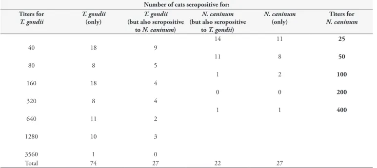
IgG antibodies against T. gondii and N. caninum were detected in 101 (50.5%) and 54 (27%) sampled cats, respectively. IgG antibody titers against T. gondii ranged from 40 (cutoff) to 2560. On the other hand, IgG antibody titers against N. caninum ranged from 25 (cutoff) to 400. Twenty-seven cats (13.5%) showed IgG antibodies for both T. gondii and N. caninum. Seventy-four cats
Table 1. Number of cats seropositive for T. gondii and N. caninum, according to IgG antibody titers.
Number of cats seropositive for: Titers for
T. gondii
T. gondii
(only)
T. gondii
(but also seropositive to N. caninum)
N. caninum
(but also seropositive to T. gondii)
N. caninum
(only)
Titers for
N. caninum
14 11 25
40 18 9
11 8 50
80 8 5
1 2 100
160 18 4
0 0 200
320 8 4
1 1 400
640 11 2
1280 10 3
3560 1 0
(34%) were seropositive only for T. gondii. Twenty-two cats (11%) were seropositive only for N. caninum (Table 1). Seventy-seven cats (38.5%) were seronegative for both parasites.
Discussion
The incidence of toxoplasmosis and neosporosis in urban areas is closely related to environmental contamination with oocysts. Direct measurement of environmental contamination by means of oocyst counting is unfeasible for technical reasons (MEIRELES et al., 2004). One interesting alternative for measuring T. gondii and
N. caninum dissemination is by analyzing the seroprevalence in free-living urban animals, which are thus used as sentinels. In this regard, the seroprevalence of T. gondii and N. caninum in animals could be an indirect indicator of the degree of parasite spreading in urban and rural areas.
The present study showed that cats in São Luís, state of Maranhão, are exposed to T. gondii and N. caninum. The prevalence of T. gondii antibodies (50.5%) in the cats of the present study was higher than what was found in previous studies conducted in Brazil, in the states of Santa Catarina (DALLA ROSA et al., 2010); São Paulo (SOGORB et al., 1972; LUCAS et al., 1998; SILVA et al., 2002; MEIRELES et al., 2004; PENA et al., 2006; BRESCIANI et al., 2007); Rio Grande do Sul (PINTO et al., 2009); and Rio de Janeiro (GONÇALVES NETO et al., 2003). On the other hand, the prevalence found in our study was lower than what was found among cats in Paraná (GARCIA et al., 1999) and Rondônia (CAVALCANTE et al., 2006). However, because different techniques and cutoff values were used, comparisons may generate misunderstandings (BRESCIANI et al., 2007).
The majority of the positive cats showed T. gondii antibodies at titers of 1:40. Lower titers may suggest latent infection (LUCAS et al., 1998). It is difficult to draw conclusions from interpreting a single serological test on cats and, therefore, paired serological tests are desirable. Titers higher than 1,024 usually represent strong determinants of toxoplasmosis with or without clinical signs of disease (BRESCIANI et al., 2007). Here, 11 cats showed antibody titers higher than 1,024. Furthermore, the level of T. gondii antibodies is not associated with oocyst shedding levels (OMATA et al., 1990).
The prevalence of antibodies against N. caninum found in the present study was similar than what was reported among cats in Araçatuba, state of São Paulo (cutoff > 1:16; BRESCIANI et al., 2007), and in Italy (cutoff > 1:80; FERROGLIO et al., 2005). On the other hand, it was higher than what was found by Dubey et al. (2002) in Brazil (11.9%; cutoff > 1:40), and by Hornok et al. (2008) in Hungary (0.6%; cutoff > 1:40). The possibility of cross-reactions with T. gondii was ruled out, given that serological cross-reactivity often occurs with soluble antigens; in the case of IFAT, it does not occur (DUBEY et al., 1996). Also, considering the fact that the lesions caused by N. caninum in cats are similar than those found in cats with toxoplasmosis (DUBEY et al., 1990), and the moderately high prevalence found in the present study, neosporosis should be a differential diagnosis for cats with neurological clinical signs.
Diet and access to the outdoor environment have been incriminated as important factors for cat infection (LUCAS et al., 1998). In our study, the sampled cats had free access to the outdoor environment, and probably had the opportunity to hunt small prey, thus becoming more susceptible to infection by T. gondii
than are cats that are exclusively kept indoors. Small birds, like pigeons and sparrows, or rodents that live in the synanthropic environment, could have been the possible prey hunted by cats. Recently, it was found that pigeons (Columba livia) and sparrows (Passer domesticus) can act as intermediate hosts for T. gondii and
N. caninum (MINEO et al., 2009; GONDIM et al., 2010). Rodents are part of the life cycle of T. gondii, acting as prey containing
T. gondii cysts for cats (HUTCHISON; DUNACHIE, 1971). Huang et al. (2004) found from PCR that 5.8% of their rat sample were positive for N. caninum and suggested that rats could serve as a reservoir of infection. On the other hand, Rattus norvegicus,
a common synanthropic rat species, could be considered to be a difficult prey for cats. Moreover, garbage food is more available than birds in urban areas (LUCAS et al., 1998).
The sampled cats must have had access to food found in domestic garbage, which is usually food similar to what is prepared for human consumption. Toxoplasma gondii and N. caninum cysts may be found in leftovers of meat for human consumption that is available in garbage. However, such meats are under sanitary control, and hence, neosporosis and toxoplasmosis from these sources were not investigated in the present survey. The seroprevalence of T. gondii among chickens in Brazil ranges from 39% to 66% (DA SILVA et al., 2003; DUBEY et al., 2003; DUBEY et al., 2006; DE OLIVEIRA et al., 2009); and among cattle, from 1% (GONDIM et al., 1999) to 71% (SANTOS et al., 2009). On the other hand, the seroprevalence of N. caninum among cattle in Brazil ranges from 14.3% (GUIMARÃES et al., 2004) to 91.2% (GUEDES et al., 2008). Intake of water contaminated by oocysts of N. caninum and T. gondii may also have played a role in transmission of these coccidia among the sampled cats.
The present study showed that cats in São Luís, Maranhão, with outdoor access, are exposed to T. gondii and N. caninum. While the role of cats as the definitive hosts in the epidemiology of toxoplasmosis is already well defined, the importance of these animals in the epidemiology of neosporosis in Brazil has not been determined yet.
Acknowledgements
The authors would like to thank Conselho Nacional de Desenvolvimento Científico e Tecnológico (CNPq) for the financial support (number 479162/2007-7).
References
Camargo ME. Improved technique of indirect immunofluorescence for serological diagnosis of toxoplasmosis. Rev Inst Med Trop São Paulo 1964; 6(3): 117-118. PMid:14177810.
Cavalcante GT, Aguiar DM, Chiebao D, Dubevt JP, Ruiz VLA, Dias RA, et al. Seroprevalence of Toxoplasma gondii antibodies in cats and pigs from rural Western Amazon, Brazil. J Parasitol 2006; 92(4): 863-864. PMid:16995406. http://dx.doi.org/10.1645/GE-830R.1
Da Silva DS, Bahia-Oliveira LMG, Shen SK, Kwok OCH, Lehman T, Dubey JP. Prevalence of Toxoplasma gondii in chickens from an area in southern Brazil highly endemic to humans.
J Parasitol 2003; 89(2): 394-396. http://dx.doi.org/10.1645/0022-3395(2003)089[0394:POTGIC]2.0.CO;2
Dalla Rosa L, Moura AB, Trevisani N, Medeiros AP, Sartor AA, Souza AP, et al. Toxoplasma gondii antibodies on domiciled cats from Lages municipality, Santa Catarina State, Brazil. Rev Bras Parasitol Vet. 2010; 19(4): 268-9. http://dx.doi.org/10.1590/S1984-29612010000400017
De Oliveira LN, Costa Junior LM, Melo CF, Ramos Silva JC, Bevilaqua CML, Azevedo SS, et al. Toxoplasma gondii isolates from free-range chickens from the northeast region of Brazil. J Parasitol 2009; 95(1): 235-237. PMid:18578589. http://dx.doi.org/10.1645/GE-1730.1
Dubey JP, Lindsay DS. A review of Neospora caninum and neosporosis.
Vet Parasitol 1996; 67(1): 1-59. http://dx.doi.org/10.1016/S0304-4017(96)01035-7
Dubey JP, Carpenter JL, Speer CA, Topper MJ, Uggla A. Newly recognized fatal protozoan disease of dogs. J Am Vet Med Assoc 1988a; 192(9): 1269-1285. PMid:3391851.
Dubey JP, Gennari SM, Labruna MB, Camargo LMA, Vianna MCB, Marcet PL, et al. Characterization of Toxoplasma gondii isolates in free-range chickens from Amazon, Brazil. J Parasitol 2006; 92(1): 36-40. PMid:16629312. http://dx.doi.org/10.1645/GE-655R.1
Dubey JP, Graham DH, Da Silva DS, Lehmann T, Bahia-Oliveira LMG. Toxoplasma gondii isolates of free-ranging chickens from Rio de Janeiro, Brazil: Mouse mortality, genotype, and oocyst shedding by cats. J Parasitol 2003; 89(4): 851-853. PMid:14533703. http://dx.doi. org/10.1645/GE-60R
Dubey JP, Hattel AL, Lindsay DS, Topper MJ. Neonatal Neospora caninum
infection in dogs: isolation of the causative agent and experimental transmission. J Am Vet Med Assoc 1988b; 193(10): 1259-1263.
Dubey JP, Lindsay DS, Adams DS, Gay JM, Baszler TV, Blagburn BL, et al. Serologic responses of cattle and other animals infected with
Neospora caninum. Am J Vet Res 1996; 57(3): 329-336. PMid:8669764.
Dubey JP, Lindsay DS, Hill D, Romand S, Thulliez P, Kwok OC, et al. Prevalence of antibodies to Neospora caninum and Sarcocystis neurona in sera of domestic cats from Brazil. J Parasitol 2002; 88(6): 1251-1252. PMid:12537122.
Dubey JP, Lindsay DS, Lipscomb TP. Neosporosis in cats. Vet Pathol
1 9 9 0 ; 2 7 ( 5 ) : 3 3 5 - 3 3 9 . P M i d : 2 2 3 8 3 8 6 . h t t p : / / d x . d o i . org/10.1177/030098589002700505
Ferroglio E, Guiso P, Pasino M, Accossato A, Trisciuoglio A. Antibodies to Neospora caninum in stray cats from north Italy. Vet Parasitol 2005; 131(1-2): 31-34. PMid:15919155. http://dx.doi.org/10.1016/j. vetpar.2005.04.012
Furuta PI, Mineo TW, Carrasco AO, Godoy GS, Pinto AA, Machado RZ. Neospora caninum infection in birds: experimental infections in
chicken and embryonated eggs. Parasitology 2007; 134(14): 1931-1939. PMid:17686190. http://dx.doi.org/10.1017/S0031182007003344
Garcia JL, Navarro IT, Ogawa L, Oliveira RC. Soroepidemiologia da toxoplasmose em gatos e cães de propriedades rurais do município de Jaguapitã, estado do Paraná, Brasil. Ciênc Rural 1999; 29(1): 99-104. http://dx.doi.org/10.1590/S0103-84781999000100018
Gonçalves Netto E, Munhoz AD, Albuquerque GR, Lopes CWG, Ferreira AMR. Ocorrência de gatos soropositivos para Toxoplasma gondii
Nicolle e Manceaux, 1909 (Apicomplexa: Toxoplasmatinae) na cidade de Niterói, Rio de Janeiro. Rev Bras Parasitol Vet 2003; 12(4): 145-149.
Gondim LF, McAllister MM, Pitt WC, Zemlicka DE. Coyotes (Canis latrans) are definitive hosts of Neosporacaninum. Int J Parasitol 2004; 34(2): 159-161. http://dx.doi.org/10.1016/j.ijpara.2004.01.001
Gondim LFP, Barbosa Junior HV, Ribeiro Filho CHA, Saeki H. Serological survey of antibodies to Toxoplasma gondii in goats, sheep, cattle and water buffaloes in Bahia State, Brazil. Vet Parasitol 1999; 82(4): 273-276. http:// dx.doi.org/10.1016/S0304-4017(99)00033-3
Gondim LS, Abe-Sandes K, Uzêda RS, Silva MS, Santos SL, Mota RA, et al.
Toxoplasma gondii and Neospora caninum in sparrows (Passer domesticus) in the Northeast of Brazil. Vet Parasitol 2010; 168(1-2): 121-124. http:// dx.doi.org/10.1016/j.vetpar.2009.09.055
Guedes MH, Guimarães AM, Rocha CM, Hirsch C. Frequency of anti-Neospora caninum antibodies in cows and fetuses from municipalities of southern Minas Gerais. Rev Bras Parasitol Vet 2008; 17(4): 189-194. PMid:19265576.
Guimarães Junior JS, Souza SL, Bergamaschi DP, Gennari SM. Prevalence of Neospora caninum antibodies and factors associated with their presence in dairy cattle of the north of Paraná state, Brazil. Vet Parasitol 2004; 124(1-2): 1-8. PMid:15350656.
Hornok S, Edelhofer R, Joachim A, Farkas R, Berta K, Répási A, et al. Seroprevalence of Toxoplasma gondii and Neospora caninum infection of cats in Hungary. Acta Vet Hung 2008; 56(1): 81-8. PMid:18401958. http://dx.doi.org/10.1556/AVet.56.2008.1.8
Huang CC, Yang CH, Watanabe Y, Liao YK, Ooi HK. Finding of
Neospora caninum in the wild brown rat (Rattus norvegicus). Vet Res
2004; 35(3): 283-90. http://dx.doi.org/10.1051/vetres:2004010
Hutchison WM, Dunachie JF, Work K, Siim JC. The life cycle of the coccidian parasite, Toxoplasma gondii, in the domestic cat. Trans R Soc Trop Med Hyg 1971;65(3): 380-99. http://dx.doi.org/10.1016/0035-9203(71)90018-6
Jackson MH, Hutchison WM. The prevalence and source of Toxoplasma infection in the environment. Adv Parasitol 1989; 28: 55-105. http:// dx.doi.org/10.1016/S0065-308X(08)60331-0
King JS, Slapeta J, Jenkins DJ, Al-Qassab SE, Ellis JT, Windsor PA. Australian dingoes are definitive hosts of Neospora caninum.
Int J Parasitol. 2010; 40(8): 945-50. http://dx.doi.org/10.1016/j. ijpara.2010.01.008
Lindsay DS, Dubey JP, Duncan RB. Confirmation that the dog is a definitive host for Neospora caninum. Vet Parasitol 1999; 82(4): 327-33. http://dx.doi.org/10.1016/S0304-4017(99)00054-0
McAllister MM, Dubey JP, Lindsay DS, Jolley WR, Wills RA, McGuire AM. Dogs are definitive hosts of Neospora caninum. Int J Parasitol 1998a; 28(9): 1473-8. http://dx.doi.org/10.1016/S0020-7519(98)00138-6
McAllister MM, Jolley WR, Wills RA, Lindsay DS, McGuire AM, Tranas JD. Oral inoculation of cats with tissue cyst of Neospora caninum. Am J Vet Res 1998b; 59(4): 441-4. PMid:9563628.
Meireles LR, Galisteo Junoir AJ, Pompeu E, Andrade Junior HF.
Toxoplasma gondii spreading in an urban area evaluated by seroprevalence in free-living cats and dogs. Trop Med Int Health 2004; 9(8): 876-81. http://dx.doi.org/10.1111/j.1365-3156.2004.01280.x
Mineo TW, Carrasco AO, Marciano JA, Werther K, Pinto AA, Machado RZ. Pigeons (Columba livia) are a suitable experimental model for
Neospora caninum infection in birds. Vet Parasitol 2009; 159(2): 149-53. PMid:19027237. http://dx.doi.org/10.1016/j.vetpar.2008.10.024
Omata Y, Oikawa H, Kanda M, Mikazuki K, Nakabayashi T, Suzuki N. Experimental feline toxoplasmosis: humoral immune responses of cats inoculated orally with Toxoplasma gondii cysts and oocysts. Nihon Juigaku Zasshi 1990; 52(4): 865-7. http://dx.doi.org/10.1292/jvms1939.52.865
Pena HF, Soares RM, Amaku M, Dubey JP, Gennari SM. Toxoplasma gondii infection in cats from São Paulo state, Brazil: Seroprevalence,
oocyst shedding, isolation in mice, and biologic and molecular characterization. Res Vet Sci 2006; 81(1): 58-67. PMid:16289158. http:// dx.doi.org/10.1016/j.rvsc.2005.09.007
Pinto LD, Araújo FAP, Stobbe NS, Marques SMT. Soroepidemiologia de
Toxoplasma gondii em gatos domiciliados atendidos em clínicas particulares de Porto Alegre, RS, Brasil. Ciênc Rural 2009; 39(8): 2464-2469.
Santos TR, Costa AJ, Toniollo GH, Luvizotto MC, Benetti AH, Santos RR, et al. Prevalence of anti-Toxoplasma gondii antibodies in dairy cattle, dogs, and humans from the Jauru micro-region, Mato Grosso state, Brazil. Vet Parasitol 2009; 161(3-4): 324-6. http://dx.doi.org/10.1016/j. vetpar.2009.01.017
Silva JC, Gennari SM, Ragozo AM, Amajones VR, Magnabosco C, Yai LE, et al. Prevalence of Toxoplasma gondii antibodies in sera of domestic cats from Guarulhos and São Paulo, Brazil. J Parasitol 2002; 88(2): 419-20. http://dx.doi.org/10.2307/3285606
Sogorb F, Jamra LF, Guimarães EC, Deane MP. Toxoplasmose espontânea em animais domésticos e silvestres, em São Paulo. Rev Inst Med Trop Sao Paulo 1972; 14(5): 314-20.