Instituto de Tecnologia Química e Biológica
Universidade Nova de Lisboa
Functional studies on BolA and
related genes:
increasing the understanding
of a protein with pleiotropic effects
inFonte Viva, Francisco Cândido Xavier, ditado pelo Espírito Emmanuel, FEB “Mas ainda que o nosso homem exterior se corrompa,
o interior, contudo, se renova, de dia para dia.”
Paulo (II CORÍNTIOS, 4:16) Observa o espírito de sequência e gradação que prevalece nos mínimos setores da Natureza. Nada se realiza aos saltos e, na pauta da Lei Divina, não existe privilégio em parte alguma.
Enche-se a espiga de grão em grão. Desenvolve-se a árvore milímetro a milímetro. Nasce a floresta de sementes insignificantes. Levanta-se a construção peça por peça. Começa o tecido nos fios. As mais famosas páginas foram produzidas letra a letra. A cidade mais rica é edificada palmo a palmo. As maiores fortunas de ouro e pedras foram extraídas do solo, fragmento a fragmento. A estrada mais longa é pavimentada metro a metro. O grande rio que se despeja no mar é conjunto de filetes líquidos.
Não abandones o teu grande sonho de conhecer e fazer, nos domínios superiores da inteligência e do sentimento, mas não te esqueças do trabalho pequenino, dia a dia (…) é indispensável saibamos perseverar com o esforço de
auto-aperfeiçoamento, em vigilância constante, na atividade que nos ajude e enobreça. (…)
Há ensejo favorável à realização? Age com regularidade de alma voltada para a meta. Há percalços e lutas, espinhos e pedrouços na senda? Prossegue mesmo assim.
O tempo, implacável dominador de civilizações e homens, marcha apenas com sessenta minutos por hora, mas nunca se detém.
Guardemos a lição e caminhemos para diante, com a melhoria de nós mesmos.
Functional Studies on BolA and related
genes: increasing the understanding of a
protein with pleiotropic effects.
Inês Batista Guinote
Dissertation presented to obtain a Doctoral degree in
Biology by Instituto de Tecnologia Química e Biológica
Instituto de Tecnologia Química e Biológica Universidade Nova de Lisboa
ii
Inês Batista e Guinote was the recipient of a Doctoral Fellowship from Fundação para a Ciência e Tecnologia (FCT): PhD grant – SFRH/BD/ 31758/2006. The work
was suspended for 5 months for maternity leave.
Work performed at:
Control of Gene Expression Laboratory Instituto de Tecnologia Química e Biológica Av. da República (Estação Agronómica Nacional) 2781-901 Oeiras – Portugal
Telf: +351 214469562 Fax: +351 214469549
iii Professora Doutora Cecília Maria Pais de Faria de Andrade Arraiano – Investigadora Coordenadora, Instituto de Tecnologia Química e Biológica, Universidade de Lisboa.
(Head of the Control of Gene Expression Laboratory, where the work of this Dissertation was performed)
Co-Supervisor:
Doutor Patrick de Oliveira Freire – Investigador do Laboratório Nacional de Investigação Veterinária, Instituto Nacional de Recursos Biológicos.
President of the Jury:
Professora Doutora Maria Helena Dias dos Santos – Investigadora Principal, Instituto de Tecnologia Química e Biológica, Universidade de Lisboa.
(Head of the Cell Physiology and NMR Laboratory,)
Examiners:
Professor Doutor Arsénio do Carmo Sales Mendes Fialho – Professor Associado
do Instituto Superior Técnico, Universidade Técnica de Lisboa (Principal Examiner).
Doutor Jorge Miguel Pereira de Oliveira da Silva Santos – Investigador EcBio Cell Biotechnologies Chief Operations Officer (COO) (Principal Examiner).
.
Professor Doutor Jorge Humberto Gomes Leitão –Professor Auxiliar do Instituto
iv
Group photo with examiners. Professor Doutor Arsénio Fialho, Professor Doutor JorgeLeitão, Doutor Patrick Freire, Inês Batista Guinote, Professora Doutora Cecília Maria, Doutor Jorge Miguel Santos, Professora Doutora Helena Santos.
v To all of my Family:
mamã, papá, mano, madrinha, padrinho, afilhada…
Hugo, Gonçalo, Clara, G.
vi
“Happiness demands bravery (…)
To be happy… is to stop being a victim of external problems and to become the author of your own story (…)
Stones in the way?
I save them all… One day I will build a castle!”
ix I would like to thank all the conditions given to me and even more importantly,
all the people that directly or indirectly contributed and supported my PhD
thesis, in the absence of which it would not exist:
First of all, I would like to acknowledge Instituto de Tecnologia Química e Biológica, for its excellence, for the fantastic opportunity of working where most instruments are available and updated, something that is vital for a good
workflow, and where so many groups interact and help each other in knowledge
transfer, equipment lending or even specific reagents supply; where facilities
have both space and light, and even geographic proximity to the sea, visited by
our group for several times throughout this dissertation work, usually celebrating
so many personal as well as professional achievements… I would like to thank
particularly to the former Director Professor Doutor Miguel Teixeira for accepting me in the institution and providing the conditions to work and to the
present Director Professor Doutor José Artur Martinho Simões for the effort to
improve the facilities, equipment and management, namely accepting Doutora Cláudia Almeida as a new and great asset revealed Lab Manager, and for allowing the presentation and discussion of the present dissertation. Finally, I
would like to recognize Doutora Susana Neves, our dearest librarian for her help
in getting so many and vital literature for protocols and overall data.
Also vital for the work here performed was the contribution of Fundação para a
Ciência e Tecnologia (FCT) in the individual grant provided in my name SFRH / BD / 31758 / 2006, but also crucial for the implement and work development
x
Leading my introduction in this institute, writing the grant project for my PhD
application, giving all the means ever request for work to proceed, providing the
chance to rotate between so many different lab tasks and learn them all, always
attentive for tiredness or family responsibilities I had, I hereby present homage to
Professora Doutora Cecília Maria Arraiano, the leader of Control of Gene Expression Laboratory. More than an extraordinary awards winning motivational lab leader, Doutora Cecília was a PhD supervisor truly decisive for
my life plans, pushing hard on me to speedily come up with a dissertation to
present in due time, avoiding to endanger any family commitments. My most
strongly, deeply, truly thanks.
To all of my lab colleagues: Ana Barbas and Patrick Freire, allocated with a merited position, the post-docs José Andrade, Sandra Viegas, Susana Domingues, Michal Malecki, and Clèmentine Dressaire, the PhD students Rute Margarida Matos, Ricardo Neves Moreira, Inês Silva, Ana Filipa Reis, Ana Margarida Saramago, Cátia Bárria, and Joana Pissarra, the former master colleague Ana Margarida Matos, that is now happily working in the industry, and our dearest and most effusive technician Andreia Aires… a special thank you!!! For the patience when I would drink coffee and completely accelerate, for
caring when my baby girl was born and when me or my owns were sick, for
being there, even if only just to give support, when things would go bad in work -
when enzymes or tests or methodologies would not cooperate; when tests would
finally work and results were nevertheless negative, again leading to dead ends!!-
More recently, for your interest in the completion of the work and writing; for
your belief in the awareness of the results; for proposing to help in which ever I
would need, spending our most precious treasure – time; and for your leap of
xi More particularly involved in my life and work here developed: a thank you kiss
to Patrick for guiding the work here described, for teaching, for providing the protocols to use but, even more, for making me search for them and become
independent, for analyzing results and sometimes put things in perspective
reducing the load; the warmest hug to Rute, luckily, my companion ever since university throughout all the degree, and until today, again we meet “by chance” if we would believe in such a thing… for being my pal, my confident, my hands
and my head (when mine was so tired that my body was more driven by the feet
then it), for giving me the best advice ever, for urging me to go to the bench if I
was sinking in the obscurities of the work, or to stop creating new experiments to
run and instead write, to submit papers in due time; for hearing ALL of my
problems and difficulties without complaint or revealing annoyed, for always
being there… I’ll be eternally yours… you will always be in my heart; Ricas, fingers crossed for this ibaG paper… I will never forget you, and I hope that your thesis does not have to involve the amount of running and stress mine did… best
wishes; light breeze, fresh air, big teen, the joy of this lab nowadays, our darling
Andreia, came in the end to “partially substitute” me from being the “lab technician” – yet I really enjoyed the possibility of helping others, and to facilitate anyone’s work… give a little bit of myself to all, even if only in really small lab
stuff – you have additionally involved yourself in my latest work with the sole
purpose of helping me in my time of need… I hope these results become the best and most promising I’ve been involved so far, for you to add it to you CV and
gain your Licenciatura in pharmacy (?), one paper ahead of all others! Cátia, a saddening event made our encounter too brief… I truly hope that our path joins
again in the future! Although we have only been together the last few months I
fell as if I have known you forever… the shared laughter and cheerful moments
xii
To all the other ITQB lab’s colleagues, especially those on the 5th floor and of the
glicobiology lab with whom we have shared difficult as well as extremely happy
moments, and that were always available to give a hand, particularly Ana Jorge, Magda Atilano, James Yates, Tânia Ribeiro, Marta Abrantes, Luciana Pereira, Isabel Correia, and Ricardo Gouveia… To the washing room ladies Pilar Campos, Carmen Fernandes, Helena Vilaranda, Sónia Moita, Alice Ferreira and their responsible Teresa Batista da Silva, for their fine work and availability when an excess of demands were asked (and those happened quite sometimes).
To the security guards, for their company when working late nights or weekends,
and joking words to maintain the joy… To all the accounting department, the
academic office, maintenance and storage services, particularly João Rodrigues, Carlos Martins and Bruno Gouveia with whom contact was constant! All the best!
To Instituto Gulbenkian Ciência (IGC), particularly the cell imaging unit, in the
xiii To all of my friends outside ITQB and science, thank you for worrying, for caring,
for knowing and want to know! My heart is yours… forever… whenever…
To my family… Gabrieis, Batistas (Alves), Guinotes for your eternal support and unconditional love…
To Luisa and Tó for your help with the kids and at home…
To Mum, Dad, and Baby Brother, for your presence in my life… past, present, and future… you are everything!...
To Hugo, for your patience… for our life… for our love… for our offspring…
To Gonçalo, Clara, G. for existing… for the shared laughter and fun…
xvii
Acknowledgements...vii
Table of contents………...xv
List of publications………...xix
Dissertation Outline……….………..xxiii
Abbreviations………...xxvii
Abstract...1
Resumo………...7
Chapter 1 General Introduction………...…..13
References………..………51
Chapter 2 BolA affects growth, binds to the promoters of Penicillin-Binding Proteins 5 and 6 and regulates their expression...69
Chapter 3 BolA can contribute to dormancy, protecting cells against external stresses in E. coli: is bolA a new persister gene?....97
Chapter 4 Characterization of the BolA homologue ibaG: a new gene involved in acid resistance...123
Chapter 5 General Discussion...157
References...163
Chapter 6 Perspectives/Future Work...165
References...170
xxi Scientific articles in international peer reviewed Journals:
GUINOTE I. B., Matos R. G., Freire P., and Arraiano C. M., BolA affects growth
and binds to the promoters of Penicillin-Binding Proteins 5 and 6 regulating
their expression, Journal of Microbiology and Biotechnology, in press.
Arraiano C. M., Andrade J. M., Domingues S., GUINOTE I. B., Malecki M., Matos
R. G., Moreira R. N., Pobre V., Reis F. P., Saramago M., Silva I. J., Viegas S. C., The
critical role of RNA processing and degradation in the control of gene
expression, FEMS Microbiology Reviews (2010), 34(5):883-923.
GUINOTE I. B., Moreira R. N., Freire P., and Arraiano C. M., Characterization of
the BolA homologue ibaG: a new gene involved in acid resistance, to be submitted.
GUINOTE I. B., Moreira, R. M., Freire P., and Arraiano C. M., Gram-negative cell
wall regulation and BolA mediated protection against stresses in E. coli, to be submitted.
GUINOTE I. B., Freire P., and Arraiano C. M., BolA can contribute to dormancy,
xxv This dissertation is divided into seven chapters.
In the first chapter is an introduction about several of the aspects that may
concern the study of BolA. This introduction incorporates all of the aspects
already known about BolA.
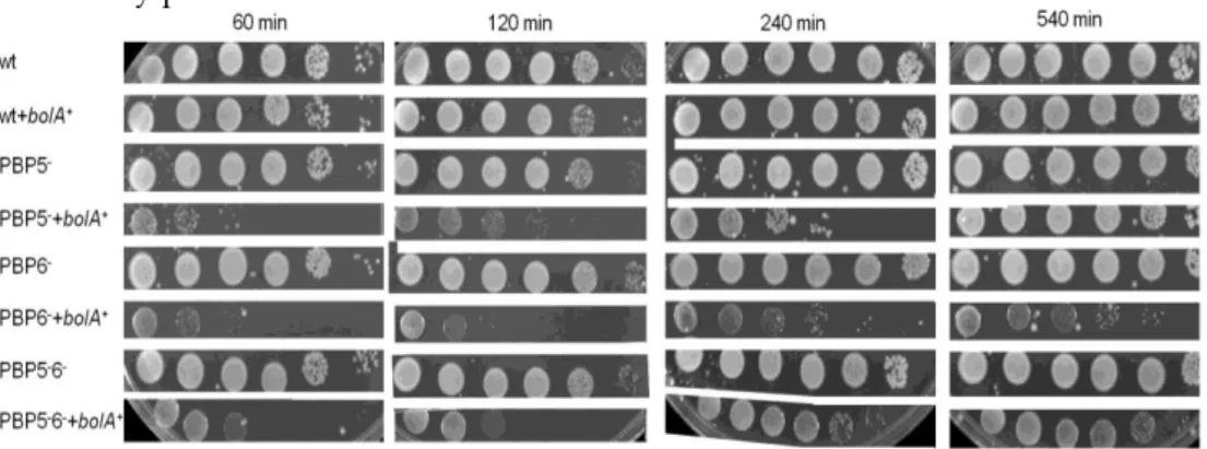
The second chapter shows that overproduction of BolA affects bacterial growth
differently depending on whether the cells are inoculated directly from plate or
from overnight batch cultures. It furthermore demonstrates that BolA is a
transcriptional regulator of the dacA and dacC genes, thus regulating both
DD-carboxypeptidases PBP5 and PBP6. It is shown that some BolA cellular effects
depend on PBP5 or PBP6.
In the third chapter we have evaluated BolA as a putative persistence protein,
inducing a decline in the multiplication potential but increasing toleration against
different stresses imposed.
In the fourth chapter the BolA homologue protein YrbA (renamed as IbaG) was
studied as a possible BolA protein homologue. Although these two proteins did
not induce similar phenotypes, they both induced a decrease in growth and
viability when overexpressed, and increased ODs and cell counts in the respective
deletion strains. IbaG did not change the morphologies of cells in any of the
conditions tested. This gene is in an operon with the essential murA gene. IbaG is
induced in acid stress conditions, and therefore was renamed as induced by acid
gene. It has furthermore conferred advantages for growth upon mild acid
xxvi
In the fifth chapter a General Discussion puts in perspective the new information
presented throughout the above chapters.
In the sixth chapter perspectives are provided regarding the future work on BolA,
taking into account several other mechanisms that BolA might interfere with.
General perspectives give suggestions that can be developed by other lab
colleagues currently working on BolA.
The seventh chapter is an appendix presenting some preliminary work related to
BolA, that has been started but is still incomplete. In Appendix I the role of Hfq
and glucose are evaluated in bolA regulation. In Appendix II we have developed
tools to evaluate E. coli bolA in pathogenic species belonging to the Burkholderia
cenocepacia complex.
A lot more work needs to be implemented into true knowledge about BolA; it is
my conviction that this molecule has still a lot to offer. But, like a precious
untouched diamond, it needs a gigantic amount of ability, work and most of all
patience to set it free.
xxix Amp ampicillin
Bcc Burkholderia cepacia complex
bp base pair
bolA1p promoter P1 of bolA gene
bolA2p promoter P2 of bolA gene
BSA bovine serum albumin
ºC degree Celsius
Cam chloramphenicol
cfu colony forming units
CF cystic fibrosis Δ deletion
Da dalton
DTT dithiothreitol
dATP 2’-deoxyadenosine
5’-triphosphate
DNA deoxyribonucleic acid
DNase deoxyribonuclease
E.coli Escherichia coli
EDTA EthyleneDiamineTetraAcetic
acid
EPS exopolysaccharide
h hour
HMM High Molecular Mass
IPTG IsoPropyl-β-D- thiogalactopyranoside
Kan kanamycin
kb kilobase
KD dissociation constant
kDa kilodalton
L liter
LB Luria-Bertani Broth
LMM Low Molecular Mass
log logarithm
M molar/molarity(mol/L)
M9 M9 minimal medium
mg milligram
µg microgram
µl microliter
µM micromolar
min minute
ml milliliter
mM millimolar
mmol millimole
mol mole
mRNA messenger RNA
MW molecular weight
Nal nalidixic acid
ng nanogram
nM nanomolar
nm nanometer
nmol nanomole
nt nucleotide
OD optical density
o.n. overnight
OM outer membrane
xxx
32P phosphorus 32 radionucleotide
PAA polyacrylamide
PAP I Polyadenilation Polymerase I
PAGE polyacrylamide gel
electrophoresis
PBP penicillin-binding-protein
PBS phosphate buffered saline
buffer
PCR polymerase chain reaction
PG peptidoglycan
pmol picomole
PNPase PolyNucleotide
Phosphorilase
psi pressure unit
RNA ribonucleic acid
RNase ribonuclease
RNaseII ribonuclease II
RNaseIII ribonuclease III
rpm rotations per minute
rRNA ribosomal RNA
RT reverse transcriptase
s second
SDS sodium dodecyl sulphate
SOB Super Optimal Broth
SOC SOB with glucose
SOS stress response pathway
triggered by DNA injuries
sRNA small RNA
SSC sodium chloride and sodium
citrate buffer/washing solution σ70 sigma 70 subunit of RNApol
(encoded by the rpoD gene)
σS sigma S subunit of RNApol
(encoded by the rpoS gene)
Tet tetracycline
Thy thymine
Tmp trimethropim
Tris trishydroxymethyl
aminomethane
[2-amino-2-(hydroxymethyl) propane-1,3-diol]
tRNA transfer RNA
UTP uracil triphosphate
UV ultraviolet radiation
V volt
v/v volume/volume
wt wild-type
w/v weight/volume
Z-ring ring of polymerized FtsZ,
that forms at the midcell in the
3 BolA is a protein that is able to change bacterial shape, confer resistance against
large antibiotic molecules and detergents, reduce permeability, change the
equilibrium of the outer membrane porins, and it is even involved in biofilm
formation. This protein has such pleiotropic effects, that its function has been
very difficult to unravel. This was the starting point for the work of this
dissertation. If bolA is responsible for global cellular changes that confer resistance to a multitude of stresses, it is imperative to obtain more molecular insights to
increase the understanding of the role of BolA in cell physiology and survival.
The first aim of this Doctoral work was to further investigate the relation of BolA
with cell wall protein intervenients and cytosqueletal elements. The fact that the
levels of BolA in cells can change considerably and the inexistence of proper
antibodies for its quantification was an obstacle. Another difficulty was that PBP5
and PBP6 share characteristics: they are quite similar in aminoacidic composition,
molecular weight and even isoelectric point. Therefore, there are substancial
methodological constraints to separate and evaluate their levels. However we
could establish that at least one of them is required for the effectiveness of BolA in
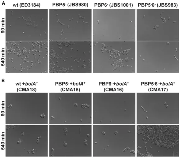
cells. In fact, the double deletion mutant for PBP5 and PBP6 was the only strain in
which the overexpression of BolA did not lead to the occurrence of spheres.
Additionally it was possible to ascertain, by Surface Plasmon Resonance analysis,
that BolA is a transcriptional regulator that negatively affects mreB and positively affects dacA and dacC. Although interaction occurs preferentially in the promoter region of these genes (where transcriptional regulation actually takes place) it was
reported to occur as well (to a lesser extent) in the Open Reading Frame regions
of these genes.
A second objective of this dissertation work was to understand the effects of the
4
increasingly expressed along the growth curve, starting the growth at different
time points/ODs was expected to correspond to starting growth with different
intracellular BolA levels. A work plan was established where starting from
OD620nm=0 (directly from colonies), a series of ODs were defined after the first
liquid cultures to initiate growth in fresh media. BolA is important enough to be
maintained in genomes throughout the living world, nevertheless, when it is
present in high levels, it prevents cultures from growing and dividing, strongly
reducing their viabilities. This work led to some unexpected results. Even when
overexpressed, the BolA-induced slowing of division rate does not seem to
represent a killing program or senescence process, given that the strong viability
reduction is not progressive along time. Even though BolA seems to prevent
division, when bolA overexpressing cultures are started with the inoculi at lag or early exponential phase they grow even better than wild type ones. In this way it
seems that BolA creates an enhanced growth potential, released upon its
clearance from the system. That could be an important mechanism to deal with
stress imposition, reducing short term growth but favoring long term resistance
and maintenance. This lead us to evaluate BolA potential as a persistence protein,
in the way those proteins have been recently studied.
A third objective came after genome wide sequence/structure prediction analysis
detected a protein homologous to BolA in E. coli. The possibility of a functional substitution by a similar protein projected that a double bolA/yrbA deletion would not be viable but this work has refuted this idea. In fact, not only the single yrbA
deletion presents a better growth and viabilities than the wild type, but the
double deletion mutant could be constructed and grows even better than the
single deletant. Conversely, an increase in the amounts of yrbA (as tested through
5 have effects on cells morphologies, again differently of BolA. The mRNA
expression pattern was quite low without overexpression but in the presence of
the pBGA01 plasmid it could be seen that this transcript is much more present in
mid exponential phase than in lag or stationary phase. The response of yrbA to stress was studied by Northern Analysis. It was shown that yrbA was induced in acid stress conditions. Furthermore the overexpression of yrbA led to increased growth upon acid challenge, therefore we named this gene ibaG, for induced by acid gene. In comparison with the wild type, the ibaG deletant strain grew the best in neutral medium but the worst in acid. The ibaG overexpressing strain grew worst in pH7 but was the strain which grew the best in pH5. The possibility
of ibaG levels being controlled by BolA was tested by Surface Plasmon Resonance analysis of interaction, and by transcriptional analysis using GFP protein as
signal. Both these methods ruled out of direct interference although it seemed
that a minimal bolA expression was necessary for proper yrbA transcription.
This work proved to be enlightening in the evaluation of BolA as a transcriptional
factor, and involved in persistence mechanisms: reduction of metabolism,
division, stress protection and boost in growth potential after system clearance.
Furthermore, the discovery of IbaG, a BolA related protein, that facilitates
survival under acid stress, is quite significant and it opens new perspectives for
9 A proteína BolA Escherichia coli induz a alteração da forma de bacillus a coccus,
confere resistência a detergentes e antibióticos de elevado peso molecular, reduz a permeabilidade, altera o equilíbrio de porinas da membrana externa e ainda induz o desenvolvimento de biofilmes. Apresentando efeitos tão abrangentes e induzindo resistência a uma multitude de stresses, o estudo da função do gene/proteína BolA constituiu o objectivo geral do trabalho que conduziu a esta Dissertação. É imperativo obter conhecimentos de nível fundamental sobre a biologia molecular de BolA, seus alvos, formas de regulação e consequências globais na fisiologia e sobrevivência celulares.
O primeiro objectivo deste trabalho de Doutoramento foi investigar uma eventual relação de BolA com proteínas de membrana e elementos do citosqueleto. A inexistência de bons anticorpos anti-BolA constituiu um obstáculo. Outra dificuldade deveu-se à similitude das proteínas PBP5 e PBP6 - em sequência de aminoácidos, peso molecular e até de ponto isoeléctrico – impossibilitando a determinação dos seus níveis isoladamente. No entanto, foi possivel verificar que os efeitos da presença excessiva de BolA só ocorrem na presença de pelo menos uma das duas DD-carboxipeptidases. De facto, apenas no delectante duplo PBP5/PBP6 com sobreexpressão de bolA se conseguiram obter curvas e
morfologias semelhantes às das estirpes não transformadas com pMAK580. Além disto, foi possível verificar por Surface Plasmon Resonance que o BolA é um
regulador transcricional, , actuando como activador para com os genes dacA e dacC, e como repressor para com mreB. Muito embora as interacções tenham sido
preferenciais com a região promotora dos genes que regula, verificou-se também interação (menor) com as grelhas abertas de leitura respectivas.
10
aprofundar os resultados do primeiro estudo, obtidos para crescimentos de estirpes com sobreexpressão de bolA. Tendo em conta que ainda não é possível
quantificar os níveis proteicos de BolA, e sabendo-se que a expressão deste é aumentada progressivamente com a curva de crescimento, desenvolveu-se um plano de trabalho em que a estirpe com sobreexpressão de bolA, CMA10, seria
avaliada quanto ao crescimento, viabilidades e morfologias, quando iniciada a partir de diferentes densidades ópticas (ODs). O crescimento líquido desta estirpe seria iniciado a partir de placa (OD620nm=0) e à medida que a cultura atingisse as
diferentes ODs predefinidas (0.5, 1.5, 3 e 6) seria utilizada sucessivamente como pré-inóculo para lançamento de novas culturas. Verificou-se que quando em níveis elevados o gene bolA dificulta o crescimento das culturas e reduz o número
de células viáveis. Ainda assim, este decréscimo de viabilidades não corresponde a um processo de senescência ou morte, tendo em conta que ao longo do tempo as unidades formadoras de colónias não diminuem. Outro resultado surpreendente foi o facto de as mesmas culturas iniciadas após um pré-inóculo em fase lag ou início de exponencial crescerem ainda melhor que as selvagens. A presença de BolA aparenta originar uma repressão temporária de crescimento celular, mas simultaneamente um potencial de crescimento rápido despoletado com o seu desaparecimento do sistema. Esta redução de crescimento a curto prazo e favorecimento a sobrevivência a longo prazo levou-nos também a avaliar o potencial do BolA enquanto hipotética proteína de persistência.
O terceiro objectivo surgiu por análise informática na qual se detectou em E. coli
uma hipotética proteína homóloga a BolA, designada por YrbA. A possibilidade de substituição funcional do BolA pelo YrbA postulava que o delectante duplo
bolA/yrbA não seria viável, o que foi refutado no âmbito deste trabalho. O
delectante simples yrbA tem melhor crescimento e viabilidades associadas que a
11 que ainda apresenta melhores resultados que o delectante simples. Em conformidade com estes resultados, a expressão aumentada de yrbA (testada
através de construção de pBGA01, um plasmídeo derivado de pBr325, sobreexpressando o yrbA) originou um decréscimo tanto nas curvas de
crescimento, como nas viabilidades. A expressão de yrbA aumentada não
originou alterações de morfologia às células em qualquer das condições testadas. A expressão endógena de mRNA mostrou-se demasiado fraca para ser detectada, mas na presença de pBGA01 foi possivel verificar que o transcrito é muito mais expresso a meio da fase exponencial que em fase lag ou estacionária. A resposta do yrbA ao stress também foi avaliada por Northern Blot e verificou-se que este é
induzido em pH ácido, pelo que foi designado de ibaG, traduzindo, gene
induzido por ácido. Confirmou-se por SPR e fluorescência (análise transcricional usando a Green Fluorescent Protein como sinal) que o BolA não interage com a
região promotora do gene em estudo, muito embora a sua presença interfira positivamente para a transcrição do yrbA.
Chapter 1
15 INDEX
COVER ...17
ABSTRACT...18
BRIEF BOLA OVERVIEW ...19
GRAM-NEGATIVE SURVIVAL UNDER STRESS CONDITIONS...20
Cellular and physiological responses to stress...20
The universal stress response regulator S sigma-factor...21 Finely tuned regulation of bolA ...23
BolA, a stress response effector protein...25 BolA “MORPHOGENETIC DETERMINISM”...26
Peptidoglycan synthesis and properties……….27
Cell wall elongation progress...29
The hierarchical, highly regulated and time controlled divisional process...30
Cell wall maturation/plasticity/protection by Low Molecular Weight PBPs...33
BolA regulation over the DD-carboxypeptidases PBP5 and PBP6...34
The bacterial cytosqueleton...35
BolA and the cytosqueleton elements ...38
PERMEABILITY MODULATION AND BIOFILM DEVELOPMENT AS STRESS
ADAPTATION MECHANISMS...39
SYNOPSIS AND FUTURE PERSPECTIVES ...41
BolA homologues ...41
Function on persistence ...44
17
BolA, a central
E. coli
regulator:
Impact on Gram-negative cell wall regulation
and adaptation to stresses
Inês Batista Guinote1, Ricardo N. Moreira1, Patrick Freire1,2 and Cecília Maria
Arraiano1
1Instituto de Tecnologia Química e Biológica/Universidade Nova de Lisboa,
Apartado 127, 2781-901 Oeiras, Portugal
2Present address: Laboratório Nacional de Investigação Veterinária - INRB,
Estrada de Benfica 701, 1549-011 Lisboa, Portugal
Correspondence to: Cecília M. Arraiano
Phone: +351 214469547
Fax: +351 214469549
E-mail: [email protected]
Running title: BolA mediated protection to stresses
Keywords: BolA, transcriptional regulator, phenotypical differentiation, stress
survival, MreB, LMW PBP, gram-negative, cell division
18
ABSTRACT
Microorganisms have continuously evolved to divide and persist in harsh
conditions. Bacteria deal with external stresses on a regular basis, many times
through major restructuration such as biofilms formation. The adaptation
mechanisms usually imply changes in cell shape, roughness, dimensions, protein
content, as well as alterations of cell wall structure, thickness and permeability.
The E. coli morphogene bolA ismainly expressed in stress conditions and when
overexpressed induces rod bacteria to become spherical. Moreover, BolA is able
to induce biofilm formation and to make changes in the outer membrane, which
gets less permeable to detrimental agents. Although there is some understanding
about the activity of BolA there is no real comprehension of its function on global
cell physiology. Does it actually protect from stress? Is that achieved by
regulation of other genes? Does it happen in complement with BolA homologues?
BolA family of proteins are found throughout the living world with the
remarkable exception of the Gram-positive bacteria. This may be due to structural
differences in their cell walls (absence of outer membrane, thicker murein layer,
presence of teichoic acids), or be somehow related to their exclusive ability to
resist stresses through differentiation of spores. Several new questions arise as
investigation proceeds into more profound understanding of this interesting
19 BRIEF BOLA OVERVIEW
Bacteria are able to activate several adaptive responses when facing any form of
stress. Alterations of cellular morphology and membrane characteristics are
important to regulate the internal exchanges with the environment. Plastic
membrane configuration, structure and regulation are beneficial to facilitate
solutes and proteins interchange, thus maximizing environmental interactions in
optimal growth conditions, and on the other hand promote a differentiation into a
more rigid structure, with less porins to ensure permeability reduction, under
unfavourable conditions. Thegene bolA was first described as a stationary phase
gene (Aldea et al., 1990; Aldea et al., 1989). Accordingly, the expression of bolA is
growth phase-regulated and controlled by the sigma factor S (rpoS gene) (Lange
and Hengge-Aronis, 1991). However, bolA has also been established as a general
stress response gene induced during early logarithmic growth in response to
several stresses, in a partially S-independent manner (Santos et al., 1999). Greater
amounts of bolA mRNA and a rounder cell morphology were found after
application of different sources of stress, such as carbon starvation, osmotic stress,
heat shock, acidic stress and oxidative stress (Santos et al., 1999). The bolA gene is
therefore an E. coli morphogene whose product induces spherical shape of the
cells when overexpressed, leading to a reduction of the exposed surface (Aldea et
al., 1988). This effect of bolA on cell morphology is mediated both by the induction
of PBP5 and PBP6 D,D-carboxypeptidases (Aldea et al., 1988; Guinote et al., 2010;
Santos et al., 2002)and the repression of the expression of MreB (Freire et al.,
2009), a bacterial homologue of actin responsible of the formation of an helical
cytosqueleton structure required for rod shape configuration in E. coli
(Carballido-Lopez, 2006). Escherichia coli 13.5 kDa BolA protein contains one
defined BolA/YrbA domain, essentially formed by a helix-turn-helix (HTH) motif
20
1989; Freire et al., 2009; Guinote et al., 2010) (Fig.1). It was proven that BolA
efficiently binds to the mreB, dacA and dacC promoter regions. It downregulates
mreB and upregulates dacA and dacC promoters, expanding its potential as a
transcriptional regulator, with repressor and activator abilities. Moreover, the
increased expression of the bolA gene was shown to be an important inducer of
biofilm formation (Vieira et al., 2004) and to modulate cell permeability (Freire et
al., 2006b). As a result, BolA constitutes a privileged target to study molecular
mechanisms of adaptation of Escherichia coli when facing adverse growth
conditions.
GRAM-NEGATIVE SURVIVAL UNDER STRESS CONDITIONS
Cellular and physiological responses to stress
E. coli cells possess several other mechanisms to cope with nutritional or toxic
stresses, besides the abilities to grow in aerobic conditions, perform anaerobic
fermentation and even respirate in the absence of oxygen. While many
Gram-positive bacteria differentiate long-lasting highly resistant spores, Gram-negative
bacteria survive for prolonged periods of starvation or multiple stresses through
physiological state adaptations designated as stationary phase. Challenged cells
show a variety of adaptations in cellular structure, morphology and physiology,
like flagella, pilli or fimbriae development, variations in diameter of the pores of its
outer membrane to accommodate larger molecules (nutrients) or to exclude
inhibitory substances, synthesizing enzymes to metabolize available compounds
or either expel or degrade toxic ones (Halsey et al., 2004). The enhanced stress
resistance exhibited by starved bacteria represents a central feature of virulence,
since nutrient depletion is regularly encountered by pathogens in their natural in
vivo and ex vivo environments (Frenkiel-Krispin et al., 2001).
Upon sensing an impending saturation level of their population density,
21 modulating the nucleoid, the transcription apparatus, the translation and
proteolytic machinery. Their cytoplasm is condensed and storage compounds and
protective substances are synthesized such as glycogen, polyphosphate or
trehalose. The volume of the periplasm increases, changes in peptidoglycan
composition and structure occur, that result in a higher resistance to autolysis
induced by penicillin or chaotropic agents. Cells tend to become smaller, develop
an ellipsoid to spherical rather than a rod morphology, and have an increased
tendency to form aggregates (Hengge-Aronis, 1999; Hengge-Aronis, 2002). The
major DNA-binding proteins, Fis, HU and Hfq, in the exponential-phase nucleoid
are replaced by a single stationary-phase protein Dps, and cellular content of the
histone-like protein H-NS increases, thereby compacting the nucleoid and
ultimately leading to silencing of the DNA functions (Ali Azam et al., 1999)
(Ishihama, 1999). The transcription apparatus is modified by replacing the major
promoter recognition subunit σ70 by σS factor in RNA polymerase complex. Hfq
becomes mainly accumulated at the cytoplasm, dynamically shifting the
transcripts’ stabilities. The translation machinery is also modulated by the
stationary-phase RMF (Ribosome Modulation Factor) that mediates direct 30S
dimer contacts between 70S ribosome monomers at the P site (Azam et al., 2000)
(Ishihama, 1999). Those translationaly incompetent 100S ribosomes may consist of
inactive storage structures that remain available for future fast resumption of
growth, once conditions change to favourable – ribosomal “hibernation” stage
(Yoshida et al., 2002). Proteolysis by the ClpXP and ClpAP complexes appears to
be mandatory to extended stationary-phase bacterial cultures viability,
controlling the levels of growth–phase regulated proteins (Weichart et al., 2003).
The universal stress response regulator σS sigma-factor
Transition into stationary phase induces the formation of a core set of several
22
stationary-phase cells are more resistant to several stresses (heat shock, H2O2,
NaCl, alkylating agents, ethanol, acetone, toluene, deoxycholate, acidic or basic
pH). Among those is the σS subunit of RNA polymerase, a major regulator of
many stationary-phase-inducible genes, that controls several morphological and
physiological processes in survival to prolonged starvation periods
(Hengge-Aronis, 1996) (Loewen et al., 1998). A reduced growth rate results in increased
rpoS transcription whereas high osmolarity, low temperature, acidic pH, and
some late log-phase signals stimulate the translation of already present rpoS
mRNA. In addition, carbon starvation, high osmolarity, acidic pH, and high
temperature result in stabilization of σS protein, which otherwise is degraded
(Hengge-Aronis, 2002) (Hengge-Aronis, 1996). Important cis-regulatory
determinants as well as trans-acting regulatory factors involved at all levels of σS
regulation have been identified: rpoS mRNA stability and translation are
controlled by several proteins (Hfq, H-NS, HU, LeuO, CspC, CspE, cAMP-CRP
complex and even DnaK) and by small regulatory RNAs (dsrA RNA and OxyR
RNA). The molecular mechanisms of signal transduction that trigger the
stationary-phase response and the induction of σS have yet to be completely
elucidated, but seem to involve molecular signals as ppGpp, UDP-glucose, and
homoserine lactone. Polyadenylation decreases the recognition of σS specific by
the specific factor RssB (also designated by SprE (Carabetta et al., 2009)), essential
for σS proteolysis by ClpXP protease. After translation, sigma S activity is still
positively modulated by trehalose and glutamate (Phadtare and Inouye, 2001)
(Hengge-Aronis, 1996) (Hengge-Aronis, 2002) (Loewen et al., 1998).
Sigma Scontrols the expression of genes that respond to general stresses, usually
involved in the uptake and metabolism of aminoacids, sugars, iron and
production of indole, among others. It also regulates several regulatory proteins,
23 and Landini, 2004), the Dps protein (DNA-binding protein) and CsgD,
homologous to the DNA binding domains of the LuxR family; and BolA.
FINELY TUNNED REGULATION OF bolA
bolA has been described as an ftsZ dependent gene, which is required to produce
bolA-mediated round cells. By analysis of bolA open reading frame (ORF), it was
showed that it encodes a protein with a predicted molecular weight of 13.5 kDa,
transcribed in a clockwise direction on the E. coli chromosome. This gene is
regulated by two different promoters, a P1 promoter, under the control of σS and an upstream P2 promoter, controlled by σD (Fig. 2). Moreover, bolA mRNA
initiated by P2 is always detected along bacterial growth, in low amounts, being
classified as a weak and constitutive promoter (Aldea et al., 1989; Lange and
Hengge-Aronis, 1991). However, transcripts originated by P1 promoter are
primarily detected when cells enter late-exponential phase of growth, beginning
of stationary phase or upon imposition of stress conditions with a concomitant
decrease of bolAp2 mRNA levels (Aldea et al., 1989). This observation gave rise to
an hypothesis of promoter occlusion, stating that the physical interaction of the
transcription machinery by σS at the bolA1p promoter, could interfere with the
ability of RNA polymerase to either interact or proceed from bolA2p (Santos et al.,
1999).
Figure 1. BolA protein structure.
2DHM PDB was taken from
http://www.pdb.org/ and the
cartoon picture created using pymol
24
Figure 2. bolA1p (P1) promoter is mainly regulated by the S transcription factor and induced in
stationary phase and stress conditions. bolA2p (P2) promoter is 70-dependent and is constitutively
expressed. OmpR, H-NS, and CRP-cAMP downregulate while ppGpp upregultes bolA transcription. Both ribonuclease III and Hfq stabilize bolA1p transcript. Question marks indicate regions of interaction or cut yet to be confirmed.
P1 promoter is growth phase and growth rate regulated and contains a gearbox
element characteristic of several other structural genes, some of which are also
induced in stationary phase. These type of promoters are induced inversely to the
growth rate (Aldea et al., 1989). Additionally, it was also observed that the bolA
P1 promoter, similarly to rpoS (encoding σS) can be downregulated by cAMP
levels (Lange and Hengge-Aronis, 1991). At the transcriptional level, ribonuclease
III has also an important role in the regulation of the expression of the bolA gene
(Freire et al., 2006a; Santos et al., 1997). RNase III is a double-stranded
endoribonuclease widely distributed among prokaryotic and eukaryotic
organisms. Moreover, it is encoded by the rnc gene, and is active as a 52 kDa
homodimer (Arraiano et al., 2010). RNase III is described as responsible for rpoS
mRNA regulation, acting as modulator of S levels in glucose starvation, ensuring σS normal levels of expression (Freire et al., 2006a; Santos et al., 1997). Since RNase III regulates σS levels, it indirectly regulates the levels of bolA mRNA.
Another factor controlling bolA mRNA is polyadenylation, which inversely
25 RssB-mediated ClpXP σS proteolysis, increasing RpoS protein levels, thus
contributing for the transcription of σS dependent genes like bolA (Santos et al.,
2006). Additionally, Yamamoto and co-workers, saw that bolA1p is negatively
regulated in vitro by phosphor-OmpR (Yamamoto, 2000).
At the post-transcriptional level, under carbon starvation, RNase III is also
involved in the bolA1p stability. The bolA1p mRNA is induced nine fold in a wt
strain while in an rnc mutant strain is fourfold induced, showing a decrease in
bolA1p RNA in the absence of RNase III (Freire et al., 2006a). It has been shown
that the stationary phase messengers such as rpoS and bolA are polyadenylated
(Cao and Sarkar, 1997). Therefore, polyadenylation probably interferes in their
post-transcriptional control.
BolA, a stress response effector protein
Similarly to rpoS, bolA induced gene expression occurs at the transition to
stationary phase related to the decrease in cells growth rate, but also in
exponential phase after imposition of virtually any stress condition (Aldea et al.,
1989; Lange and Hengge-Aronis, 1991; Santos et al., 1999).
In all stress trials developed, BolA was overexpressed and its cellular effects were
noticeable, in spite of some differences. Sudden carbon starvation and increased
osmolarity, respectively, resulted in about 17-fold and 22-fold increase in mRNA
levels derived from bolA1p 1 h after stress imposition on exponential phase of
growth (more than three and fourfold of what happened in stationary phase). A
threefold increase of bolA1p mRNA levels is registered in response to both heat
shock and acidic stress. Heat shock induction of bolA1p is immediate (maximum
after 15 min), and mRNA levels reduction along time relate to significant increase
in optical densities and moderate recovery in viabilities (possibly due to favoring
elongation to division). The acid induction of bolA1p expression is more gradual,
26
contrast, the bolA1p mRNA gradual increase, up to eight times, 2 hours after the
onset of H2O2 oxidative stress, and inhibits both growth and viabilities (Santos et
al., 1999). bolA1p stress induction overrides the normal regulation imposed by
growth rate, which is strictly the result of σS-directed transcription. In fact, there
is differential dependence on σS for induction of bolA1p mRNA levels under
different stress conditions, and certain basal stress response operate in the
absence of σS. The stress-dependent activation of bolA1p should confer E. coli
Gram-negative bacteria protection against a variety of stresses, based on the
reduction of surface area exposed to damaging agents while decreasing the
surface to volume ratio and promoting biofilm formation (Santos et al., 1999).
BolA “MORPHOGENETIC DETERMINISM”
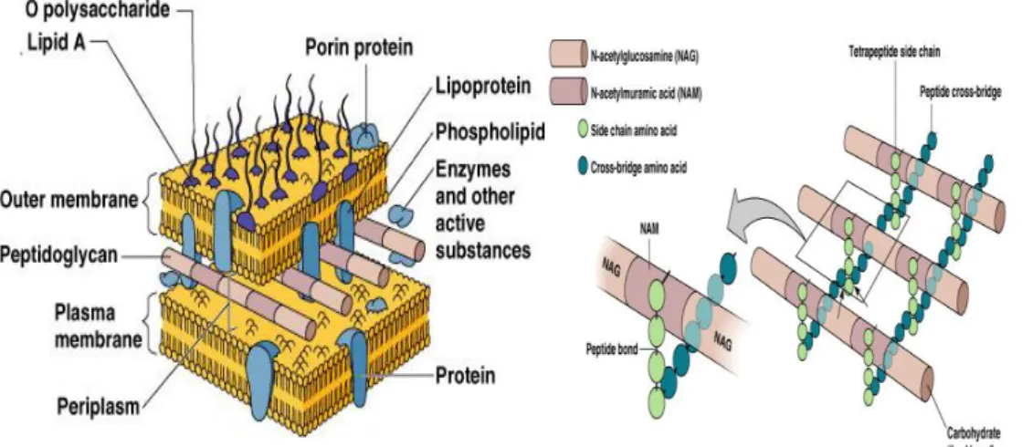
BolA effects are pleiotropic, but a special interest goes to cell wall related
mechanisms (Fig. 3), since bacteria morphology is profoundly affected by an
increase in bolA levels (Fig. 4).
BolA can act over elongation, peptidoglycan synthesis and maturation or cell
division mechanisms, or even regulating cytosqueletal elements.
Figure 3. Gram-negative bacteria membrane and cell wall organization with particular incidence on
27
Peptidoglycan synthesis and properties
Peptidoglycan is a vital bacterial macromolecule, around 6 nm thick (Matias et al.,
2003), that provides a structure that maintains cell shape, mediating interactions
and regulating exchanges between the internal cellular media and their
environment (Hughes et al., 1975; Vollmer et al., 2008). It constitutes the sacculus,
that presents a flexibility due to the ability of changing cell length while
maintaining the diameter virtually unaltered (van den Bogaart et al., 2007). Even
though murein contains and maintains bacterial size and shape, those are not
merely determined based on the chemical composition or structure of the
peptidoglycan polymer, but depends also on cellular morphogenetic apparatus
since restoration of murein in spheroplasts leads to the formation of round sacculi
(Schwarz and Leutgeb, 1971; Weidel et al., 1960). Polar murein is not differentially
composed from the lateral one, even though it is metabolically inert and
significantly less deformable, eventually due to predominant alignment of the
(more flexible) peptides in the direction of the long axis with the (more rigid)
glycan strands perpendicularly arranged (Boulbitch et al., 2000). When septation
was prevented, patches of stable murein were also observed at regular intervals,
corresponding to future poles of filaments (Rothfield, 2003). Polar inert
peptidoglycan influences outer membrane proteins mobility, the free movement
of non-reacting protein species, and secretion generating asymmetries (den
Blaauwen et al., 2008).
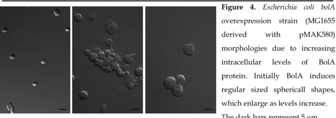
Figure 4. Escherichia coli bolA
overexpression strain (MG1655
derived with pMAK580)
morphologies due to increasing
intracellular levels of BolA
protein. Initially BolA induces
regular sized sphericall shapes,
which enlarge as levels increase.
28
During exponential growth, there is a fast and progressive enrichment of the
murein sacculus in newly synthesized trimers and dimers of lipoprotein-bound
muropeptides at the expense of the respective monomers, at the tetra to tripeptide
side chains and not at the oligosaccharide glycan chains of the peptidoglycan
precursors (Burman and Park, 1983). Particularly in resting cells, and
progressively when cultures are reaching stationary phase, murein globally
changes its structure becoming hypercross-linked and richer in covalently bound
lipoprotein, while reducing the mean length of the glycan chains. These
modifications may provide extra resistance, hardening the damaging agents
entering into cells, and the eventual peptidoglycan hydrolysis, although they
might also complicate the cell wall metabolism and thus require structural
changestooccur when in transition to active growth (Pisabarro et al., 1985).
Duplication of bacterial cells is quite demanding in terms of processes that need
to occur in synchrony. Before septation, cells seem to require a minimum of
previous lateral elongation, a process which demands not only membrane and
cytosqueleton expansion and rearrangements, but also extension of the murein
sacculus (den Blaauwen et al., 2008; Donachie and Begg, 1989; Grover et al., 1977).
In fact, when rod cells are imposed spherical forms (using rodA or pbpA sensitive
mutants) they maintain cell length and the ratio of DNA/mass, in a way that cocci
increase both their volume and DNA contents four to six times, thus suggesting a
minimum cell length for nucleoid separation (Donachie and Begg, 1989). The
peptidoglycan extension occurs by addition and polymerization, into the
pre-existent sacculus, of new alternating acetylglucosamine (GlcNAc) and
N-acetylmuramic acid (MurNAc) nucleotide sugar-linked –
disaccharidepentapeptide units - precursor strands, that later on become
29 Polymerization of the peptidoglycan precursors is operated by transglycosylases
– the High Molecular Weight (HMW) PBPs PBP1a, PBP1b, PBP1c, PBP2 and PBP3
- and cross-linking is catalysed by transpeptidases (Ghuysen, 1991; Holtje, 1998;
Sauvage et al., 2008). At least the remodelling of the murein sacullus is mediated
by carboxypeptidases (which remove the terminal D-ala residues from the
pentapeptide) and endopeptidases (which hydrolyse the glycan chains’ cross
-links) and eventually hydrolases like amidases and LD-carboxypeptidases
(Holtje, 1998; Nicholas et al., 2003; Priyadarshini et al., 2007; Templin, 2004).
Cell wall elongation progress
Bacterial elongation occurs by simultaneous incorporation of precursors in small
number - since some of the components of the elongation machinery are scarce
(20 to 40 molecules of PBP2 per cell) (Den Blaauwen et al., 2003; Spratt, 1975;
Spratt, 1977). They locate along MreB filaments (interacting with MreC and D
(Dye et al., 2005; Figge et al., 2004)) and FtsZ spirals (Daniel and Errington, 2003;
den Blaauwen et al., 2008). Synthesis occurs by insertion of cross-linked glycan
strands (Bertsche et al., 2005; Born et al., 2006; den Blaauwen et al., 2008) thus
creating “mosaic structures made up of (…) all-new and all-old peptidoglycan”
with fast turn-over (De Pedro et al., 2003a; den Blaauwen et al., 2008; Koch and
De Pedro, 2006). Elongation may either be a continuous process along cell cycle or
stall while PBP2 is relocated to the constriction site helping in the septation
process, in a way that cell poles would be synthesized both by lateral and septal
mode, given that its blockage inhibits peptidoglycan synthesis in a constant value
(60%). Blocking PBP3 in turn reflects accordingly to the constriction processes:
about 35%, periodically (Thibessard et al., 2002; Wientjes and Nanninga, 1991;
30
The hierarchical, highly regulated and time controlled divisional process
Upon polymerization of the Z-ring (see below “The bacterial cytosqueleton”),
several other proteins concur to the midcell contributing to the septation process.
This is a very well defined hierarchic, constitutionally interdependent and time
controlled event (Fig. 5).
Initially FtsZ filaments interact with FtsA (47KDa) in a short conserved
C-terminal end site that overlaps the one of ZipA where they both modulate the
protofilaments stability by tethering them to the membrane (Addinall and
Lutkenhaus, 1996; den Blaauwen et al., 2008; Hale and de Boer, 1999; Lowe et al.,
2004; Pichoff and Lutkenhaus, 2007). ZapA or YshA is other 10KDa non-essential
protein that interacts with FtsZ rendering the filaments more stable and
enhancing bundle (Gueiros-Filho and Losick, 2002). Specifically in high
osmolarity conditions FtsZ-ring requires the interaction with the ABC transporter
FtsE/X to be assembled (Corbin et al., 2007; Schmidt et al., 2004).
S-adenosylmethionine transferase (SAM) is also essential to the divisome assembly,
eventually due to the inability of FtsQ, W, I and/or N to locate properly at the
Z-ring (Wang et al., 2005). FtsK is the first enzyme to be recruited, after the FtsZ Z-ring
is polymerized. It is a multifunctional and multidomain protein whose
cytoplasmic C-terminal domain is involved in DNA segregation and N-terminal
transmembrane domain is responsible for cell division (Bigot et al., 2007; Dorazi
and Dewar, 2000). Although ftsK null mutants are compensated when FtsA or
FtsQ are overexpressed, they tend to generate multiseptated filament cells
suggesting an additional role in final closure of the newly formed poles (Geissler
and Margolin, 2005). In addition, the deletion of dacA which encodes for PBP5 can
also reverse to rods the filaments formed due to a single mutation in one of the
transmembrane helices of ftsK44, while its increase induces the formation of oval
31 Figure 5. Schematic representation of the assembly of the components of the divisome. The boxed
proteins represent subcomplexes: (a) the Z-ring with FtsZ-FtsA-ZipA-ZapA, which interacts with
FtsE/X; (b) the subcomplex FtsQLB, which contains a heterodimer of FtsL and FtsB; (c) the subcomplex
FtsW-PBP3; (d) PBP1B and FtsN interact with PBP3 and could be part of the subcomplex FtsW-PBP3.
MurG, PBP2 and PBP5, AmiC and EnvC are located at the division and are part of the cell division
machinery. Dashed lines: interaction detected using a two-hybrid system; Solid lines: interactions
detected using different techniques. Copied from (den Blaauwen et al., 2008) by courtesy of Benoît
Wolff.
FtsQ, L and B are membrane proteins bearing a small N-terminal intracellular
region connected to the C-terminal periplasmic domain through a
transmembrane helix (Chen et al., 2002; D'Ulisse et al., 2007; Scheffers et al., 2007;
van den Ent et al., 2008). FtsQ interacts with PBP3, FtsW and FtsN (D'Ulisse et al.,
2007; Di Lallo et al., 2003; Karimova et al., 2005) and is furthermore able to back
recruit FtsK, making it a good candidate for the regulation of new pole synthesis
(Goehring et al., 2005). FtsL and B complex is enough to recruit FtsW and PBP3 to
the septasome. It thus seems that later division proteins group into complexes
previously to their final arrangement, perhaps allowing to overcome eventual
flaws and ensuring survival (Goehring et al., 2006). Similarly to the elongation
machinery, these complexes are limited in numbers- there are about 20-40 FtsQ
32
PBP3 is targeted to the septa by its first 56 N-terminal amino acid residues shown
to interact with FtsW, the first 70 with FtsQ, the periplasmic non-catalitic module
with FtsL, apart from the C-terminal penicillin-binding transpeptidase activity
module (Adam et al., 1997; Karimova et al., 2005; Piette et al., 2004). This protein
is additionally responsible for the localization of PBP1b at the division site
independently of its activity and the complex interacts with FtsN (Derouaux et
al., 2008; Karimova et al., 2005). PBP1c then interacts with PBP3 and PBP1b, which
also interacts with FtsW (Derouaux et al., 2008; Schiffer and Holtje, 1999). Finally,
MipA, a structural scaffolding protein for murein, interacts with PBP1b and MltA,
an outer membrane lipoprotein and lytic transglycosylase that hydrolyses murein
(Lommatzsch et al., 1997); and MgtA, a monofunctional peptidoglycan
glycosyltransferase that catalyzes glycan chain elongation of the bacterial cell
wall, interacts with PBP3, FtsW, and FtsN (Derouaux et al., 2008). In terms of
localization, all the PBP1 bifunctional transglycosylase-transpeptidase family of
proteins seem to participate indifferently in the lateral and septal murein
synthesis (Bertsche et al., 2006).
AmiC, a periplasmic N-acetylmuramyl-L-alanine amidase, and EnvC, a murein
hydrolase, which locate at the Z-ring during constriction, are suggested to be
responsible for final cleavage of the septal peptidoglycan, consequentially
separating the daughter cells. At least AmiC requires FtsN for proper localization
and the enzymatic performance (Bernhardt and de Boer, 2004); (Bernhardt and de
Boer, 2003; Heidrich et al., 2001).
The peptidoglycan and some of the respective division machinery seem to be
essential for bacterial cell growth even in cells known to have no cell wall like
L-form-like E. coli and Chlamidial species (Joseleau-Petit et al., 2007; McCoy and
Maurelli, 2006). Cell division is truly inseparable of cell wall synthesis at
mid-cell/future poles (Joseleau-Petit et al., 2007). Besides the peptidoglycan and