Mem Inst Oswaldo Cruz, Rio de Janeiro, Vol. 112(11): 785-789, November 2017 785
online | memorias.ioc.fiocruz.br
Construction of Mycobacterium tuberculosis cdd knockout and
evaluation of invasion and growth in macrophages
Anne Drumond Villela1/+, Valnês S Rodrigues-Junior1, Antônio Frederico Michel Pinto1,
Virgínia Carla de Almeida Falcão1,2, Zilpa Adriana Sánchez-Quitian1,2, Paula Eichler1,
Cristiano Valim Bizarro1,2, Luiz Augusto Basso1,2,3, Diógenes Santiago Santos1,2
1Pontifícia Universidade Católica do Rio Grande do Sul, Centro de Pesquisas em Biologia Molecular e Funcional, Instituto Nacional de
Ciência e Tecnologia em Tuberculose, Porto Alegre, RS, Brasil
2Pontifícia Universidade Católica do Rio Grande do Sul, Programa de Pós-Graduação em Biologia Celular e Molecular, Porto Alegre, RS, Brasil 3Pontifícia Universidade Católica do Rio Grande do Sul, Programa de Pós-Graduação em Medicina e Ciências da Saúde, Porto Alegre, RS, Brasil
Cytidine deaminase (MtCDA), encoded by cdd gene (Rv3315c), is the only enzyme identified in nucleotide biosyn-thesis pathway of Mycobacterium tuberculosis that is able to recycle cytidine and deoxycytidine. An M. tuberculosis
knockout strain for cdd gene was obtained by allelic replacement. Evaluation of mRNA expression validated cdd deletion and showed the absence of polar effect. MudPIT LC-MS/MS data indicated thymidine phosphorylase expression was decreased in knockout and complemented strains. The cdd disruption does not affect M. tuberculosis growth both in Mid-dlebrook 7H9 and in RAW 264.7 cells, which indicates that cdd is not important for macrophage invasion and virulence.
Key words: Mycobacterium tuberculosis - cdd gene - cytidine deaminase
doi: 10.1590/0074-02760170105
Financial support: INCT-TB (Brazil), Decit/SCTIE/MS-MCT-CNPq-FNDCT-CAPES, CNPq (441720/2014-5 to ADV), BNDES (14.2.0914.1).
+ Corresponding author: [email protected] Received 15 March 2017
Accepted 30 May 2017
Tuberculosis is an infectious disease caused mainly by Mycobacterium tuberculosis. The World Health Or-ganization estimated that 10.4 million people developed tuberculosis in 2015, resulting in 1.8 million deaths (WHO 2016). Although an efficient chemotherapy ex-ists, affordable, short, effective and well-tolerated treat-ments for coadministration with anti-HIV agents, latent, drug-susceptible and drug-resistant tuberculosis are still needed to decrease the global incidence of the disease (Mdluli et al. 2015). The first step towards the search for novel therapeutic strategies is to better understand im-portant metabolic pathways of the pathogen. Pyrimidine biosynthesis pathway provides pyrimidine nucleotides that are essential components of many biomolecules. Py-rimidine nucleotides in M. tuberculosis may be synthe-tised de novo from simple precursors, or may be obtained by the salvage pathway from preformed pyrimidine bases and nucleosides (Warner et al. 2014). While the de novo
pathway is a high energy demanding process, the salvage pathway might be preferentially utilised under restricted energy availability (Villela et al. 2011).
Cytidine deaminase (MtCDA, EC 3.5.4.5) is encoded by cdd gene (Rv3315c, Gene ID: 887975), and is within
cdd-add operon with deoA (Rv3314c, thymidine phos-phorylase, MtTP, EC 2.4.2.4) and add (Rv3313c, adenos-ine deaminase, MtAD, EC 3.5.4.4) genes (Roback et al. 2007). The cdd gene was predicted to be non-essential by Himar1-based transposon mutagenesis in H37Rv
(Sas-setti et al. 2003, Griffin et al. 2011). MtCDA is an impor-tant enzyme from the pyrimidine salvage pathway in M. tuberculosis that recycles cytidine and 2’-deoxycytidine for uridine and 2’-deoxyuridine synthesis, respectively (Sánchez-Quitian et al. 2010), and is the only enzyme identified by sequence homology in nucleotide biosyn-thesis pathway of M. tuberculosis that is able to rescue cytidine and deoxycytidine (Villela et al. 2011). Deoxy-uridine can be converted to uracil by pyrimidine nucleo-side phosphorylase (EC 2.4.2.4, deoA, Rv3314c) enzyme. Then, uracil phosphoribosyltransferase (EC 2.4.2.9, upp, Rv3309c) enzyme catalyses the conversion of uracil to uridine monophosphate (UMP), which is the precursor of all pyrimidine nucleotides (Villela et al. 2011). Uridine nucleosidase (EC 3.2.2.3) and uridine phosphorylase (EC 2.4.2.3) that convert uridine into uracil, and uridine ki-nase (EC 2.7.1.48) that catalyses the conversion of uridine into UMP, were not identified by sequence homology in
M. tuberculosis genome (Cole et al. 1998).
Functional and structural studies of MtCDA enzyme were described previously (Sánchez-Quitian et al. 2010, 2015); however, no information about the direct essenti-ality of cdd gene and its role in M. tuberculosis infection is available. Here, we describe the construction of an M. tuberculosis knockout strain for cdd gene (KO), evalua-tion of mRNA expression of cdd, deoA and add genes, assessment of protein expression by MudPIT LC-MS/ MS, in vitro growth studies, and analysis of cdd deletion in M. tuberculosis invasion and growth in a macrophage model of infection. To evaluate the effects of cdd dis-ruption on M. tuberculosis growth, the KO strain was compared with M. tuberculosis H37Rv wild-type (WT) and complemented (CP) strains.
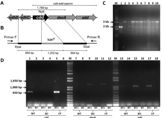
(PCR) from M. tuberculosis H37Rv genomic DNA, using primers forward (5’-tttttctagacccagcgttgggcaacgaagt-3’) and reverse (5’-tttttctagagcaccctcagccagcttcttg-3’), both containing XbaI restriction sites (in bold). The 1,788 bp fragment was subsequently cloned into pUC19 using the
XbaI restriction site. The cdd gene was disrupted by the in-sertion of a kanamycin cassette from pUC4K into unique internal enzyme restriction site NotI (Fig. 1B). Insert was released from pUC19 derivative vector by digestion with
XbaI, and subcloned into XbaI linearized pPR27xylE
vector (pPR27xylE::cdd kan) (Fig. 1B). The pPR27xylE
plasmid contains a thermosensitive origin of replica-tion, the xylE reporter gene, and the sacB counterselect-able marker (Pelicic et al. 1997). M. tuberculosis H37Rv strain was transformed by electroporation with ~ 2 µg of pPR27xylE::cdd kan plasmid. Possible KO clones were se-lected in two steps, as described previously (Villela et al. 2017). Genomic DNA was isolated and PCRs were carried out using gene-specific screening primers forward (5’-gt-gtctttgcggctgtagtc-3’) and reverse (5’-gggcagttcatctcc-gtca-3’) to determine whether the WT or the KO strain was present in the targeted chromosomal region (Fig. 1B). Among the nine clones screened for the KO of cdd gene, all amplified a band of 3,293 bp compatible with a double-crossover gene replacement event (Fig. 1C).
To obtain the CP strain, a fragment containing the cdd
gene, its upstream (183 bp) region containing the natural promoter, and 101 bp downstream to cdd was amplified by PCR from M. tuberculosis H37Rv genomic DNA using primers forward (5’-ggggtctagattgtcgccgttgtattcacc-3’) and reverse (5’-ggggtctagagtcggtataggccttgacga-3’), both containing XbaI restriction sites (in bold). Next, the amplicon was cloned into XbaI linearised pNIP40/b (pNIP40::cdd), a mycobacteriophage Ms6-derived inte-grative vector (Freitas-Vieira et al. 1998), and the KO strain was transformed by electroporation with pNIP40::cdd, as described previously (Villela et al. 2017). The stability of the mutation introduced by gene replacement in M. tuber-culosis was evaluated by plating KO and CP strains on me-dia with and without antibiotics. The difference between the colonies obtained on plates containing antibiotics was not significant when compared with the ones obtained on plates without antibiotic, which indicates that the intro-duced mutation is stable (data not shown).
As mentioned previously, the cdd together with deoA
and add genes are predicted to form an operon. Thus, to investigate the effect of cdd disruption on deoA and add
genes, levels of mRNA expression of cdd, deoA, and add
genes were monitored in WT, KO and CP strains. Ten milliliters of M. tuberculosis cultures were grown up to
Fig. 1: genomic environment of cdd gene in Mycobacterium tuberculosis (A), regions cloned into pPR27xylE vector (B), agarose gel electrophoresis of polymerase chain reaction (PCR) products from knockout clones (C), and mRNA expression of cdd, deoA, and add genes (D). (A) Genomic region of cdd gene (402 bp) containing unique internal NotI site and flanking genes; the operon cdd-add is indicated. (B) The cdd gene and flanking regions were amplified by PCR from M. tuberculosis H37Rv genomic DNA, and the cdd gene was disrupted by the insertion of a kanamycin cas-sette (kanR) into NotI site (cdd::kanR). The cdd::kanR fragment was cloned into pPR27xylE vector using XbaI restriction site. Annealing regions of
787 Mem Inst Oswaldo Cruz, Rio de Janeiro, Vol. 112(11), November 2017
an OD600 of 0.6 - 0.8. The cell pellet was suspended in 1 mL of TRIzol (Invitrogen, Carlsbad, CA, USA), and dis-rupted in 2 mL screw-cap tubes containing 0.1 mm silica spheres using a L-Beader 3 (Loccus Biotecnologia, Cotia, Brazil). The aqueous phase was extracted with 200 μL of chloroform, and RNA was precipitated with 500 µL of isopropanol. Remaining DNA in RNA samples was digested with DNAse (RNAse free DNAse set, QIAgen, Hilden, Germany) and RNA samples were purified by us-ing an RNA purification kit (RNeasy mini kit, QIAgen, Hilden, Germany). Synthesis of the first strand of cDNA was performed using 0.5 μg RNA from M. tuberculosis
H37Rv, KO, and CP strains as template and random hex-amers as primers, following the instructions in the Super-Script III First-Strand Kit protocol (Invitrogen, Carlsbad, CA, USA). An aliquot of cDNA synthesis reaction was used to amplify cdd gene with primers cdd F (5’-ggggtc-tagattgtcgccgttgtattcacc-3’) and cdd R (5’-ggggtctagagtc-ggtataggccttgacga-3’), deoA gene with primers deoA
F (5’- cgcatatgaccgacttcgcattcgacgcccc-3’) and deoA R (5’-agaagctttcagacgatccgatcgacgattagc-3’), and add gene with primers add F (5’-gtcatatgaccgctgcgccgaccctgcag-3’) and add R (5’- ctggatcctcactcgctgtgacccatgagc -3’). Am-plification products were analysed in 1 % agarose gels. As shown in Fig. 1D, no cdd expression was observed in KO strain (lane 3), but was detected in WT (lane 1) and CP (lane 5) strains. The deoA and add mRNA expression were observed in all strains (Fig. 1D), which indicated the disruption of cdd by insertion of a kanamycin cassette did not exert a polar effect on the expression of these genes. No or minor DNA contamination was observed on nega-tive controls, in which the cDNA synthesis was performed without the reverse transcriptase enzyme (Fig. 1D).
Although the transcription of deoA and add genes were not affected by cdd deletion in KO strain, the ex-pression of downstream genes to cdd might be affected at the protein level if the translation is coupled. Coupled translation was observed in Escherichia coli, where the translation efficiency of one gene affects indirectly the translational level of downstream genes within an oper-on, potentially causing a strong phenotype (Levin-Karp et al. 2013). The fact that the start and stop codons of
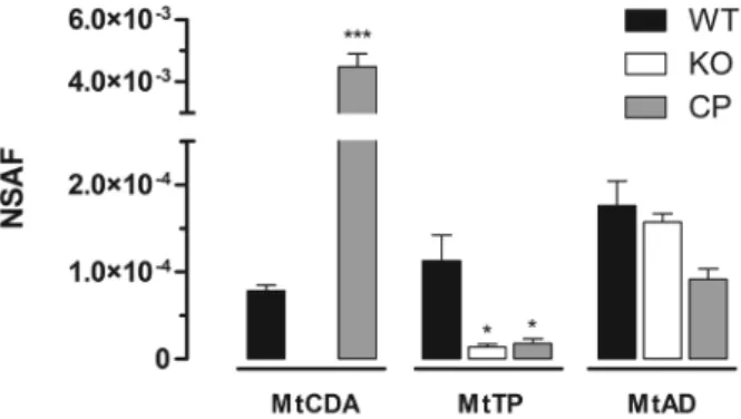
cdd, deoA and add genes overlap strengthens the pos-sibility of a coupled expression. Therefore, Liquid Chro-matography and Tandem Mass Spectrometry (LC-MS/ MS) and Multidimentional Protein Identification Tech-nology (MudPIT) analyses were performed to evaluate MtCDA, MtTP, and MtAD expression levels in WT, KO and CP strains. Cytoplasmic fractions of mycobacterial protein extracts from WT, KO and CP strains were ob-tained by ultracentrifugation, as described (Gunawar-dena et al. 2013). Chloroform/methanol protein precipi-tation was performed in WT, KO and CP cytoplasmic fraction (200 µg) according to (Wessel & Flügge 1984). Protein pellets were resuspended in 100 mM Tris HCl pH 8.5 containing 8 M urea, and digested according to Klammer and MacCoss (2006). Chromatographic separations were performed on a nanoLC Ultra 1D plus with autosampler (Sciex, Framingham, MA, USA) con-nected to a LTQ-XL Orbitrap Discovery hybrid instru-ment (Thermo Fisher Scientific, Waltham, MA, USA) through a nanoeletrospray ion source (Thermo Fisher
Scientific, Waltham, MA, USA). Biphasic MudPIT col-umns and capillary analytical colcol-umns were prepared in house (Villela et al. 2015). MudPIT analysis was carried out according to Wolters (Wolters et al. 2001). Analyses were performed in technical triplicates. Data was col-lected with one MS1 full-scan in the Orbitrap (400-1600 m/z range; 30,000 resolution) followed by data depen-dent CID MS/MS spectra of the eight most intense ions in the ion trap, with dynamic exclusion applied. Mass spectra were searched for candidate peptides with the software Comet (Eng et al. 2012) in the platform Pat-ternLab for Proteomics (Carvalho et al. 2016). The da-tabase contained a non-redundant M. tuberculosis ref-erence proteome (ID UP000001584, www.uniprot.org) and the reverse sequences of all entries. The validity of the peptide spectra matches (PSMs) was assessed us-ing the module Search Engine Processor (SEPro) from Patternlab for Proteomics, with a false discovery rate of 1%. Normalised spectral abundance factor (NSAF) was calculated according to Zybailov et al. (2006), and data were evaluated with the one-way ANOVA analy-sis, followed by Bonferroni’s post-test, using GraphPad Prism 5.0. Differences were considered significant at the 95% level of confidence. With average outputs of 76,880 spectra and 12,700 unique peptides per MudPIT run, the proteomic pipeline applied here identified a total of 2075 mycobacterial proteins in the cytoplasmic fraction of the three strains, considering only proteins identified con-sistently in three technical replicates (data not shown). Spectra matching peptides from MtAD and MtTP were identified in WT, KO and CP protein extracts. A sig-nificant decrease in the level of MtTP (encoded by deoA
gene) was observed in both KO and CP strains when comparing with WT strain (Fig. 2). This result suggests the occurrence of translational coupling between the neighboring genes cdd and deoA. On the other hand, the levels of MtAD, encoded by the third gene in the operon (add) (Fig. 1A), was not affected by the disruption of cdd
gene (Fig. 2). This difference in translation interdepen-dency might be explained by the fact that the disrupted
cdd gene is closer to deoA than to add (Fig. 1A). MtCDA
peptides were identified in both WT and CP but not in KO protein extracts (Fig. 2), indicating that the disrup-tion of cdd gene abolishes the production of MtCDA protein in the KO strain. MudPIT LC-MS/MS results are in agreement with mRNA expression evaluation, in which cdd gene expression was identified in WT and CP strains but was absent in KO strain (Fig. 1D). MtCDA peptides were only detected in WT strain using ultra-centrifugation for protein fractionation and MudPIT for peptide fractionation and identification. Seven spectral counts of two MtCDA unique peptides were identified in the WT strain technical triplicates. In the CP strain technical triplicates, 464 spectral counts of 10 MtCDA unique peptides were identified, a 66-fold difference in the levels of MtCDA in WT and CP strains. The dif-ference in MtCDA levels between WT and CP strains could be explained by the fact that, though expressed by MtCDA natural promoter, the complemented copy of
cdd gene is integrated in a different genome region. The growth rate of the WT, KO, and CP strains were compared to determine whether cdd disruption leads to alterations during in vitro cultivation of M. tuberculosis. Growth curves were started at an optical density at 600 nm (OD600) of 0.01 in Middlebrook 7H9 10% OADC 0.05% tween-80 containing proper antibiotics, in triplicate, at 37ºC, 80 rpm. Aliquots were removed from each culture at different time points and the OD600 was determined. As shown in Fig. 3A, the three strains have a similar pattern of growth when grown in Middlebrook 7H9 medium.
To examine whether the cdd gene was important for invasion and growth in phagocytic cells, we determined the bacterial loads of the WT, KO, and CP strains by us-ing the macrophage model of infection. RAW 264.7 mac-rophage cell line was cultured and infected with WT, KO or CP M. tuberculosis strains as described previously (Villela et al. 2017), with minor modifications. Briefly, infection of macrophages was performed at a multiplic-ity of infection of 1:1 (bacteria/macrophage) at 37ºC with 5% CO2. After 18 h, infection was terminated, and at this time point, 3, 7, 14 and 21 days of incubation, infected macrophages were lysed (Rodrigues-Junior et al. 2014), and plated on Middlebrook 7H10 agar supplemented with
10% OADC. Colony-forming unit (CFU) was evaluated after incubation of plates for three weeks at 37ºC. This experiment was performed in triplicate, and the results were expressed as mean numbers of the logarithms of CFU per well, and were evaluated with the two-way ANOVA analysis, followed by Bonferroni’s post-test, using GraphPad Prism 5.0. Differences were considered significant at the 95% level of confidence. As shown in Fig. 3B, no significant differences were observed in intracellular growth among WT, KO, and CP strains in macrophages after 18 h, three, seven and 21 days after infection. A decrease in bacterial load of KO strain was observed after 14 days of macrophage infection; how-ever, compared to WT, it was not statistically significant (Fig. 3B). Although the decrease in bacterial load of KO strain after 14 days of macrophage infection was shown to be statistically significant (p < 0.05) when compared with CP strain, the analysis of the area under the curve revealed very similar intracellular bacterial growth for WT (total area mean ± standard error of the mean: 23.25 ± 0.47), KO (23.32 ± 0.27), or CP (23.06 ± 0.11) strains. Therefore, these data suggest that the disruption of cdd
gene does not affect the M. tuberculosis growth in RAW 264.7 cells in the experimental conditions employed here.
In conclusion, a M. tuberculosis KO strain for cdd
gene was constructed by allelic replacement. The CP strain was obtained by transforming the KO with pNIP40::cdd plasmid that expresses cdd gene from its natural promoter. The cdd deletion was validated at the RNA level, and was confirmed at the protein level by MudPIT LC-MS/MS. The disruption of cdd gene did not affect the mRNA expression of deoA and add genes, both located downstream of cdd on the same operon. However, MudPIT LC-MS/MS data indicated the MtTP protein level was decreased in both KO and CP strains, when compared with its level in WT strain, which could be explained by a translational coupling between cdd
and deoA genes. The M. tuberculosis growth kinetics in Middlebrook 7H9 medium was not affected by the disruption of cdd gene. The results for RAW 264.7 cells suggest that the cdd gene product plays no role in M. tuberculosis invasion, growth and virulence in
789 Mem Inst Oswaldo Cruz, Rio de Janeiro, Vol. 112(11), November 2017
phages. Even though the recycling of nucleotides and nucleosides might represent a significant energy sav-ing for the cell, the MtCDA activity is not required in the context of in vitro growth and macrophage invasion and infection by M. tuberculosis under the experimental conditions employed here. These findings could be ex-plained by the redundancy found in nucleotide metabo-lism of M. tuberculosis. Although MtCDA is the only enzyme identified by sequence homology in M. tuber-culosis nucleotide biosynthesis pathway that is able to rescue cytidine and deoxycytidine (Villela et al. 2011), there are enzymes from de novo pathway that synthesize all pyrimidine nucleotides from simple precursors (War-ner et al. 2014). These enzymes might compensate for the absence of cdd gene in KO strain. However, it should be pointed out that studies under hypoxic and/or nutrient limitation conditions that likely mimic the environment in which latent bacilli survive should be pursued to pro-vide a solid experimental basis for the role of cdd gene product. Different results of gene essentiality and impor-tance for M. tuberculosis growth and survival might be obtained under different experimental conditions.
ACKNOWLEDGEMENTS
To Dr Mary Jackson and Dr Brigitte Gicquel for providing the pNIP40/b and pPR27xylE plasmids.
AUTHORS’ CONTRIBUTION
ADV constructed and validated the Mycobacterium tu-berculosis knockout and complemented strains, performed the growth curve, and wrote the manuscript; ADV and VSRJ performed macrophage infection experiments; AFMP, PE and CVB carried out MudPIT LC-MS/MS experiments and per-formed data analysis; VCAF participated in all biosafety level 3 experiments; ZASQ constructed the plasmids; LAB contributed in the analysis of results and manuscript revision; and DSS con-ceived the study and participated in its design and coordination.
REFERENCES
Carvalho PC, Lima DB, Leprevost FV, Santos MD, Fischer JS, Aqui-no PF, et al. Integrated analysis of shotgun proteomic data with PatternLab for proteomics 4.0. Nat Protoc. 2016; 11(1): 102-17. Cole ST, Brosch R, Parkhill J, Garnier T, Churcher C, Harris D, et
al. Deciphering the biology of Mycobacterium tuberculosis from the complete genome sequence. Nature. 1998; 393(6685): 537-44. Eng JK, Jahan TA, Hoopmann MR. Comet: an open source tandem
mass spectrometry sequence database search tool. Proteomics. 2012; 13(1): 22-4.
Freitas-Vieira A, Anes E, Moniz-Pereira J. The site-specific recom-bination locus of mycobacteriophage Ms6 determines DNA inte-gration at the tRNA(Ala) gene of Mycobacterium spp. Microbiol-ogy. 1998; 144(12): 3397-406.
Griffin JE, Gawronski JD, Dejesus MA, Ioerger TR, Akerley BJ, Sassetti CM. High-resolution phenotypic profiling defines genes essential for mycobacterial growth and cholesterol catabolism. PLoS Pathog. 2011; 7(9): e1002251.
Gunawardena HP, Feltcher ME, Wrobel JA, Gu S, Braunstein M, Chen X. Comparison of the membrane proteome of virulent My-cobacterium tuberculosis and the attenuated Mycobacterium bo-vis BCG vaccine strain by label-free quantitative proteomics. J Proteome Res. 2013; 12(12): 5463-74.
Klammer AA, MacCoss MJ. Effects of modified digestion schemes on the identification of proteins from complex mixtures. J Pro-teome Res. 2006; 5(3): 695-700.
Levin-Karp A, Barenholz U, Bareia T, Dayagi M, Zelcbuch L, An-tonovsky N, et al. Quantifying translational coupling in E. coli
synthetic operons using RBS modulation and fluorescent report-ers. ACS Synth Biol. 2013; 2(6): 327-36.
Mdluli K, Kaneko T, Upton A. The tuberculosis drug discovery and development pipeline and emerging drug targets. Cold Spring Harb Perspect Med. 2015; 5(6): a021154.
Pelicic V, Jackson M, Reyrat JM, Jacobs Jr WR, Gicquel B, Guilhot C. Ef-ficient allelic exchange and transposon mutagenesis in Mycobacte-rium tuberculosis. Proc Natl Acad Sci USA. 1997; 94(20): 10955-60. Roback P, Beard J, Baumann D, Gille C, Henry K, Krohn S, et al. A
predicted operon map for Mycobacterium tuberculosis. Nucleic Acids Res. 2007; 35(15): 5085-95.
Rodrigues-Junior V, dos Santos A, Villela AD, Belardinelli JM, Mor-bidoni HR, Basso LA, et al. IQG-607 abrogates the synthesis of mycolic acids and displays intracellular activity against Myco-bacterium tuberculosis in infected macrophages. Int J Antimi-crob Agents. 2014; 43(1): 82-5.
Sánchez-Quitian ZA, Rodrigues-Junior V, Rehm JG, Eichler P, Triv-ella DBB, Bizarro CV, et al. Functional and structural evidence for the catalytic role played by glutamate-47 residue in the mode of action of Mycobacterium tuberculosis cytidine deaminase. RSC Adv. 2015; 5: 830-40.
Sánchez-Quitian ZA, Schneider CZ, Ducati RG, de Azevedo Jr WF, Bloch Jr C, Basso LA, et al. Structural and functional analyses of
Mycobacterium tuberculosis Rv3315c-encoded metal-dependent ho-motetrameric cytidine deaminase. J Struct Biol. 2010; 169(3): 413-23. Sassetti CM, Boyd DH, Rubin EJ. Genes required for mycobacte-rial growth defined by high density mutagenesis. Mol Microbiol. 2003; 48(1): 77-84.
Villela AD, Eichler P, Pinto AFM, Rodrigues-Junior V, Yates III JR, Bizarro CV, et al. Gene replacement and quantitative mass spectrometry approaches validate guanosine monophosphate synthetase as essential for Mycobacterium tuberculosis growth. Biochem Biophys Rep. 2015; 4: 277-82.
Villela AD, Rodrigues-Junior VS, Pinto AFM, Wink PL, Sánchez-Quitian ZA, Petersen GO, et al. Characterisation of iunH gene knockout strain from Mycobacterium tuberculosis. Mem Inst Oswaldo Cruz. 2017; 112(3): 203-8.
Villela AD, Sánchez-Quitian ZA, Ducati RG, Santos DS, Basso LA. Pyrimidine salvage pathway in Mycobacterium tuberculosis. Curr Med Chem. 2011; 18(9): 1286-98.
Warner DF, Evans JC, Mizrahi V. Nucleotide metabolism and DNA replication. Microbiol Spectrum. 2014; 2(5): doi: 10.1128/micro-biolspec.MGM2-0001-2013.
Wessel D, Flügge UI. A method for quantitative recovery of protein in dilute solution in the presence of detergents and lipids. Anal Biochem. 1984; 138(1): 141-3.
WHO - World Health Organization. Global tuberculosis report 2016. Geneva: WHO; 2016.
Wolters DA, Washburn MP, Yates 3rd JR. An automated multidimen-sional protein identification technology for shotgun proteomics. Anal Chem. 2001; 73(23): 5683-90.