Pedro Luiz de Camargo1, Simone Akemi Uenaka1, Maitê Bette Motta1, Cristina Harumi Adania2, Letícia Yamasaki3, Amauri A. Alfieri3, Ana Paula F. R. L. Bracarense3*
1
Departamento de Clínicas Veterinárias, Universidade Estadual de Londrina, Londrina, PR, Brasil; 2Centro Brasileiro para
Conservação de Felinos Neotropicais, Associação Mata Ciliar, Jundiaí, SP, Brasil; 3Departamento de Medicina Veterinária
Preventiva, Universidade Estadual de Londrina, Londrina, PR, Brasil.
Submitted: March 15, 2010; Returned to authors for corrections: April 26, 2010; Approved: June 21, 2010.
ABSTRACT
Ten captive neotropical Brazilian feline were submitted to gastroscopic examination and samples of gastric
mucosa from fundus, corpus and pyloric antrum were evaluated for the presence of Helicobacter species.
Warthin-Starry (WS) staining and PCR assay with species-specific primers and enzymatic cleavage were
applied for bacterial detection and identification. Histological lesions were evaluated by haematoxylin and
eosin staining. All animals showed normal gross aspect of gastric mucosa. Helicobacter heilmannii was
confirmed in 100% of the samples by WS and PCR assay. Mild lymphocytic infiltrate in the lamina propria
was observed in eight animals, mainly in the fundus region. Small lymphoid follicles were seen in three
animals. No significant association between Helicobacter infection and histological findings was verified.
These observations suggest that gastric Helicobacter spp. could be a commensal or a eventual pathogen to
captive neotropical feline, and that procedures, way life, and stress level on the shelter apparently had no
negative repercussion over the integrity of the stomach.
Key words: Helicobacter, feline, gastric diseases, PCR, Leopardus.
INTRODUCTION
Helicobacter pylori infection in human beings has been
associated with gastritis, gastric and duodenal ulcer, gastric
adenocarcinoma, and MALT (mucosa associated-lymphoid
tissue) lymphoma. While H. pylori is accepted as the dominant
human gastric bacterial pathogen, a small percentage of human
infections have been associated with H. heilmannii, which is
more prevalent in pet and wild animals (3). The high
prevalence of this species in animals has been suggested to
have implication for public health (5). However, the subtypes
of H. heilmannii strain that colonizes humans and animals are
different, indicating that animals represent a lower risk to man
(17).
In several studies gastritis was associated with
Helicobacter spp. infection in tigers, lions and cheetahs.
Helicobacter acinonyx was implicated in gastritis in tigers
(19). In a retrospective survey conducted with 69 captive
cheetahs, 40% of the deaths were associated with severe
gastritis induced by Helicobacter spp. infection (13).
Moreover, the infection was also associated with
gastroduodenal adenocarcinoma and rectal adenoma in a
cougar (25). By contrast, others researches found no
association between infection and gastric mucosal
inflammatory infiltrate, suggesting that the bacteria are
commensal or opportunist pathogens (7, 23). Also, the study of
28 semi captive cheetahs found out mild or no lesions in 27,
concluding that gastritis was uncommon in these animals and
proposing that stress could play a major role in the
development of Helicobacter-associated gastritis (12).
In captive wild feline animals stress and host factors might
be important issues in the development of gastritis associated
with Helicobacter infection (23). The presence of Helicobacter
spp. in the stomach of wild or captive feline has been reported
in several countries (6, 10, 13); however the relationship
between infection and gastric diseases remains controversial.
Gross and histological analysis, and molecular characterization
of Helicobacter species in captive neotropical Brazilian feline
have not been previously described. Therefore, the purpose of
the current report was to examine the presence of Helicobacter
in gastric mucosa, and evaluate the association between
infection, gross and histological findings in neotropical
Felidae.
MATERIALS AND METHODS
Specimens
Samples of gastric mucosa from four female little spotted
cat (Leopardus tigrinus), five female ocelots (Leopardus
pardalis), and one male cougar (Puma concolor) were
evaluated. The animals were housed at Brazilian Center to
Conservation of Neotropical Feline – Mata Ciliar Association,
São Paulo, southern Brazil. None of the feline had any previous
signs of gastric disease. The animals were fed with fresh meet
of chicken, cattle, and viscera. The study was approved by the
institutional Ethics Committee for Animal Experimentation.
Food and water were withheld for 12 h before gastroscopy
and gastric biopsy. The animals were anaesthetized with
xylazine (1 mg/kg, im) and ketamine (10 mg/Kg, im). The
biopsy procedure was performed with a flexible paediatric
endoscope (2.3 mm diameter biopsy) and two samples of
gastric mucosa from pyloric antrum, corpus and fundus were
collected. The mucosal aspect, presence of content, friability
and bleeding at biopsy were evaluated at gastroscopy.
Gastric samples were fixed in Bouin’s solution for 24 h,
embedded in paraffin wax and processed routinely. Sections (3
m) were stained with haematoxylin and eosin (HE) and
Warthin-Starry (WS) method for histological evaluation and
Helicobacter detection, respectively. For molecular analysis a
further sample from each gastric region was placed into a tube
DNase and RNase-free and frozen at –20ºC until DNA
extraction.
Histological analysis
Location and colonization density of Helicobacter were
assessed. Colonization density was scored based in the number
of bacteria per field (1000x magnification) as 0 (no infection);
1 (1 to 10 bacteria per field); 2 (11 to 30); 3 (31 to 50); and 4
(more than 50 bacteria). Histological changes were evaluated
and scored as described elsewhere (20). Briefly, the number of
lymphoid aggregates, the number of inflammatory cells and
epithelial changes were evaluated. Gastritis was diagnosed
when more then four inflammatory cells per field in the lamina
propria were observed.
Polymerase Chain Reaction
Gastric biopsies were digested with 25 l proteinase K (20
mg/ml) in 200 l of lysis buffer pH 8.0 (100 mM NaCl, 100
mM EDTA, and 0.5% dodecyl sodium sulphate), agitated for
30 sec and then incubated at 56°C for 3 h. The DNA was
extracted with a combination of phenol/chloroform/isoamyl
alcohol and silica/guanidinium isothiocyanate methods as
described by Alfieri et al. (1). Sterile ultrapure water was
included as negative control in all DNA extraction.
The genes targeted for Helicobacter genus and
Helicobacter species, the primer sequences, the annealing
temperatures, and the size (base pairs) of amplified fragments
were described in a previous study (2). All gastric samples
were tested for the species H. pylori, H. heilmannii, H. felis,
performed in a final volume of 25 l containing 1 l of
extracted DNA, 0.4 mM of each deoxynucleotide, 0.5 l (20
pmol) of each primer, 1× PCR buffer (20 mM Tris HCl pH 8.4
and 50 mM KCl), 1.5 mM MgCl2, 1.25 U Platinum Taq DNA
polymerase (InvitrogenTM Life Technologies, USA), and
ultra-pure water. A negative control was included in every
amplification reaction. Amplifications were performed in a
PTC-100TM thermocycler (MJ Research Inc, USA). The
enzymes VspI (7) and Hinf I (4) (Life Technologies®) were
applied in the material amplified with specific pair primers, and
the enzyme Hinf I in the species specific primers. Seventeen
microliters from amplified material were added to 2 µl of
specific buffer and 1 µl of enzyme, and maintained at 37 °C for
1 hr. Products from PCR and from enzymatic cleavage were
analyzed by electrophoresis in 2% agarose gel with ethidium
bromide in Tris-Borate-EDTA (TBE) buffer and visualized
under UV light. The expected lengths products from enzymatic
cleavage were disposed in Table 1.
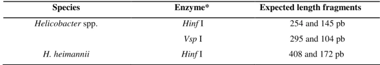
Table 1. Species and expected length (in base pairs) from fragments according to the enzyme employed.
Species Enzyme* Expected length fragments
Helicobacter spp. Hinf I 254 and 145 pb
Vsp I 295 and 104 pb
H. heimannii Hinf I 408 and 172 pb
* Invitrogen Life Technologies ®
Statistical Analysis
The association between colonization density,
inflammatory infiltrate and Helicobacter infection in the
gastric regions was evaluated by the Kruskal-Wallis test. The
Spearman test was performed to verify the correlation between
colonization density and inflammation (18). Significance was
set at p < 0.05.
RESULTS
All animals showed a normal macroscopic aspect of
esophagus mucosa, except in one Leopardus tigrinus that had
no herring bone pattern at the distal segment of the esophagus.
In three animals small patches of mucosal erythema and bilious
fluid were observed in gastric mucosa. Plant fragments and hair
coat were seen in the stomach of all animals.
Tightly coiled bacteria, 5 to 12
µ
m long, were detected in100% of the samples stained by WS method (Figure 1). The
bacteria were seen isolate or in clusters in superficial mucus
layer and gastric pits. Scores of bacterial colonization ranged
from 1 to 4 (Table 2). Although scores 3 and 4 were more
frequently observed in the corpus region, there was no
significant difference in colonization density between the
gastric regions (p = 0.54).
Histological evaluation showed mild lymphocytic
infiltrate in fundus. Inflammatory infiltrate is significantly
higher in this region than in corpus and pyloric antrum
(p<0.001). Two animals showed no inflammatory cells in
gastric mucosa even with high colonization density (Table 2).
No association was found between bacterial colonization and
mucosal inflammation (p = 0.330). Epithelial alterations were
rarely seen and when present were sparse and mild in
magnitude.
In three animals lymphoid follicles were observed in the
lamina propria. The lymphocyte aggregates appeared small and
fewer reactive. No more than one lymphoid follicle per sample
was observed. The frequency and distribution of lymphoid
follicles are summarized in Table 2.
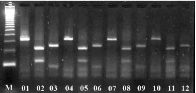
Samples from all animals generated a 399 bp amplicon
with Helicobacter genus reaction. Cleavage of the amplified
products by genus specific primer (399 bp) using Hinf I and
with 295 and 104 bp in length, respectively (Figure 2). The
reactions with species specific primers were positive only for
H. heilmannii,represented bya 580 pb amplicon. Cleavage of
H. heilmannii amplicon with Hinf I enzyme resulted in
fragments with 408 and 172 bp in length (Figure 3). No mixed
infection was detected.
Figure 1. The gastric mucosa of a Leopardus tigrinus.H. heilmannii colonizing gastric pits. Warthin-Starry staining. Bar, 5 µm.
Table 2. Scores of bacterial colonization and cellular inflammatory infiltrate observed in gastric mucosa samples of captive neotropical Brazilian feline.
Bacterial and inflammatory cell infiltrate scores in gastric regions
Species Animal Fundus Corpus Pyloric antrum
Bacterial Cell infiltrate Bacterial Cell infiltrate Bacterial Cell infiltrate
Leopardus tigrinus 01 3* 1 4 0 2 0
02 2 1 3 0 2 1
03 2 1 3 0 4 0
04 3* 1 3* 0 3 0
Leopardus pardalis 05 4 0 3 0 4 0
06 1 0 4 0 1 0
07 2 1 2 0 2 0
08 3 1 2* 0 2 0
09 2 1 2 0 3 0
Figure 2. Ethidium bromide stained 2% agarose gel electrophoresis of restriction fragment length polymorphism (RFLP) with
Hinf I and Vsp I enzymes of Helicobacter spp. genus amplicons. Lane M: 123 bp ladder (Invitrogen Life Technologies, USA);
Lanes 1, 4, 7, and 10: Helicobacter spp. genus amplicons (399 bp) from captive neotropical Brazilian feline gastric mucosal
samples; Lanes 2, 5, 8, and 11: Helicobacter spp. genus amplicons digested with Hinf I (254 and 145 bp). Lanes 3, 6, 9, and 12:
Helicobacter spp. genus amplicons digested with Vsp I (295 and 104 bp).
Figure 3. Ethidium bromide stained 2% agarose gel electrophoresis of restriction fragment length polymorphism (RFLP) with
Hinf I enzyme of Helicobacter heimannii amplicons. Lane M: 123 bp ladder (Invitrogen Life Technologies, USA); Lanes 1, 3, 5,
7, 9, and 11: H. heilmannii amplicons (580 bp) from captive neotropical Brazilian feline gastric mucosal samples; Lanes 2, 4, 6, 8,
DISCUSSION
No previous studies of gastric endoscopic pattern in
Brazilian neotropical feline were reported. The findings were
compared to the patterns registered for domestic cats. On the
endoscopic evaluation the most unexpected alteration was the
absence of transversal rings observed upon distal esophagus in
an adult Leopardus tigrinus. Others animals of the same
species that were evaluated, showed a herring bone pattern.
Thispattern is typically seen in domestic cats, and is expected
in Felidae family (21). The ingestion of grass and plants is
considered a normal pattern between feline, and bilious reflux
probably is related to pyloric relaxation mediate by anesthetic
drugs (22). Mucosal erythema could be seen in domestic cats
and dogs as a result of local blood flow variations in
physiologic conditions (8). The endoscopic aspects observed
were not associated with gastric disease, as animals presented
normal behavior and activity level, good nutritional status and
showed no alterations on physical examination. Furthermore,
the feline had no previous signs of gastric disease, such as
weight loss, variable appetite or vomit.
This study had characterized that Brazilian neotropical
captive feline were commonly infected by Helicobacter, as
free-ranging and captive cheetahs from South Africa and North
America (14). Gastritis accompanied by vomit and weight loss
was reported as a major cause of death in captive cheetahs,
meanwhile the association with Helicobacter infection still
controversial (23). Differently from cheetahs, captive
Leopardus tigrinus, L. pardalis and Puma color showed no
signs of gastric disease, supporting the hypotheses of
commensal bacteria in these species.
In none mucosal sample bacteria resembling H. pylori or
H. acinonyx were observed as previously reported (6, 19, 24).
The bacterial morphology and distribution on the mucosa was
similar to previously observed in Brazilian pet cats (20). The
results of PCR and enzymatic cleavage confirmed the infection
by H. heilmannii which was the most frequent species detected
in cheetahs (23) and cats (16). Nevertheless, to the best of the
authors’ knowledge, this report represents the first cases of
Helicobacter infection in captive neotropical feline confirmed
by PCR assay in Brazil.
Gastritis has been associated with Helicobacter infection
in a variety of wild carnivores (9, 10). In the present study,
mild lymphocytic infiltrate was frequently observed, mainly in
the corpus region. However, no association between
colonization density and the degree of inflammation was
verified. Furthermore, mucosal samples with no sign of
inflammation were taken from two infected animals. It has
been suggested that gastric pathology develops when the
bacteria acquire characteristics such as the cag pathogenicity
island. The cagA gene is a marker for virulence factors in H.
pylori, inducing a neutrophilic inflammation (15), however this
genomic region was not identified in samples from cheetahs
with gastritis (23). Although it is possible that other
pathogenicity factors could be involved in Helicobacter spp.
gastritis, it is more likely that H. Heilmannii in captive
neotropical feline are commensals or opportunistic organisms.
The presence of lymphoid aggregates was reported as the
main histological change associated with the Helicobacter spp.
infection in semi or captive feline (10, 19). However, in our
study small lymphoid follicles were seen in three animals,
despite the presence of Helicobacter in all samples. These
structures are normally seen on gastrointestinal tract,
increasing in number and dimension in response to antigenic
stimulation. Thus, apparently no relationship exists between
the presence of bacteria and inflammatory infiltrate or
lymphoid aggregates in wild feline.
Extrinsic factors, stress and genetic impoverishment had
an important role in the pathogenesis of gastritis (23). The fact
that captive feline had a greater occurrence of gastritis than
wild animals (14) strengthen the effect of stress in this lesion.
Our results had demonstrated that, differently from captive
cheetahs, captive feline from the Atlantic Forest (São Paulo,
Brazil) had no significant gastritis. Differences in conservation
management and adaptability to environmental changes could
explain the absence of gastric disease in this captive
population.
neotropical Brazilian feline are commonly infected by H.
heilmannii. The infection was not associated with clinical
signs, gross or histological changes, suggesting that these
gastric bacteria are just commensals or occasional pathogens.
Also, the practices employed to maintain the animals seem
adequate to minimize stressful conditions, contributing to
gastric mucosal homeostasis.
ACKNOWLEDGMENTS
The authors wish to thank CNPq-Brazil for awarding a
research fellowship to A.P.F.R.L. Bracarense and A.A. Alfieri.
REFERENCES
1. Alfieri, A.A.; Parazzi, M.E.; Takiuchi, E.; Médici, K.C.; Alfieri, A.F. (2006). Frequency of group A rotavirus in diarrhoeic calves in Brazilian cattle herds, 1998-2002. Trop. Anim. Health Prod. 38, 521-526. 2. Camargo, P.L.; Alfieri, A.A.; Bracarense, A.P.F.R.; Menoli, R.; Spinosa,
S.R.; Hagiwara, M.K. (2003). Use of polymerase chain reaction and enzymatic cleavage in the identification of Helicobacter spp. in gastric mucosa of human beings from North Paraná, Brazil. Mem. Inst. Oswaldo Cruz 98, 265-268.
3. Cattoli, G.; Bart, A.; Klaver, P.S.; Robijn, R.J.; Beumer, H.J.; Van Vugt, R.; Pot, R.G.; Van der Gaag, I.; Vanderbroucke-Grauls, C.M.; Kuipers, E.J.; Kusters, J.G. (2000). Helicobacter acinonychis eradication leading to the resolution of gastric lesions in tigers. Vet. Rec. 147, 164-165. 4. Clayton, C.L.; Kleanthous, H.; Coates, P.J.; Morgan, D.D.; Tabaqchali,
S. (1992). Sensitive detection of Helicobacter pylori by using polymerase chain reaction. J. Clin. Microbiol. 30, 192-200.
5. Dieterich, C.; Wiesel, P.; Neiger, R.; Blum, A.; Corthesy-Theulaz, I. (1998). Presence of multiple “Helicobacter heilmanni” strains in an individual suffering from ulcers and in his two cats. J. Clin. Microbiol. 36, 1366-1370.
6. Eaton, K.A.; Radin, M.J.; Kramer, L.; Wack, R.; Sherding, R.; Krakowka, S.; Fox, J. G.; Morgan, D.R. (1993). Epizootic gastritis associated with gastric spiral bacilli in cheetahs (Acinonyx jubatus). Vet. Pathol. 30, 55-63.
7. Germani, Y.; Dauga, C.; Duval, P.; Huerre, M.; Levy, M.; Pialoux, G.; Sansonetti, P.; Grimont, P.A. (1997). Strategy for the detection of Helicobacter species by amplification of 16S rRNA genes and identification of H. felis in a human gastric biopsy. Res. Microbiol. 148, 315-326.
8. Guilford, W.G. (2005). Upper Gastrointestinal Endoscopy. In: McCarthy, T.C. (ed). Veterinary Endoscopy for the Small Animal Practioner.
Elsevier Saunders, Missouri, USA, p.279-321.
9. Hill, J.E.; Khanolkar, S.S.; Stadtländer, C.T.K.H. (1997). Gastric ulcer associated with a Helicobacter-like organism in a cougar (Felis concolor). Vet. Pathol. 34, 50-51.
10. Jakob, W.; Stolte, M.; Valentin, A.; Schröder, H.D. (1997). Demonstration of Helicobacter pylori-like organisms in the gastric mucosa of captive exotic carnivores. J. Comp. Pathol. 116, 21-33. 11. Kinsel, M.J.; Briggs, M.B.; Venzke, K.; Forge, O.; Murnane, R.D.
(1998). Gastric spiral bacteria and intramuscular sarcocysts in African lions from Namibia. J. Wildl. Dis. 34, 317-324.
12. Lobetti, R.; Picard, J.; Kriek, N.; Rogers, P. (1999). Prevalence of helicobacteriosis and gastritis in semicaptive cheetahs (Acinonyx jubatus). J. Zoo Wildl. Med. 30, 492-496.
13. 13 Munson, L.; Nesbit, J.W.; Meltzer, D.G.; Colly, L.P.; Bolton, L.; Kriek, N.P. (1999). Diseases of captive cheetahs (Acinonyx jubatus jubatus) in South Africa: a 20-year retrospective survey. J. Zoo Wildl. Med. 30, 342-347.
14. Munson, L.; Terio, K.; Werley, M.; Jago, M.; Bagot-Smith, A.; Marker, L.(2005). Extrinsic factors significantly affect patterns of disease in free-ranging and captive cheetah (Acinonyx jubatus) populations. J. Wildl. Dis. 41, 542–548.
15. Nilsson, C.; Sillén, A.; Eriksson, L.; Strand, M.L.; Enroth, H.; Normark, S.; Falk, P.; Engstrand, L. (2003). Correlation between cag Pathogenicity Island composition and Helicobacter pylori-associated gastroduodenal disease. Infect. Immun. 71, 6573-6581.
16. Norris, C.R.; Marks, S.L.; Eaton, K.A.; Torabian, S.Z.; Munn, R.J.; Solnick, J.V. (1999). Healthy cats are commonly colonized with “Helicobacter heilmannii” that is associated with minimal gastritis. J. Clin. Microbiol. 37, 189-194.
17. Priestnall, S.L.; Wiinberg, B.; Spohr, A.; Neuhaus, B.; Kuffer, M.; Wiedmann, M.; Simpson, K.W. (2004). Evaluation of "Helicobacter heilmannii" subtypes in the gastric mucosas of cats and dogs. J Clin Microbiol. 42, 2144-2151.
18. Shot, S. (1990). Statistics for health professionals. WB Saunders, Philadelphia.
19. Schröder, H.D.; Ludwig, C.; Jakob, W.; Reischl, U.; Stolte, M.; Lehn, N. (1998). Chronic gastritis in tigers associated with Helicobacter acinonyx. J. Comp. Pathol. 119, 67-73.
20. Takemura, L.S.; Camargo, P.L.; Alfieri, A.A.; Bracarense, A.P.F.R.L. (2009). Helicobacter spp. in cats: association of infecting species and epithelial proliferation within the gastric lamina propria. J. Comp. Pathol. 141, 127-134.
21. Tams, R.T. (1999). Small animal endoscopy. Mosby Company, St. Louis.
22. Tams, R.T. (2003). Handbook of Small Animal Gastroenterology. Saunders Company, St. Louis.
24. Wack, R.F.; Eaton, D.A.; Kramer, L.W. (1997). Treatment of gastritis in cheetahs (Acinonyx jubatus). J. Zoo Wildl. Med. 28, 260-266.
25. Yamazaki, Y.; Aono, I.; Ohya, T.; Shibahara, T. Kadota, K. (2002).