online | memorias.ioc.fiocruz.br
Helicobacter are helicoidal, flagellated, Gram-neg-ative and microaerophilic bacteria that include a large number of species isolated from the gastrointestinal tracts of humans and other animal species, such as Old and New World primates (Dubois et al. 1994, Lundström et al. 2001, Tamashiro et al. 2005).
Baskerville and Newell (1988) have described the clinical association of chronic gastritis due to the natu-ral infection of Old World primates (Macaca mulatta) by the bacterial species Helicobacter pylori. Other studies have described how rhesus monkeys also develop gas-tric pathologies, such as peptic ulcers and even stomach cancer, when infected by H. pylori (Dubois et al. 1991, 1994, 1999). Therefore, these animals have been used as models for comparative studies with humans. The in-duced infection of the New World Saimiri sp. resulted in a slight and temporary inflammation of the mucosa in a great majority of animals (Stadtländer et al. 1998). Thus, the results from studies focused on understanding Helicobacter spp infections and associated diseases in various hosts might help to increase our understanding of pathogenic mechanisms (Atherton 2005).
A new species of Helicobacter was isolated and as-sociated with chronic colitis in the New World monkey species Saguinus oedipus (Saunders et al. 1999). In 2005, Mello et al. (2005) described the presence of these bacte-ria in the gastric environment of marmosets (Callithrix jacchus) and suggested that a natural bacterial colonisa-tion could occur among these primates. More recently, a new species of Helicobacter (Helicobacter callitrichis) was isolated from C. jacchus (Tamashiro et al. 2005).
Additionally, polymorphism in the ABO blood groups of humans and other primates, determined by the expres-sion of the A, B or H(O) antigens, is made up of fucosy-lated and terminal oligosaccharides linked to proteins and lipids, whose main function responsible for and capable of promoting their conservation throughout evolution still remains unknown (Fox et al. 2008). These antigens are distributed in tissues and are primarily localised in epithe-lial cells that function as a barrier to the external environ-ment; hence, it is believed that these antigens could either serve as ligands to specific pathogens or inhibit pathogen-ic interactions at the cell surface (Henry 2001).
The direct interactions of H. pylori with ABH anti-gen structures initially demonstrated that individuals ex-pressing fucosylated antigens (H and Lewis b) were more susceptible to disease because the microorganism utilises these epitopes to adhere to host cells (Borén et al. 1993). The presence of such receptors has been investigated in the gastric mucins of rhesus monkeys (Lindén et al. 2004) and the results are consistent with previous evidence. Financial support: FINEP, UFPA, IEC, CENP
+ Corresponding author: delia@ufpa.br Received 21 March 2011
Accepted 13 September 2011
Immunodetection of
Helicobacter
sp. and the associated expression
of ABO blood group antigens in the gastric mucosa
of captive and free-living New World primates in the Amazon region
Délia Cristina Figueira Aguiar1/+, Vera Lúcia de Souza Barros1,2, Washington Luiz Assunção Pereira3,
rosane do Socorro Pompeu de Loiola4, Gyselly Cássia Bastos de Matos2,
João Valsecchi5, Tereza Cristina Oliveira Corvelo6
1Faculdade de Medicina Veterinária 6Instituto de Ciências Biológicas, Universidade Federal do Pará, Belém, PA 2Seção de Patologia,
Instituto Evandro Chagas, Ananindeua, PA, Brasil 3Instituto de Saúde e Produção Animal, Universidade Federal Rural da Amazônia,
Belém, PA, Brasil 4Seção de Microbiologia, Laboratório Central do Estado, Belém, PA, Brasil 5Diretoria Técnico Científica,
Instituto de Desenvolvimento Sustentável Mamirauá, Tefé, AM, Brasil
The histo-blood group ABH antigens were first described in humans. These antigens are only present on eryth-rocytes from great apes and humans, while in more primitive animals they are found in tissues and body fluids. The ABH antigens are mainly distributed in tissues exposed to the external environment and potentially serve as ligands for pathogens or inhibitors of tissue connections. The objective of this paper was two-fold: (i) to determine the presence of Helicobacter sp. in the gastric mucosa of 16 captive and 24 free-living New World monkeys and (ii) to evaluate the presence of histopathological alterations related to bacterial infection and the associated expression of ABH antigens in the tissue. Stomach tissues from 13 species of monkey were assessed using haematoxylin-eosin and modified Gram staining (Hucker) methods. An immunohistochemical analysis of the tissue revealed the presence of infectious bacteria that were characteristic of the genus Helicobacter sp. The results demonstrate that various species of monkey might be naturally infected with the Helicobacter sp. and that there is an increased susceptibility to infection. This study serves as a comparative analysis of infection between human and non-human primates and indicates the presence of a new species of Helicobacter.
Studies in human adults and children in Brazil (Martins et al. 2006, Rodrigues et al. 2007) and other
countries (Brigić et al. 2002, Kanbay et al. 2005) indi -cate an association of ABO blood types with H. pylori infection. Individuals with O and Lewis b phenotypes demonstrated an increased susceptibility to infection and colonisation, which resulted in the development of gastric pathologies.
Therefore, it is important to understand the epide-miology and zoonotic potential of helicobacteriosis and to identify new Helicobacter spp from non-human primates because their susceptibility to many species-specific pathogens is similar to humans and, thus, they serve as valuable models for studying human infectious diseases (Bennett et al. 1998, Tamashiro et al. 2005).
In this study, we identified the presence of Helico-bacter sp. in different species of captive and free-living New World primates and examined the occurrence of histopathological changes related to this infection. Moreover, we examined the expression of these antigens in the gastric mucosa as markers for genetic predisposi-tion to bacterial infecpredisposi-tion.
MATErIALS AND METHODS
Animals - Wild monkeys were obtained from the fol-lowing distinct geographical areas in the Amazon Re-gion, Brazil: Amazon National Park, a municipality of Itaituba located in the southwestern area of the state of Pará (PA), Mamirauá Sustainable Development Reserve, state of Amazonas (AM), located in the middle of the Solimões River region, Amanã Sustainable Develop-ment Reserve, Central Amazon, AM, located in the low-er Rio Negro region, and forest areas in the municipality of Juriti, PA. The captive animals were obtained from the National Centre of Primates (CENP) located in the city of Ananindeua, PA (Table I).
A total of 16 samples of gastric tissue were obtained from captive monkeys that died at different time periods throughout the duration of the study. These animals did not present a history of stomach disease and were fed a balanced diet of fresh fruit, roots, insects and other items in accordance with the natural diet for each spe-cies. Some of the samples were obtained from the CENP collection (2004-2005), while others came from autop-sies conducted during the study period (2005-2007). The organs from 24 free-living wild monkeys were obtained through donations from other projects in different geo-graphic areas of the Amazon.
Histopathological diagnosis and detection of the ge-nus Helicobacter - All of the specimens analysed were obtained from the antral region of the stomach. The gas-tric tissue from the primate species was collected in buff-ered formalin and processed in paraffin. Subsequently, 4-5 µm tissue slices were mounted onto silanised slides for use in histopathological, modified Gram staining (Hucker 1921) and immunohistochemical analyses.
Slides containing the gastric tissue from each ani-mal were stained using the haematoxylin-eosin method for the histopathological diagnosis of gastric tissue and modified Gram staining (Hucker 1921) was used to de-tect Helicobacter-type HLO. These analyses were
per-formed using conventional optical microscopy. The bac-teria were characterised by their bacillary (curved and spiralled) and coccoid characteristics.
Immunohistochemistry for detecting H. pylori - The tissue were deparaffinised in xylene, rehydrated in methanol dilutions and washed with phosphate buffer (pH 7.6) followed by antigenic exposure in a citrate buf-fer (pH 6.0) heated for 15 min in a microwave. The tis-sues were pre-incubated in blocking buffer [phosphate buffered saline (PBS) and bovine albumin at a 1:20 dilu-tion] for 10 min and subsequently incubated with anti-H. pylori antibody (polyclonal rabbit anti-H. pylori;DAKO, Denmark B0471) for 1 h at a dilution of 1:100 in block-ing buffer (Haqqani 2001). After the primary antibody incubation, the slides were again washed and blocked in blocking buffer. The secondary antibody (polyclonal swine anti-rabbit AP; DAKO D0306) was added at a di-lution of 1:80 and the slides were incubated for 1 h. After a final wash with PBS, the staining was visualised with HistoMark RED activating solution (KLP Laboratories, Maryland USA; 556900). The gastric tissue slices were additionally stained with haematoxylin. As a positive control, a sample of human H. pylori gastric tissue was used. For the negative controls, the primary antibody was replaced with blocking buffer.
Immunohistochemistry for detecting ABH antigen expression - To determine whether the A, B and H an-tigens were expressed in the primate organs, a modified
TABLE I
Primate species and origin analysed in this study
Specie n
Origin
(identification of the animal)
Alouatta nigerrima 1 PARNA (An) Aotus infulatus 2 CENP (Ai1, Ai2) Cacajao melanocephalus 2 RDSA(Cm1, Cm2) Callicebus hoffmanni 2 PARNA (Ch1-Ch2) Callithrix jacchus 5 CENP (Cj1-Cj5) Cebus albifrons 2 PARNA (Cal1, Cal2) Cebus apella 9 CENP (Cap1-Cap4); JTR (Cap5);
PARNA (Cap6-Cap9) Mico humeralifer 1 PARNA (Mh) Pithecia irrorata 2 PARNA (Pi1, Pi2) Saguinus fusicolis 1 CENP (Sf) Saguinus inustus 3 RDSA (Si1-Si3) Saimiri sciureus 8 CENP (Ss1 a Ss4);JTR (Ss5-Ss8) Saimiri vanzolinii 2 RDSM (Sv1, Sv2)
Total 40
indirect immunoperoxidase technique was used(Pedal et al. 1989, Martins et al. 2006). Antibodies produced com-mercially for the detection of human antigens were em-ployed. To detect the H antigen, Ulex europaeus lectin (Sigma, Switzerland - UEA I) was utilised. The tissue slices were processed, mounted onto histological slides, deparaffinised in xylene and then treated with metha-nol containing 0.3% H2O2. Subsequently, the sections were washed in phosphate buffer (pH 7.6) and blocked in blocking solution (PBS and bovine albumin), followed by a 1-h incubation with anti-A and anti-B monoclonal antibodies (Fresenius Hemocare Brasil) at a 1:10 dilution and peroxidase-conjugated U. europaeus lectin (Sigma) diluted 1:50 in blocking buffer at room temperature. All of the slides were washed with phosphate buffer and the slides that were incubated with anti-A and anti-B antibod-ies were treated with blocking solution followed by a sec-ond incubation with peroxidase-conjugated anti-mouse
IgM (μ-chain specific - Sigma) for 1 h. After washing in
phosphate buffer, the slides were developed in Tris buf-fer containing diaminobenzidine and hydrogen peroxide. The slides were subsequently stained with haematoxylin, dehydrated and mounted with Entellam (Sigma).
Because all primates are secretors of ABH substanc-es (Apoil et al. 2000), the characterisation of the blood groups was based on the expression of the individual an-tigens in the stomach epithelium and mucus. This clas-sification method was previously established in another study (Ito et al. 1990) because saliva or other secretions from the animal were not available.
Non-parametric statistical tests (i.e., G-test, Kappa, Fisher tests, among others) were conducted to detect dif-ferences between the variations investigated using the computing program BioEstat 5.0 (Ayres et al. 2007). Sta-tistical significance was accepted at the 95% confidence internal level (p value < 0.05).
Ethics - The protocol for the collection and sampling of wild and captive animals was approved by the Federal En-vironmental Agency (IBAMA 086/2004, 0013/2004 and 001/2008) and the Ethics in Research Committee at the Evandro Chagas Institute (069-2005) Belém, PA, Brazil.
rESULTS
Detection of Helicobacter-type HLO using modi-fied Gram staining - Of the 40 samples of monkeys analysed in this study using the modified Gram staining method, 19 were considered positive for the presence of HLO, eight being from the captive animals and 11 from free-living monkeys.
Among the positive captive primates, three samples were from the species Cebus apella,one from Saimiri sciureus,three from C. jacchus and one from Aotus in-fulatus.
In the free-living group of monkeys, three samples were from S. sciureus,two from C. apella,one from Caca-jao melanocephalus, one from Saguinus inustus,one from Saimiri vanzolinii, one from Mico humeralifer, one from Pithecia irrorata and one from Callicebus hoffmanni.
The binomial test revealed that the proportion of ani-mals that were Gram-positive for HLO did not differ be-tween the free-living and captive animals (pbi = 0.7960).
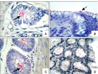
Immunohistochemical detection of H. pylori - When the samples were tested using immunohistochemistry for the specific detection of H. pylori, 12 positive sam-ples were found; five samsam-ples were from the CENP and seven were from the wild animals group (Figure).
The five CENP animals that were positive for H. pylori belonged to the species A. infulatus, C. apella, C. jacchus, S. sciureus and Saguinus fusicolis; the last two species were negative for H. pylori in the modi-fied Gram staining analysis. The wild primates were from the species S. vanzolinii, C. melanocephalus, S. inustus, C. apella, M. humeralifer, P. irrorata and C. hoffmanni.
A comparison of the different diagnostic methods used in this study (Table II) indicated that there was sig-nificant consistency between the modified Gram stain-ing and immunohistochemical techniques in detectstain-ing the Helicobacter sp. Notably, the immunohistochemical analysis detects specific epitopes of the bacteria, while the modified Gram staining technique only detects HLO. These analyses were performed using bacteria that were characterised according to their bacillary (curved and spiral) and coccoid characteristics.
TABLE II
Methods for detecting Helicobacter sp. in samples of primates investigated
Test IHQ positive IHQ negative Total
Gram-positive 10 9 19
Gram-negative 2 19 21
Total 12 28 40
MacNemar test by exact method: p (B/C) = 0.0654; IHQ: im-munohistochemistry.
The histopathological diagnosis using haematoxylin-eosin staining detected the presence of gastric inflam-mation in eight captive and 13 wild animals. The results from the statistical tests (Fisher exact) showed that the occurrence of gastritis among free-living and captive animals was not significantly different (p > 0.05). There-fore, no significant association between gastritis and the presence of HLO infection was verified through either modified Gram staining and/or immunohistochemical methods (Table III).
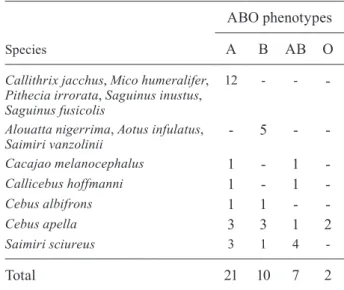
All of the animals were characterised in terms of their human-type ABO blood group phenotypes (Table IV). No significant association was found between the phenotypes and the presence of bacterial infection (G-test = 2.8924; p = 0.4085).
Expression of the ABH antigens in the gastric mu-cosa - In this study, the HLO were observed in the re-gions of the epithelium on the surface of the gastric mu-cosa, the surface mucus and in the lumen of the gastric glands. Additionally, when normal and inflamed areas of the gastric mucosa from the same individual were compared, changes in the expression pattern of ABH antigens in areas of inflamed tissue were observed as a loss and/or reduction of the staining intensity (data for the ABH antigenic activity not shown).
DISCUSSION
The results of this study are consistent with previous results showing that Helicobacter can naturally colonise different primate species (Stadtländer et al. 1998, Solnick et al. 1999, 2003). Previous studies have established infec-tions in New World primates of the genera Callithrix and Saguinus by new species of the Helicobacter genus (Saun-ders et al. 1999). Recently, Won et al. (2007) described a new species in C. jacchus, H. callitrichis, which dem-onstrated that many species of Helicobacter are capable ofcolonising the gastric mucosa of New World primates (Reindel et al. 1999, Mello et al. 2005).
Various methods of histopathological staining are employed for detecting H. pylori in humans, such as the silver (Warthin-Starry) and Giemsa stains. In this study, we used the modified Gram staining method because it is widely utilised (Araujo et al. 1993, Pereira et al. 2001, Assumpção et al. 2010) for the detection of bacteria in humans and has also been employed in studies with monkeys (Mackie & O’Rourke 2003).
Indirect immunohistochemistry revealed the presence of specific epitopes for H. pylori in 30% of the apes (12/40), while modified Gram staining detected bacteria with the same bacillary morphology as Helicobacter in almost half of the animal specimens tested (19/40); thus, new species of this genus might be present in New World primates.
Confirmation of the presence of HLOshould not be restricted to a single diagnostic technique and therefore, immunohistochemistry was performed using an antibody specific for H. pylori. However, other studies (Loffeld et al. 1991, Jonkers et al. 1997) conducted to test the speci-ficity of this antibody have shown cross-reactivity with the Campylobacter sp., a bacterium that does not colonise the gastric environment. Moreover, Reindel et al. (1999) have shown that this antibody cross-reacts with the spe-cies Helicobacter helmanii, a bacterium normally found in the stomachs of dogs, cats and pigs, which has also been detected in primates (Fox & Lee 1997). Thus, in this study, we cannot exclude the possibility of the presence of this and other species of Helicobacter in some of the cases investigated that were detected by the anti-H. py-lori antibody in the immunohistochemical analysis. Fur-thermore, other common epitopes that might be shared between H. pylori and new species of Helicobacter, al-though not yet identified systematically, could also be recognised by the antibody (Mello et al. 2005).
It was not possible to establish any relationship be-tween inflammation and Helicobacter sp. infection be-cause some of the monkeys that were negative by modified
TABLE III
Detection of inflammation of the gastric mucosa and of the Helicobacter sp. bacteria by means of Gram-modified and immunohistochemistry methods
in the primate study groups
Animals Gastritis
Detection methods
Total Gram+/
IHQ+
Gram+/
IHQ-Gram-/ IHQ+
Gram-/
IHQ-VL Present 3 3 0 7 13
Absent 4 1 0 6 11
CAT Present 2 2 2 2 8
Absent 1 3 0 4 8
Total - 10 9 2 19 40
CAT: captivity; IHQ: immunohistochemistry; VL: wild-living.
TABLE IV
Phenotypes of the ABO blood groups in the species studied
Species
ABO phenotypes
A B AB O
Callithrix jacchus, Mico humeralifer, Pithecia irrorata, Saguinus inustus, Saguinus fusicolis
12 - -
-Alouatta nigerrima, Aotus infulatus, Saimiri vanzolinii
- 5 -
-Cacajao melanocephalus 1 - 1 -Callicebus hoffmanni 1 - 1
-Cebus albifrons 1 1 -
-Cebus apella 3 3 1 2
Saimiri sciureus 3 1 4
Gram staining and/or immunohistochemical analysis had gastric inflammation. Therefore, it is rational to conclude that the helicobacteriosis in the gastric mucosa of these primates is not a primary factor for inducing an inflam-matory response. Furthermore, in the stomach lesions of the monkey species investigated, an infiltrate of mono-nuclear cells and the presence of lymphoid follicles was not common. Thus, the observed inflammation might be associated with other etiological factors (Khanolkar-Gaitonde et al. 2000) because gastritis origins are of a multifactorial nature. For example, both wild and captive primates suffer from stress induced by the maintenance of the colony’s social hierarchy, a known factor in the on-set of gastritis (Tamashiro et al. 2005).
There was no association detected between the genet-ic markers for the ABO blood group system phenotypes and bacterial infection or the inflammatory process. However, previous studies have demonstrated that vari-ous H. pylori strains might be associated with specific fucosylated ABH antigens in the gastric tissue of both human and non-human primates (Mahdavi et al. 2002, Aspholm-Hurtig et al. 2004, Lindén et al. 2004, Styer et al. 2010). However, the expression of these ABH an-tigens was reduced in the gastric mucosa of some of the animals in this study, which suggests that the inflamma-tion and other associated infecinflamma-tions might affect the tis-sue densities of specific ABH antigens, which H. pylori uses as receptors and therefore influence susceptibility to this infection. Notably, the small sampling number reduced the efficacy of the test and the relationship be-tween the susceptibility of the host and the pathogen was statistically insignificant.
Non-human primates, especially those from the Old World, have been used as models for the study of vari-ous infectivari-ous diseases in humans because they are also susceptible to many pathogens. This susceptibility is associated with the evolutionary relationship within the primate order. The results of this study seem to indicate that various species of New World monkeys have simi-lar risks for natural infection by the Helicobacter sp. Thus, this study provides a comparative analysis of the Helicobacter infection between human and non-human primates that results in the identification of new species in these hosts and increases our understanding of the pathogenic mechanisms of helicobacteriosis.
ACKNOWLEDGEMENTS
To the various participating institutions represented by their researchers and technicians, to RDSAM, for ceding pri-mate tissue samples mediated by the technician, Mr. Arlindo de Souza Junior (in memoriam), an employee of UFPA.
rEFErENCES
Apoil PA, Roubinet F, Despiau S, Mollicone R, Oriol R, Blancher A 2000. Evolution of alpha 2-fucosyltransferase genes in primates: relation between an intronic Alu-Y element and red cell expres-sion of ABH antigens. Mol Biol Evol17: 337-351.
Araujo MTF, Alves-Junior JM, Araujo RM, Eiko KS 1993. Frequency of Helicobacter pylori in gastric cancer patients in Belém, Pará, Brazil. Arq Bras Cir Dig8: 92-95.
Aspholm-Hurtig M, Dailide G, Lahmann M, Kalia A, Ilver D, Roche N, Vikström S, Sjöström R, Lindén S, Bäckström A, Lundberg
C, Arnqvist A, Mahdavi J, Nilsson UJ, Velapatiño B, Gilman RH, Gerhard M, Alarcon T, López-Brea M, Nakazawa T, Fox JG, Correa P, Dominguez-Bello MG, Perez-Perez GI, Blaser MJ, Normark S, Carlstedt I, Oscarson S, Teneberg S, Berg DE, Borén T 2004. Functional adaptation of BabA, the H. pylori ABO blood group antigen binding adhesin. Science305: 519-522.
Assumpção MB, Martins LC, Barbosa HPM, Barile KAS, Almeida SS, Assumpção PP, Corvelo TCO 2010. Helicobacter pylori in dental plaque and stomach of patients from northern Brazil.
World J Gastroenterol 16: 3033-3039.
Atherton JC 2005. The pathogenesis of Helicobacter pylori-induced gastro-duodenal diseases. Annu Rev Pathol1: 63-96.
Ayres M, Ayres Jr M, Ayres AL, Santos AS 2007. BioEstat 5.0. Apli-cações estatísticas nas áreas das ciências biológicas e médicas, Instituto de Desenvolvimento Sustentável Mamirauá - IDSM/ MCT/CNPq, Belém, 324 pp.
Baskerville A, Newell DG 1988. Naturally occurring chronic gastritis and C. pylori infection in the rhesus monkey: a potential model for gastritis in man. Gut29: 465-472.
Bennett BT, Abee CR, Henrickson R 1998. Bacterial and mycotic diseases. In SV Gibson (ed.), Nonhuman primates in biomedical research: diseases, Academic Press, New York, p. 59-102.
Borén T, Falk P, Roth KA, Larson G, Normark S 1993. Attachment of Helicobacter pylori to human gastric epithelium mediated by blood group antigens. Science262: 1892-1895.
Brigić E, Terzić S, Iljazović E, Cickusić E 2002. Association between
chronic gastritis in childhood, Helicobacter pylori and ABO blood groups. Med Arh 56: 57-58.
Dubois A, Berg DE, Incecik ET, Fiala N, Heman-Ackah LM, Del Valle J, Yang M, Wirth HP, Perez-Perez GI, Blaser MJ 1999. Host specificity of Helicobacter pylori strains and host respons-es in experimentally challenged nonhuman primatrespons-es. Gastroen-terology116: 90-96.
Dubois A, Fiala N, Heman-Ackah LM, Drazek ES, Tarnawski A, Fishbein WN, Perez-Perez GI, Blaser MJ 1994. Natural gastric infection with Helicobacter pylori in monkeys: a model for spi-ral bacteria infection in humans. Gastroenterology106: 1405-1417.
Dubois A, Tarnawski A, Newell DG, Fiala N, Dabros W, Stachura J, Krivan H, Heman-Ackah LM 1991. Gastric injury and inva-sion of parietal cells by spiral bacteria in rhesus monkeys. Are gastritis and hyperchlorhydria infectious diseases? Gastroen-terology 100: 884-891.
Fox JG, Lee A 1997. The role of Helicobacter species in newly rec-ognized gastrointestinal tract diseases of animals. Lab Anim Sci 47: 222-255.
Fox LC, Gigli E, De La Rasilla M, Fortea J, Rosas A, Bertranpetit J, Krause J 2008. Genetic characterization of the ABO blood group in Neandertals. BMC Evol Biol8: 342.
Haqqani MT 2001. Acridine orange stain in the histological identifi-cation of Helicobacter pylori. J Clin Pathol 54: 734.
Henry M 2001. Molecular diversity in the biosynthesis of GI tract glycoconjugates. A blood-group-related chart of microorganism receptors. Transfus Clin Biol8: 226-230.
Hucker GJ 1921. A new modification and application of the Gram stain. J Bacteriol6: 395-397.
Jonkers D, Stobberingh E, De Bruine A, Arends JW, Stockbrugger R 1997. Evaluation of immunohistochemistry for the detection of Helicobacter pylori in gastric mucosal biopsies. J Infect35: 149-154.
Kanbay M, Gür G, Arslan H, Yilmaz U, Boyacioglu S 2005. The rela-tionship of ABO blood group, age, gender, smoking and Helico-bacter pylori infection. Dig Dis Sci 50: 1214-1217.
Khanolkar-Gaitonde SS, Reubish GK JR, Lee CK, Stadtländer CT 2000. Isolation of bacteria other than Helicobacter pylori from stomachs of squirrel monkeys (Saimiri spp) with gastritis. Dig Dis Sci45: 272-280.
Lindén S, Borén T, Dubois A, Carlstedt I 2004. Rhesus monkey gas-tric mucins: oligomeric structure, glycoforms and Helicobacter pylori binding. Biochem J79: 765-775.
Loffeld RJ, Stobberingh E, Flendrig JA, Arends JW 1991. Helico-bacter pylori in gastric biopsy specimens. Comparison of culture, modified giemsa stain and immunohistochemistry. A retrospec-tive study. J Pathol165: 69-73.
Lundström AM, Sundaeus V, Bölin I 2001. The 26-kilodalton, AhpC homologue, of Helicobacter pylori is also produced by other
Helicobacter species. Helicobacter6: 44-54.
Mackie JT, O’Rourke JL 2003. Gastritis associated with Helicobacter -like organisms in baboons. Vet Pathol40: 563-566.
Mahdavi J, Sondén B, Hurtig M, Olfat FO, Forsberg L, Roche N, Ang-strom J, Larsson T, Teneberg S, Karlsson KA, Altraja S, Wadström T, Kersulyte D, Berg DE, Dubois A, Petersson C, Magnusson KE, Norberg T, Lindh F, Lundskog BB, Arnqvist A, Hammarström L, Borén T 2002. Helicobacter pylori SabA adhesin in persistent infection and chronic inflammation. Science297: 573-578.
Martins LC, Corvelo TCO, Oti HT, Loiola RSP, Aguiar DC, Barile KAS, Amaral RK, Barbosa HP, Fecury AA, Souza JT 2006. ABH and Lewis antigen distributions in blood, saliva and gastric mucosa and H. pylori infection in gastric ulcer patients. World J Gastroenterol12: 1120-1124.
Mello MF, Monteiro AB, Fonseca EC, Pissinatti A, Ferreira AM 2005. Identification of Helicobacter sp. in gastric mucosa from captive marmosets (Callithrix sp.; Callitrichidae, primates). Am J Primatol66: 111-118.
Pedal IV, Reichert W, Corvelo TCO 1989. Das Samenbläschenepithel Lewis-positiver individuen sezerniert lea in sialylierter form.
In Beiträge zur gerichtlichen medizin Wien, vol. 42,Institut für Rechtsmedizin der Universität Heidelberg, Heidelberg, p. 154-158.
Pereira LPLB, WaisbergJ, André EA, Zanoto A, Mendes JP, Soares HP 2001. Detection of Helicobacter pylori in gastric cancer. Arq Gastroenterol38: 240-235.
Reindel JF, Fitzgerald AL, Breider MA, Gough AW, Yan C, Mysore JV, Dubois A 1999. An epizootic of lymphoplasmacytic gastritis attributed to Helicobacter pylori infection in cynomolgus mon-keys (Macaca fascicularis). Vet Pathol 36: 1-13.
Rodrigues RV, Corvelo TC, Ferrer MT 2007. Seroprevalence of He-licobacter pylori infection among children of different socioeco-nomic levels in Porto Velho, state of Rondônia. Rev Soc Bras Med Trop40: 550-554.
Saunders KE, Shen Z, Dewhirst FE, Paster BJ, Dangler CA, Fox JG 1999. Novel intestinal Helicobacter species isolated from cot-tontop tamarins (Saguinus oedipus) with chronic colitis. J Clin Microbiol37: 146-151.
Solnick JV, Canfield DR, Yang S, Parsonnet J 1999. The rhesus mon-key (Macaca mulatta) model of Helicobacter pylori: noninvasive detection and derivation of specific pathogen free monkeys. Lab Anim Sci 49: 197-201.
Solnick JV, Chang K, Canfield DR, Parsonnet J 2003. Natural acqui-sition of Helicobacter pylori in newborn rhesus macaques. J Clin Microbial41: 5511-5516.
Stadtländer CT, Gangemi JD, Stutzenberger FJ, Lawson JW, Lawson BR, Khanolkar SS, Elliott-Raynor KE, Farris HE JR, Fulton LK, Hill JE, Huntington FK, Lee CK, Monath TP 1998. Experimen-tally induced infection with Helicobacter pylori in squirrel mon-keys (Saimiri spp): clinical, microbiological and histopathologic findings. Lab Anim Sci48: 303-309.
Styer CM, Hansen LM, Cooke CL, Gundersen AM, Choi SS, Berg DE, Benghezal M, Marshall BJ, Peek RM JR, Borén T, Solnick JV 2010. Expression of the BabA adhesion during experimental infection with Helicobacter pylori. Infect Immun78: 1593-1600.
Tamashiro KLK, Nguyen MMN, Sakai RR 2005. Social stress: from rodents to primates. Front Neuroendocrin 26: 27-40.