Vector Competence of
Culex quinquefasciatus
Say from
Different Regions of Brazil to
Dirofilaria immitis
Silvia Maria Mendes Ahid/+, Pádua Suely da Silva Vasconcelos,
Ricardo Lourenço-de-Oliveira*
Departamento de Patologia, Curso de Medicina Veterinária, Universidade Estadual do Maranhão, Campus Paulo VI, Tirirical, 65054-970 São Luís, MA, Brasil *Laboratório de Transmissores de Hematozoários, Departamento
de Entomologia, Instituto Oswaldo Cruz, Rio de Janeiro, RJ, Brasil
The vector competenceof Culex quinquefasciatus from five localities in Brazil to Dirofilaria immitis
was evaluated experimentally. Females from each locality were fed on an infected dog (~ 6 microfi-lariae/µl blood). A sample of blood fed mosquitoes were dissected approximately 1 h after blood meal. These results demonstrated that all had ingested microfilariae (mean, 4.8 to 24.6 microfilariae/mos-quito). Fifteen days after the infected blood meal, the infection and infective rates were low in all populations of Cx. quinquefasciatus. The mean number of infective larvae detected in the head and proboscis of these mosquitoes was 1-1.5. The vector efficiency, the number of microfilariae ingested/ number of infective larvae, was low for all populations of Cx. quinquefasciatus. However, the survival rate for all populations was high (range 50-75%). The survival rate of Aedes aegypti assayed simulta-neously for comparison was low (24.7%), while the vector efficiency was much higher than for Cx. quinquefasciatus. These data suggest that the vector competence of all assayed populations of Cx. quinquefasciatus to D. immitis in Brazil is similar and that this species is a secondary vector due to its low susceptibility. Nevertheless, vector capacity may vary between populations due to differences in biting frequency on dogs that has been reported in Brazil.
Keys words: Dirofilaria immitis - Culex quinquefasciatus - mosquito - vector competence - Culicidae - Brazil
Dirofilaria immitis (Leidy) is a nematode para-site that infects the right ventricle and pulmonary artery of dogs and other carnivores, and is trans-mitted by mosquitoes (Diptera, Culicidae). The parasite has a wide geographical distribution, with dogs serving as the principle host, although infec-tion has been reported in other animals and occa-sionally in man (Robinson et al. 1977, Kasai et al. 1981). The complete development of microfilariae has been reported in species of Culex, Aedes,
Anopheles, Mansonia, Psorophora and Co-quillettidia (Ludlam et al. 1970).
In some insects there exist biochemical prod-ucts in the hemolymph and other tissues, includ-ing the Malpighian tubule cells and thoracic muscle cells, that may block the development of the para-site by mechanisms comprising sequestration, en-capsulation and melanization. The intensity of these reactions varies according to the susceptibility of the mosquito host, resulting in high survival rates
+Corresponding author. Fax: 55-98-245.1500. E-mail:
silviaahid@bol.com.br Received 2 December 1999 Accepted 19 June 2000
of both the parasite and mosquitoes of susceptible populations (Christensen 1981, Christensen & Tracy 1989, Talluri & Cancrini 1994). The migra-tion and development of large numbers of D. immitis larvae within the mosquito can produce elevated levels of host mortality (Nayar & Sauerman 1975). Thus, melanization can act as an important mechanism for vector survival by limit-ing the number of larvae that complete develop-ment and thereby guaranteeing its own survival (Christensen 1981, Christensen & Forton 1986, Christensen & Tracy 1989). In addition, some mosquito species have well developed cibarial ar-mature, the teeth of which, when numerous and/or developed, injuring ingested microfilariae and re-ducing their survival potential (McGreevy et al. 1978, Coluzzi et al. 1982).
The importance of Cx. quinquefasciatus Say as an intermediate host for Wuchereria bancrofti
is well known. In 1901, Bancroft had already noted the development of D. immitis in Cx. pipiens fatigans (= Cx. quinquefasciatus) (apud Villavaso & Steelman 1970). The vector competence of noc-turnal domestic mosquitoes (Cx. quinquefasciatus
et al. 1970, Lowrie 1991, Loftin et al. 1995). In Rio de Janeiro Cx. quinquefasciatus was consid-ered a secondary vector of D. immitis (Labarthe et al. 1998a, b). In São Luís (State of Maranhão), where the prevalence of canine dirofilariasis var-ies from 11.3 to 52.5% depending on the origin of the dogs and the proximity to the sea shore, only 0.5% of the Ae. taeniorhynchus and 0.1% of Cx. quinquefasciatus examined were found to be natu-rally infected. Cx. quinquefasciatus accounted for 54% of the total mosquitoes collected (man and dogs) and 96.7% of the total captured in dog baited traps (Ahid et al. 1999, Ahid & Lourenço-de-Oliveira 1999). The marked difference noted in the frequency of Cx. quinquefasciatus collection us-ing live dog baited traps in Maranhão and that seen in Rio de Janeiro could reflect differences in the vector capacity of this species in these two loca-tions. Moreover, there have been reports of vector competence differences between Cx. quinque-fasciatus populations isolated from regions with various levels of prevalence of bancroftian filari-asis(Janousek & Lowrie 1989, Lowrie et al 1989, Brito et al. 1997, Calheiros et al. 1998). Could Cx. quinquefasciatus from Maranhão show a differ-ent degree of susceptibility to infection with D. immitis in comparison to other parts of Brazil?
In the present study, the vector competence of
Cx. quinquefasciatus in relation to D. immitis was evaluated under laboratory conditions using mos-quitoes originating from five distinct Brazilian populations.
MATERIALS AND METHODS
The experiments were performed using female
Cx. quinquefasciatus derived from 32 to 107 wild collected females from each of five test locations: Recife, Pernambuco (PE), the city of Rio de Janeiro (RJ), Porto Velho, Rondônia (RO), São Luís, Maranhão (MA), and Florianópolis, Santa Catarina (SC). Transmission of D. immitis was previously reported in all sample locations (Alves et al. 1993, Labarthe 1997, Ahid et al. 1999), except Porto Velho, where the examination of 45 dogs for the presence of the parasite were negative (Lima et al. 1996). The progenies from each location were reared separately, but simultaneously under the same conditions with regard to feeding, tempera-ture and illumination. Larvae were reared in groups of approximately 400 in 40 x 25 cm pans and pro-vided with fish food (TetraMinTm) mixed with cat food (WhiskasTm). Pupae were transferred daily (in containers filled with tap water) to screened cages (40 x 40 x 40 cm) where adults emerged. Adult mosquitoes were daily provided with 10% sucrose solution. Three to five days-old nuliparous (F7) females were used for experimental infections
with D. immitis. Twenty-four hours prior to blood-feeding on the infected blood source, sucrose was removed. The colonies were maintained and ex-perimental infections were conducted in the labo-ratory at 29ºC ± 1ºC and 70% ± 10% relative hu-midity (RH).
To identify different mosquito populations fol-lowing feeding, the females from each location were marked with luminous powder of different colors (Luminous Powder-Bioquip products) a few minutes prior to the infection experiment. Females were separated by observing the color of adhering luminous powder when illuminated with a black fluorescent light source. Approximately 1,500 fe-male Cx. quinquefasciatus (approximately 300 from each population) and 300 Ae. aegypti (São Luís strain, as control) reared under the same con-ditions as Cx. quinquefasciatus, were placed simul-taneously in a covered cage (120 x 100 x 80 cm) along with a previously anesthetized, infected dog. All the mosquitoes were allowed to feed on the dog from 23.00 h to 3.00 h in total darkness. Dur-ing the first hour of exposure to the dog five en-gorged females from each population were col-lected using an aspirator and immediately dissected (as described below) to determine the number of ingested microfilariae.
A nine years old, male Labrador with a natural
D. immitis infection (approximately six microfi-lariae per µl of blood) was used as the source of infection for the test mosquitoes. The dog was an-aesthetized with acepramazine (0.2 ml/kg) during the entire exposure period. To determine the aver-age number of circulating microfilariae, six 20 µl blood samples were collected from the dog and examined by Giemsa staining. Three of the samples were collected 24 h prior to exposure to the mos-quitoes while the other three were collected 1 h pre-exposure.
In this study we refer to “infected mosquitoes” as those in which larvae of D. immitis were ob-served at any stage of development. The term “in-fective mosquitoes” refers to mosquitoes that had third stage larvae in their head and/or proboscis (H/p). The analysis of the average number of mi-crofilariae ingested soon after engorgement was performed using the Duncan test in order to trans-form the data into a logarithmic trans-form for analysis of variance at the 5% level. Other variables were analyzed using the χ2 test for non parametric data at the 5% level.
RESULTS AND DISCUSSION
Among the total female Cx. quinquefasciatus
(approximately 300 per population) placed in con-tact with the infected dog, 858 engorged. A vari-able percentage of engorged females was observed, ranging from 40 to 98% for different populations (Table I). In the case of Ae. aegypti 57% of the females engorged. All Cx. quinquefasciatus dis-sected immediately after feeding contained mi-crofilariae in the midgut, with rates that varied from 24 to 123 microfilariae (mf) per mosquito (mosq). The average numbers of mf/mosq were 4.8 ± 4.3
to 24.6 ± 23.3 (Tables I, II). Statistical analysis (Duncan Test) revealed a significant difference in the average number of mf/mosq for PE and RO populations of Cx. quinquefasciatus (24.6 ± 23.3; 4.8 ± 4.3, respectively). No significant differences were observed between the average number of mf/ mosq in Cx. quinquefasciatus from PE and Ae. aegypti (26.6 ± 11.5) or between RO population and the other populations of Cx. quinquefasciatus
(Tables I, II). Calheiros et al. (1998) infected Cx. quinquefasciatus with W. bancrofti and encoun-tered three to 102 larvae (average 19.8 ± 19.5) in the midgut of 97% of mosquitoes dissected imme-diately after feeding on infected blood. Loftin et al. (1995) reported average levels of mf/mosq of 34.2 ± 6.3, 29 ± 6.1 and 39.3 ± 11 in Cx. quinquefasciatus, Cx. tarsalis and Ae. vexans re-spectively, that fed on a dog infected with D. immitis. In our experiments all Ae. aegypti were found to be infected with an average mf/mosq level of 26.6 ± 11.5 (Tables I, II).
We observed a rapid coagulation of midgut contents in female Cx. quinquefasciatus which were dissected within 1 h of feeding. Here, the for-mation of crystals, a considerable reduction in the
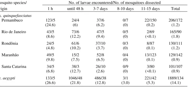
TABLE I
Distribution of infected and infective mosquitoes, number of microfilariae (mf) ingested by female
Culex quinquefasciatus of different origin and by Aedes aegypti, fed on a naturally infected dog (~6 mf/20 µl of blood)
Mosquitoes Mosquitoes with L3 Mosquitoes Mosquitoes examined from the (infective larvae) species/ Total of examined 1st to 10th day between the 11th to 15th Origins mosquitoes 1 h after feeding after feedinga day after feedingb
Engorged Dissected No. of mf/mosq. No. of X X (%) infected dissected infected mosq./ L3/dissected L3/infected
mosq./dissec. X ± S dissected mosq. mosq.
(%) (%)
Cx. quinquefasciatus
Pernambuco 295 172 5/5 24.6 ± 23.3 4/17 0.1 1.4
(98) (100) (23.5)
Rio de Janeiro 121 90 5/5 8.6 ± 4.5 6/16 <0.1 1
(40) (100) (37.5)
Rondônia 136 111 5/5 4.8 ± 4.3 12/19 0.1 1.5
(45) (100) (63.2)
Maranhão 164 142 5/5 9.8 ± 8.9 6/14 0.1 1.2
(55) (100) (42.8)
Santa Catarina 142 107 5/5 6.8 ± 6.6 6/22 <0.1 1.5
(47) (100) (27.3)
Ae. aegypti 170 134 5/5 26.6 ± 11.5 84/87 4 4.1
(57) (100) (96.5)
movement of the microfilariae and the presence of dead or injured larvae were observed. Similar ob-servations were reported by Nayar and Sauerman (1975) and Lowrie (1991), who suggested that the presence of crystals of oxyhemoglobin, resulting from the lysis of blood cells, was the principle fac-tor responsible for the reduced larval activity and death of D. immitis following ingestion by Cx. quinquefasciatus. In addition, Lowrie (1991) in a study with Cx. quinquefasciatus found that 12% of the mf of D. immitis had been damaged by the action of the cibarial armature. In the present study, the rapid coagulation and formation of crystals was not observed in Ae. aegypti, instead the microfi-lariae were active and moving freely in the midgut soon after their ingestion.
Regarding the number of larvae encountered in female Cx. quinquefasciatus at different sam-pling times, no significant differences were noted between females dissected 48 h post engorgement (χ2α5 = 2.15) and those sampled at intervals be-tween three to seven days (χ2α5 = 3.437) or be-tween 11 to 15 days (χ2 α5 = 2.956) (Table II). Similarly, differences were not observed between the mean larval development/mosquito for differ-ent test populations of Cx. quinquefasciatus (χ2α5 =3.84). During the 8th to 10th day post feeding, moribund female Cx. quinquefasciatus were dis-sected but none were found to be infected (Table II). Macêdo et al. (1998) encountered averages of 6.9 and 8.4 larvae/mosquito in Ae. scapularis and
Ae. aegypti fed using an apparatus containing blood infected with D. immitis (60 to 70mf/20 µl). In
our experiments the mean numbers of mf/mosq re-corded in Ae. aegypti were greater than those ob-served in Cx. quinquefasciatus at all sampling times (Table II). More female Ae. aegypti maintained larvae in the Malpighian tubules than Cx. quinquefasciatus, irrespective of the origin of the population (23.5 to 63.2% of infected mosquitoes) (Table I).
The data indicate that the infection during the first 48-h feeding period for Cx. quinquefasciatus
was not sufficient to induce an elevated level of mortality in those mosquitoes: MA 1.46% (2/137), PE 2.39% (4/167), SC 2.94% (3/102), RO 5.66% (6/106) and RJ 7.06% (6/85). Significant differ-ences were not noted (χ2α5 = 5.7) when we com-pared the mortality levels of different populations of Cx. quinquefasciatus. However, populations of
Cx. quinquefasciatus and Ae. aegypti showed a marked difference. During this period the level of mortality observed for Ae. aegypti was elevated (31%). These results are similar to those reported by Serrão (1998), i.e. 24.7 and 35.7% for females fed on blood with moderate microfilaraemia (3,000 to 5,000 mf/ml). Although the female Cx. quinquefasciatus from PE had ingested a signifi-cantly larger number of mf than had any other population, this did not result in a higher mortality rate in the two days following the infected blood meal. In the case of Ae. aegypti there was a corre-lation between the number of mf ingested and mortality rate. These data support the hypothesis that fewer live mf reach the Malpighian tubules in
Cx. quinquefasciatus than in Ae. aegypti, owing to
TABLE II
Number of Dirofilaria immitis larvae observed over a 15 day period in female Culex quinquefasciatus from different regions of Brazil and in Aedes aegypti, that fed on an infected dog (~6 microfilariae/20 µl blood).
Average in parenthesis
Mosquito species/ No. of larvae encountered/No. of mosquitoes dissected
Origin 1 h until 48 h 3-7 days 8-10 days 11-15 days Total
Cx. quinquefasciatus
Pernambuco 123/5 24/4 37/6 0/7 22/150 206/172
(24.6) (6) (6.2) (0) (0.2) (1.2)
Rio de Janeiro 43/5 73/6 47/5 0/5 2/69 165/90
(8.6) (12.2) (9.4) (0) (<0.1) (1.8)
Rondônia 24/5 61/6 37/10 0/3 8/87 130/111
(4.8) (10.2) (3.7) (0) (0.1) (1.2)
Maranhão 49/5 15/2 52/8 0/4 13/123 129/142
(9.8) (7.5) (6.5) (0) (0.1) (0.9)
Santa Catarina 34/5 38/3 26/10 0/9 3/80 101/107 (6.8) (12.7) (2.6) (0) (<0.1) (0.9)
the barriers encountered in the digestive tract. These barriers may include the action of the cibarial armature, blockage by rapid coagulation of the in-gested blood and the presence of crystals (Nayar & Sauerman 1975, McGreevy et al 1978, Lowrie 1991, Loftin et al. 1995).
At the end of the 15-day post feeding period no significant difference in survival was observed among the test Cx. quinquefasciatus populations (χ2α5 = 6.66), with a variation of between 50.8% to 75% (Table III). Levels of survival were greater than those reported by Brito et al. (1999), who only recorded a 30.6% survival in Cx. quinquefasciatus
from Alagoas (AL) infected with D. immitis. More-over, our data differ from the values of 17 and 63% survival reported by Lowrie (1991) using two dif-ferent strains of Cx. quinquefasciatus (Haiti and USA). Nevertheless, our values are similar to those observed by Calheiros et al. (1998) who evaluated the experimental infection of a Brazilian strain of
Cx. quinquefasciatus with W. bancrofti and re-ported 66% survival rate. A higher level of sur-vival was observed for Brazilian populations of Cx. quinquefasciatus in this study than Ae. aegypti
(24.7%) (Table III).
Third stage larvae were encountered in the Malpighian tubules only on day 11 in Cx. quinquefasciatus from PE and MA and only on day 13 in specimens from RJ, RO and SC.
Infec-tive larvae L3 were observed in the head and pro-boscis (H/p) from day 13 in Cx. quinquefasciatus
from MA and on day 14 in females from PE, RJ and SC. Females from RO were the only popula-tion that infective stage larvae were not observed in the H/p within the 15 day observation period, despite the observation of live L3 (0.1 L3/mosq) in the Malpighian tubules (Table I). These findings are in agreement with studies on Cx. quinque-fasciatus from other locations (Kartman 1953, 1954, Villavaso & Steelman 1970, Lowrie 1991, Loftin et al. 1995, Brito et al. 1999). In the case of
Ae. aegypti, L3 were detected in the Malphigian tubules on day 11 and in the H/p on day 12 after feeding on the infected dog. In general terms, three-four times as many L3 were detected in Ae. aegypti
than were found in Cx. quinquefasciatus test popu-lations (1 to 1.5 L3/infected mosquito) (Table I).
The level of infection ranged from 12 to 20.7% in the populations of Cx. quinquefasciatus (Table III), although no significant difference was ob-served (χ2α5 = 2.66). This level of infection was lower than that reported by Loftin et al. (1995), who observed the presence of L3 in 40.6% of in-fected Cx. quinquefasciatus. The infection rate found in our study with Ae. aegypti was very high (96.3%) (Table I), greatly surpassing the values of 27.6% for this species and 79.5% for Ae. scapularis
noted by Macêdo et al. (1998).
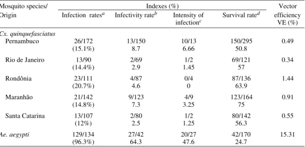
TABLE III
Results of infection with Dirofilaria immitis in Culex quinquefasciatus from five distinct regions of Brazil and in Aedes aegypti after 15 days observation
Mosquito species/ Indexes (%) Vector Origin Infection ratesa Infectivity rateb Intensity of Survival rated efficiency
infectionc VE (%)
Cx. quinquefasciatus
Pernambuco 26/172 13/150 10/13 150/295 0.49
(15.1%) 8.7 6.66 50.8
Rio de Janeiro 13/90 2/69 1/2 69/121 0.34
(14.4%) 2.9 1.45 57
Rondônia 23/111 4/87 0/4 87/136 1.44
(20.7%) 4.6 0 63.9
Maranhão 21/142 9/123 4/9 123/164 0.91
(14.8%) 7.3 3.25 75
Santa Catarina 13/107 2/80 1/2 80/142 0.55
(12%) 2.5 1.25 56.3
Ae. aegypti 129/134 27/42 20/27 42/170 15.31
(96.3%) 64.3 47.6 24.7
The level of infectivity fluctuated (2.5 to 8.7%) depending on the Cx. quinquefasciatus population (Table III). The intensity of the infection [mosq with L3 in the H/p/mosq with L3 in any other loca-tion] evaluated on day 15 post feeding also varied in relation to population origin i.e. 6.7% in PE to 1.2% in SC, without including Cx. quinquefasciatus
from RO, since no L3 were encountered in the H/p at the conclusion of the experiment. In the case of
Ae. aegypti, the level of infection was 96.3% and the intensity of infection reached 47.6%.
The vector efficiency (VE) was low (0.3 to 1.4%) for different populations of Cx. quin-quefasciatus, with no significant differences be-tween populations (χ2 α5 = 1.79). These values were similar to those obtained by Kartman (1953) of 0.8% and by Lowrie (1991) who reported val-ues of 0.3 to 1.6%. In both studies Cx. quinquefasciatus females were fed under condi-tions of microfilaraemia similar to those in our experiment. However, our values are slightly higher than that reported by Brito et al. (1999) (i.e. VE = 0.0%) and lower than that reported by Loftin et al. (1995), who determined a VE of 2.7% for the AL strain of Cx. quinquefasciatus used in their study. In the case of Ae. aegypti, the VE in our study was 15.3%, very similar to the value of 15.8% determined for this species by Brito et al. (1999).
Different Brazilian populations of Cx. quinquefasciatus are capable of supporting the development of D. immitis until the infective stage, demonstrating that this species is susceptible to infection and has vector potential. Nevertheless, this susceptibility was limited for the specimens derived from five different population suggesting that Cx. quinquefasciatus is of secondary impor-tance in the transmission of D. immitis in Brazil. Coincidentally, L3 were not detected in the H/p of
Cx. quinquefasciatus from RO, the only location where D. immitis infection of this species of mos-quito has yet to be reported. Possibly, this particu-lar population may be more refractory than the other populations examined, with the infective cycle of the helminth being retarded in mosqui-toes from RO. Irrespective of the population ori-gin, the level of survival shown by Cx. quinquefasciatus infected with D. immitis is higher than that seen for other vectors including Ae. scapularis, Ae. aegypti, Ae. taeniorhynchus and Ae. fluviatilis (Nayar & Sauerman 1975, Kasai & Wil-liams 1986, Lowrie 1991, Macêdo et al. 1998). This suggests that in areas where transmission is main-tained by primary vectors of greater susceptibility and where the microfilaraemia is high among ca-nine populations, Cx. quinquefasciatus could play an important role in maintaining the transmission of D. immitis. This is, in part, because Cx.
quin-quefasciatus is present during the entire year (Labarthe et al. 1998a, Ahid & Lourenço-de-Oliveira 1999) and the majority of the mosquitoes survive infection by D. immitis. In MA, where the incidence of biting by Cx. quinquefasciatus in dog populations was much higher than in other locali-ties such as in RJ, the vector capacity of Cx. quinquefasciatus for D. immitis may be elevated (Labarthe et al. 1998a, Ahid & Lourenço-de-Oliveira 1999). Finally, given that Cx. quin-quefasciatus is anthropophilic and occurs at high frequencies in areas where the prevalence of ca-nine dirofilariasis is high, it seems possible that man will have an increased probability of becom-ing an occasional host for this parasite.
ACKNOWLEDGMENTS
To André Luiz da Silva, for his relevant collabora-tion; Mr Joaquim Ferreira Neto, Dr Fátima dos Santos and Dr Zulmira Medeiros for providing the mosquito samples from SC, RO and PB, respectively; Mr Elizado Costa and Dr Lucy Câmara of the FNS-Entomology/ São Luís, MA; Dr Eduardo Lago, for providing the dog for use in the experiment, to Dr Daniel Praseres (UEMA) for assistance during the animal infection, and to Prof. José Roberto Soares from the Department of Mathemat-ics at UFMA for his assistance with statistical analyses.
REFERENCES
Ahid SMM, Lourenço-de-Oliveira R 1999. Mosquitos vetores potenciais da dirofilariose canina no Nordeste do Brasil. Rev Saú Públ33: 560-565.
Ahid SMM, Lourenço-de-Oliveira R, Saraiva LQ 1999. Dirofilariose canina na Ilha de São Luís, Nordeste do Brasil: uma zoonose potencial. Cad Saú Públ 15: 405-412.
Alves LC, Cole EF, Athayde ACR 1993. Prevalência da filariose canina no bairro de Dois Irmãos, Recife, PE. Rev Brasil Parasitol Vet 2: 68.
Brito AC, Fontes G, Rocha EMM, Rocha, DAM, Regis L 1999. Development of Dirofilaria immitis (Leidy) in Aedes aegypti (L.) and Culex quinquefasciatus
(Say) from Maceió, Alagoas, Brazil. Mem Inst Oswaldo Cruz 94: 575-576.
Brito AC, Williams P, Fontes G, Rocha EMM 1997. A comparison of two Brazilian populations of Culex quinquefasciatus (Say, 1823) from endemic and non-endemic areas to infection with Wuchereria bancrofti
(Cobbold, 1877). Mem Inst Oswaldo Cruz 92: 33-36.
Calheiros CML, Fontes G, Williams P, Rocha EMM 1998. Experimental infection of Culex (Culex)
quinquefasciatus and Aedes (Stegomyia) aegypti
with Wuchereria bancrofti. Mem Inst Oswaldo Cruz 93: 855-860.
Christensen BN 1981. Observations on the immune re-sponses of Aedes trivittatus against Dirofilaria immitis. Trans R Soc Trop Med Hyg 75: 439-443. Christensen BN, Forton KF 1986. Hemocyte-mediated
Christensen BN, Tracy JW 1989. Arthropod transmit-ted parasites: mechanisms of immune interaction.
Am Zoology 29: 387-398.
Coluzzi M, Concetti A, Ascoli F 1982. Effect of cibarial armature of mosquitoes (Diptera:Culicidae) on blood-meal haemolysis. J Insect Physiol 28: 885-888.
Janousek TE, Lowrie Jr RC 1989. Vector competency of Culex quinquefasciatus (Haitian strain) follow-ing infection with Wuchereria bancrofti. Trans R Soc Trop Med Hyg 83: 679-680.
Kartman L 1953. Factors influencing infection of the mosquito with Dirofilaria immitis (Leidy, 1956). Exp Parasitol 2: 27-78.
Kartman L 1954. Suggestions concerning an index of experimental filaria infection in mosquitoes. Am J Trop Med Hyg 3: 329-337.
Kasai N, Mattos EA, Costa JO 1981. Dirofilaria immitis
e Dipetalonema reconditum em cães de Vitória, Espírito Santo. Arq Esc Vet 33: 425-429.
Kasai N, Williams P 1986. Infecção experimental de
Aedes fluviatilis (Lutz, 1904) por Dirofilaria immitis
(Leidy, 1856). Rev Brasil Biol 46: 277-283. Labarthe NV 1997. Dirofilariose canina: diagnóstico,
prevenção e tratamento adulticida. Revisão de literatura. Clinica Vet 10: 10-16.
Labarthe NV, Serrão ML, Melo YF, Oliveira SJ, Lourenço-de-Oliveira R 1998a. Mosquito frequency and feeding habits in an enzootic canine dirofilariasis area in Niterói, State of Rio de Janeiro, Brazil. Mem Inst Oswaldo Cruz 93: 145-154.
Labarthe NV, Serrão ML, Melo YF, Oliveira SJ, Lourenço-de-Oliveira R 1998b. Natural potential vectors of Dirofilaria immitis (Leidy, 1856) in Itacoatiara, oceanic region of Niterói municipality, Rio de Janeiro, Brazil. Mem Inst Oswaldo Cruz 93:
425-432.
Lima DC, Melo YF, Serrão MLC, Labarthe NV 1996. Pesquisa da infecção por Dirofilaria immitis na cidade de Porto Velho, Rondônia. Anais do XVIII Cong. Brasil. Clin. Peq. Animais-Anclivepa, Pernambuco, p. 16.
Loftin KM, Byford RL, Loftin MJ, Craig ME 1995. Potential mosquito vectors of Dirofilaria immitis in Bernalillo country, New Mexico. J Am Mosq Con-trol Assoc 11: 90-93.
Lourenço-de-Oliveira R, Deane LM 1995. Presumed
Dirofilaria immitis infections in wild caught Aedes
taeniorhynchus and Aedes scapularis in Rio de Janeiro, Brazil. Mem Inst Oswaldo Cruz 90: 387-388.
Lowrie Jr RC 1991. Poor vector efficiency of Culex quinquefasciatus following infection with Dirofi-laria immitis. J Amer Mosq Control Assoc 7: 30-36. Lowrie Jr RC, Eberhard ML, Lammie PJ, Raccurt CP, Kartz SP, Duverseau YT 1989. Uptake and devel-opment of Wuchereria bancrofti in Culex quinquefasciatus fed on Haitian carriers with dif-ferent microfilaria densities. Amer J Trop Med Hyg 41: 429-435.
Ludlam KW, Jachowski LA, Otto GF 1970. Potential vectors of Dirofilaria immitis. J Am Vet Med Ass 157: 1354-1359.
Macêdo FC, Labarthe N, Lourenço-de-Oliveira R 1998. Susceptibility of Ae. scapularis (Rondani, 1848) to
Dirofilaria immitis (Leidy, 1856), an emerging zoonosis. Mem Inst Oswaldo Cruz 93: 435-437. McGreevy PB, Bryan JH, Oothuman P, Kolstrup N 1978.
The lethal effects of the cibarial and pharyngeal armatures of mosquitoes on microfilariae. Trans R Soc Trop Med Hyg 72: 361-368.
Nayar JK, Sauerman DM 1975. Physiological basis of host, susceptibility of Florida mosquitoes to Dirofi-laria immitis. J Insect Physiol 21: 1965-1975. Ramachandran CP 1970. A Guide to Methods and
Tech-niques in Filariasis Investigations, Filar Res Off Inst Med Res, Kuala Lumpur, 39 pp.
Robinson NB, Chavez CM, Conn JH 1977. Pulmonary dirofilariasis in man: A case report and review of the literature. J Thor Cardiov Surg 74: 403-408. Serrão MLC 1998. Aedes aegypti (Linnaeus 1762) como
Vetor de Dirofilaria immitis(Leidy 1856)e Ação da Ivermectina sobre Larvas do Nematóide neste Mos-quito, Thesis, Instituto Oswaldo Cruz, Rio de Janeiro, 50 pp.
Talluri VM, Cancrini G 1994. An ultrastructural study on the early cellular response to Dirofilaria immitis
(Nematoda) in the Malpighian tubules of Aedes aegypti (refractory strains). Parasite 1: 343-348. Taylor AER 1960. The development of Dirofilaria
immitis in the mosquito Aedes aegypti. J Helminthol 34: 27-38.