Methylation status of the
SOCS 1
and
JUNB
genes in chronic myeloid leukemia
patients
Padrão de metilação dos genes SOCS 1 e JUNB em pacientes com leucemia mieloide crônica
Márcia Cristina R. Pena1
Maria Inês M. C. Pardini2
Virgilio A. R. Colturato3
Nídia A. Pinheiro4
Artigo / Article
Alterations in the methylation status of genes may contribute to the progression of Chronic Myeloid Leukemia (CML). In this study, the methylation status in exon2 of SOCS-1 and promoter regions of both SOCS-1 and JUNB were evaluated in CML patients. The methylation status of these genes was analyzed using methylation-specific Polymerase Chain Reaction (MSP) in 30 samples from CML patients, 30 samples from these same patients after hematopoietic stem cell transplantation (HSCT) and 30 samples from healthy controls. The samples of CML patients presented methylation as follows: JUNB gene (3.3%), promoter region of the SOCS-1 gene (6.6%) and exon2 of the SOCS-1 gene (46.6%). The samples of the healthy individuals presented methylation (10%, P = 0.002) only in exon 2 of the SOCS-1 gene. After transplantation, patients presented alterations in the methylation status of the promoter region of the SOCS-1 gene (6.6%), exon2 of SOCS-1 (46.6%) and the promoter region of the JUNB gene (16.6%). Methylation of the promoter regions of the SOCS-1 gene and the JUNB gene is not a frequent event in CML. In contrast, SOCS-1 gene methylation in exon2 is a frequent event, susceptible to alterations in status after HSCT with possible implications for the progression of this disease. Rev. Bras. Hematol. Hemoter. 2009; 31(3):147-152.
Key words: Chronic myeloid leukemia; methylation; MSP-PCR, SOCS-1, JUNB.
1
Bióloga – Laboratório de Hematologia, Divisão Hemocentro – Faculdade de Medicina de Botucatu, Universidade Estadual Paulista, Botucatu-SP.
2
Pesquisadora – Coordenadora do Laboratório de Biologia Molecular, Divisão Hemocentro – Faculdade de Medicina de Botucatu, Universidade Estadual Paulista, Botucatu-SP.
3
Médico Hematologista/Hemoterapeuta e Oncologista Clínico, Hospital Amaral Carvalho, Jaú-SP.
4
Professora Adjunta do Centro de Ciências Naturais e Humanas (CCNH) – Universidade Federal do ABC (UFABC), Santo André-SP e Pesquisadora do Laboratório de Biologia Molecular, Divisão Hemocentro – Faculdade de Medicina de Botucatu, Universidade Estadual Paulista, Botucatu-SP.
Correspondência: Nídia Alice Pinheiro
Universidade Federal do ABC (UFABC) - Centro de Ciências Naturais e Humanas (CCNH) Rua Catequese, 242, 4° Andar – Bairro Jardim
09090-400 – Santo André-SP – Brasil
Tel: (55 11) 4437-1600 r:439 – Fax: (55 11) 4996-3166 E-mail: [email protected] / [email protected] Doi: 10.1590/S1516-84842009005000050
Introduction
Chronic myeloid leukemia (CML) is characterized by reciprocal translocation between chromosome 9 and chromosome 22 t(9;22)(q34;q11), leading to the formation of the Philadelphia chromosome (Ph1).1 This translocation is
responsible for the gene fusion of the ABL oncogene with the BCR gene resulting in the BCR/ABL2 hybrid gene. This
gene codifies the altered protein tyrosine kinase,3 which plays
an important role in the pathogenesis of this disease. Chronic myeloid leukemia is responsible for 15% to 20% of all leukemias in adults and incidence rates range from 1 to 2 cases per 100.000 inhabitants.4
see the antigens of the recipient as foreign.5 The preventive
treatment for GVHD is performed by using immuno-suppressants.6
DNA methylation in the promoter region is an event which may alter the expression of a specific gene.7 In mammals,
DNA methylation occurs by adding a methyl group on carbon-5 of cytosine. DNA methyltransferase enzymes (DNMTs) catalyze this reaction, specially where cytosine (C) lies next to guanine (G) (5' -CpG - 3').8
Alterations in the methylation status, mainly in cancer, may lead to the disease progression by either hypermethylation of the CpGs islands of tumor suppressor genes or hypometylation of protooncogenes and deacetylation of histones.7 Many genes in CML, including p53, p16, calcitonin, cadherin 13, ABL , hPER3, JUNB
and SOCS-1 have been reported to be inactivated by methylation.9-14
Proliferation and differentiation of hematopoietic pre-cursor cells are regulated by many cytokines.15 SOCS
(suppressor of cytokine signaling) proteins are related to negative regulation of many cytokine signaling pathways, including suppression of JAK-STAT (Janus tyrosine kinase-signal transducers and activators of transcription) kinase-signaling, among others.16,17 Loss of this regulation may be associated
with leukemogenesis and the progression of CML.14SOCS-1
transcript is often present in reduced levels in cells and it may rapidly be induced by several kinds of cytokines, hormones and growth factors. SOCS-1 protein has been shown to be an inhibitor of several cytokine signaling pathways, including interferons, interleukins IL-2, IL-3, IL-4, IL-6, erythropoietin, thrombopoietin, among others.16,18
Suppression of the SOCS-1 gene by aberrant hyper-methylation has been observed in several diseases such as hepatocellular carcinoma,17 multiple myeloma,19 acute myeloid
leukemia,20,21,22 ovarian and breast carcinomas,23
myelodysplastic syndrome,24,25,26 and chronic myeloid
leukemia.14,33
JUNB gene is a member of the AP-1 transcription factor complex (activating protein-1). This gene, is constitutively expressed in human mature granulocytes and its expression promotes myeloid differentiation.27 JUNB protein can
regulate transcription of several genes either in a positive or negative way.28 Mice lacking JunB protein expression in
the myeloid lineage develop a myeloproliferative disease, progressing to blast crisis that resembles human chronic myeloid leukemia.13
As mentioned previously, SOCS-1 and JUNB play an essential role on the control of proliferation and cellular differentiation. Loss or suppression of their expressions may alter the methylation status in CML.
In this study, the methylation status in exon2 of
SOCS-1 and promoter regions of SOCS-1 and JUNB were analyzed in 30 CML patients using methylation-specific (MSP)29 polymerase chain reaction (PCR).
Patients and Method
Patient samples
Peripheral blood and bone marrow samples were randomly collected from 30 CML patients (14 male, 16 female) attended at Blood Transfusion Center, in a Medical School Teaching Hospital Unesp, Botucatu/Amaral Carvalho Hospital, Jaú, Brazil from June 2001 to January 2004. After transplantation, samples were collected over a period ranging from 22 to 380 days. Peripheral blood samples from 30 healthy volunteers were used as controls. The average ages were 38.8 and 36.2 for patients and controls, respectively. At the time of transplantation, 27 patients were in the chronic phase, one patient in the accelerated phase and two patients in the blastic crisis of the disease. All patients were found to be t(9;22) positive by cytogenetics and BCR/ABL positive by molecular methods at the time of diagnosis.
The local Ethical Committee approved this study.
DNA extraction
DNA was extracted from peripheral blood and bone marrow samples using Wizard® Genomic DNA purification Kit (PROMEGA-USA), according to manufacturer's instruction and its quality was evaluated by polymerase chain reaction (PCR) amplification of the GAPDH housekeeping gene.
Methylation- specific PCR analysis (MSP) and direct sequencing of bisulfite
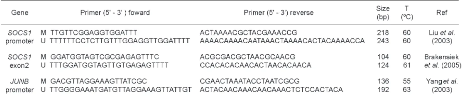
The methylation status of the promoter region of
SOCS-1 was analyzed by methylation specific polymerase chain reaction (PCR) as previously described.29 Primers for
MSP were designed to amplify both methylated (M) or unmethylated (U) alleles. SOCS-1 gene (GenBank accession number U20734) was amplified using specific primers for the promoter region and exon2.14,24 (Table 1) JUNB gene (GenBank
accession number U20734) was amplified using primers specific for the promoter region of the JUNB gene.13 (Table 1).
DNA from healthy donors was used as negative controls. Normal DNA from peripheral blood was treated in vitro with SssI methyltransferase (New England Biolabs, Beverly, MA) in order to generate positive controls for methylated alleles of SOCS-1.30 Two µg of genomic DNA
methylation-specific or unmethylation-methylation-specific primer as described previously (Table 01). MSP cycling conditions, JUNB and
SOCS-1 promoters were: 95ºC for 7 min, 35 cycles of 95°C for 1min 30 s, annealing for 1min (SOCS-1 at 60°C and JunB at 55°C), 72°C for 2 min and a final extension at 72°C for 7 min. For exon 2 of SOCS-1, cycling conditions were: 95°C for 05 min followed by 40 cycles of 95°C for 30 s, annealing at 60°C for 45 s, 72°C for 30 s, and a final extension at 72°C for 5 min. PCR products were electrophoresed on 6 % non-denaturing polyacrylamide gels, silver-stained. Results from duplicate experiments were used to determine methylation status. To confirm the efficiency of DNA modification, direct sequencing of the PCR products was done using primers methylated
SOCS1-M/JUNB-M and unmethylated SOCS1-U/JUNB-U PCR in both forward and reverse ways. PCR products were
sequenced on an ABI Prism 377 DNA Sequencer with DNA Sequencing kit (Applied Biosystem, UK) according to the manufacturer's instructions.
Statistical analysis
The Chi-square and Fischer tests were used for statistical analysis. Probability values of ≤ 0.05 were regarded as statistically significant.
Results
The analysis of the methylation status of the SOCS-1
gene and the JUNB gene was carried out by MSP (Methylation-Specific PCR) (Figure 1). The results were confirmed by direct DNA sequencing (data not shown).
(one gained methylation and one lost methylation); 14 patientes (46.6%) showed alterations of methylation status in exon2 of the SOCS-1 gene (12 gained and 2 lost methylation) (Table 3), in 16 patients (53,3%) the methylation status did not change, while 11 patients continued to show the methylation status of SOCS-1 gene exon 2 after HSCT and 5 patients was not detected the methylation; 5 patients (16.6%) showed alteration of methylation status in the
JUNB gene (they gained methylation) (Table 3).
In two patients was observed the disease recurrence. One patient showed methylation in all genes studied after HSCT, had a survival of 1020 days and died of non-Hodgkins lymphoma. The other patient showed only methylation in gene SOCS-1 exon2 after HSCT and is still alive.
The patients studied before and after the HSCT, showed a low frequency of methylation in the promoter region of genes SOCS-1 and JUNB, no statistically significant result was found. In contrast, there is a significant difference in methylation of SOCS-1 gene exon2 found in patients with CML before and after the HSCT.
Methylation was not detected in the promoter region of SOCS-1 and JUNB genes in hematopoietic stem cell donors.
After HSCT, 36.6% (11/30) of the patients developed acute graft-versus-host disease (GVHD), and 70% (21/30) of the patients developed chronic graft-versus-host disease.
Discussion and Conclusions
The methylation status in the promoter region and exon 2 of the SOCS-1 gene, and in the promoter region of the
JUNB gene was analyzed in CML patients.
The methylation status of these genes was not influenced by the age of the patients or their gender, although several studies have suggested that DNA methylation is related to patients' age.31,32
In this study, the two patients who showed methylation in the promoter region of the SOCS-1 gene were in the chronic phase of the disease. No methylation was detected in the three patients who were in the accelerated phase or blast crisis. Hartirnaz et al.33 analyzed
56 blood samples of patients with CML and no methylation was detected in the promoter region of the SOCS-1 gene. In contrast, Liu et al.14 found methylation levels of 52% (52/
100) in the same region, and they were more frequent in patients who were in the blastic crisis (67%) than in those who were in the chronic phase of the disease (46%). In the The resulting BCR-ABL mRNA research was identified
in 10% (3/30) of patients after HSCT, and 90% (27/30) had shown molecular remission. There was no significant difference between patients who had molecular remission after HSCT with the methylation of genes studied. Twenty-seven out of the 30 patients were in the chronic phase of the disease and only two patients showed methylation in the promoter region of the SOCS-1gene. No methylation was found in the three patients who were in the accelerated phase or blastic crises.
Before the hematopoietic stem cell transplantation (HSCT), methylation levels of 6.6% (2/30) in the promoter region of the SOCS-1 gene; 46.6% (14/30) (P = 0.002) in exon2 of the SOCS-1 gene and 3.3% (1/30) in the promoter region of the JUNB gene were observed (Table 2).
In the control samples without the disease, methylation of 10% (3/30) in exon2 of the SOCS-1 gene was found, and no methylation was found in the promoter regions of SOCS-1 and JUNB genes (Table 2).
present study, no methylation was detected in the control group, which is in agreement with Liu et al.,14 Johan et al.25
and Hartirnaz et al.33
Regarding the analysis of the methylation status in exon 2 of the SOCS-1 gene in patients with CML, our findings (46.6%) are similar to those found by Hartirnaz et al.33 which
detected methylation levels of 58.9% in this region. The greatest difference was the frequency of the methylation status in exon2 of the SOCS-1 gene in the control group: in our study we observed low rate of methylation similar to those observed by Johan et al.25 and Chin et al.20 while
Hartirnaz et al.33 found methylation level of 93.8% in the exon2
of the SOCS-1 gene.
It's important to point out that Hartirnaz et al.33 used
primers directed against regions slightly different from those used by the other authors.
Methylation was detected in the promoter region of the JUNB gene in only one patient who was in the chronic phase of the disease (3.3%). These results were not in agreement with those of Yang et al.13 which observed
methylation of 100% (32/32) in samples of CML patients, in which 21 patients were in the chronic phase and 11 in the blastic crises. Findings in the literature suggest that lower levels of methylation in the chronic phase are explained by a higher proportion of normal circulating cells in the peripheral blood.14
In this study, 18 gains of methylation were observed in the genes studied after HSCT. The probability of transference of such alterations to the recipients was discarded once no methylation was detected in the hematopoietic stem cell donors.
The three patients who were in the accelerated phase or blastic crises had some gain of methylation after HSCT. A higher number of patients would be necessary to evaluate the real significance of our finding.
When a possible correlation between methylation status and graft-versus-host disease was investigated, no statistically significant result was found. However, patients who acquired methylation in exon2 of the SOCS-1 gene were more prone to develop chronic GVHD after HSCT.
Whether the immunosuppressants (specially metotrexate - MTX) used had any influence on alterations of the methylation status could not be determined because 29 out of 30 patients received the same kind of treatment containing MTX.
These results showed that methylation in the promoter regions of SOCS-1 and JUNB are not a frequent event in CML patients. In contrast, methylation in exon2 of the SOCS-1 gene is a frequent event, susceptible to alterations of status after HSCT. Lack of negative regulation of cytokine signaling pathways due to methylation can lead to alterations of cell proliferation. Further studies are needed to determine the mechanisms involved in alterations of the methylation status in chronic myeloid leukemia.
Acknowledgments
We are grateful to the patients who participated in this study. We thank Paulo Eduardo de Abreu Machado and Paula Hokama for helpful discussions, Solange Franzolin for help with statistical analysis.
Resumo
Alteração no padrão de metilação gênica pode contribuir para a progressão da leucemia mielóide crônica (LMC). Neste estudo, o padrão de metilação no exon 2 do gene SOCS-1 e região promotora de ambos SOCS-1 e JUNB foram avaliadas em pacientes com LMC. O padrão de metilação desses genes foi analisado usando a técnica "methylation-specific polymerase chain reaction (MSP)" em 30 amostras de pacientes com LMC, 30 amostras desses mesmos pacientes após transplante de medula óssea (TMO) e 30 amostras controle de indivíduos saudáveis. As amostras de pacientes com LMC apresentaram o seguinte padrão de metilação: gene JUNB (3.3%), região promotora do gene SOCS-1 (6.6%) e exon2 do gene SOCS-1 (46.6%). Amostras dos indivíduos saudáveis apresentaram metilação somente no exon 2 do gene SOCS-1 (10%, P = 0.002). Após o transplante, os pacientes apresentaram alterações no padrão de metilação da região promotora do gene SOCS-1 (6.6%), no exon2 do gene SOCS-1 (46.6%) e na região promotora do gene JUNB (16.6%). Metilação das regiões promotoras dos genes SOCS-1 e JUNB não é um evento frequente em LMC. Em contraste, metilação no exon 2 do gene SOCS-1 apresenta-se como um evento frequente, suscetível a alterações no padrão de metilação após TMO. Rev. Bras. Hematol. Hemoter. 2009;31(3):147-152.
Palavras-chave: Leucemia mieloide crônica; metilação; MSP-PCR SOCS-1; JUNB.
References
1. Nowell PC, Hungerford DA. A minute chromosome in human chronic granulocytic leukemia. Science. 1960;132:1497-99.
2. Groffen J, Stephenson JR, Heisterkamp N, de Klein A, Bartram CR, Grosveld G. Philadelphia chromosomal breakpoints are clustered within a limited region, bcr, on chromosome 22. Cell. 1984;36(1):93-9.
3. Ren R. Mechanisms of BCR-ABL in the pathogenesis of chronic myelogenous leukaemia. Nat Rev Cancer. 2005;5(3):172-83. 4. Faderl S, Talpaz M, Estrov Z, O'Brien S, Kurzrock R, Kantarjian
HM. The biology of chronic myeloid leukemia. N Engl J Med. 1999;341(3):164-72.
5. Carreras E, Brunet S, Rovira M, Sierra J, Urbano IA. Manual de transplante hemopoyetico. Espana: Ediciones Antares 1998;269-290.
6. Byrne JL, Stainer C, Hyde H, Miflin G, Haynes AP, Bessell EM, et al.
Low incidence of acute graft-versus-host disease and recurrent leukaemia in patients undergoing allogeneic haemopoietic stem cell transplantation from sibling donors with methotrexate and dose-monitored cyclosporin A prophylaxis. Bone Marrow Transplant. 1998;22(6):541-5.
8. Attwood JT, Yung RL, Richardson BC. DNA methylation and the regulation of gene transcription. Cell Mol Life Sci. 2002;59 (2):241-57.
9. Malinen T, Palotie A, Pakkala S, Peltonen L, Ruutu T, Jansson SE. Acceleration of chronic myeloid leukemia correlates with calcitonin gene hypermethylation. Blood. 1991;77(11):2435-40.
10. Guinn BA, Mills KI. p53 mutations, methylation and genomic instability in the progression of chronic myeloid leukaemia. Leuk Lymphoma. 1997;26(3-4):211-26.
11. Asimakopoulos FA, Shteper PJ, Krichevsky S, Fibach E, Polliack A, Rachmilewitz E, et al. ABL1 methylation is a distinct molecular event associated with clonal evolution of chronic myeloid leukemia. Blood. 1999;94(7):2452-60.
12. Yang MY, Chang JG, Lin PM, Tang KP, Chen YH, Lin HY, et al. Downregulation of circadian clock genes in chronic myeloid leukemia: alternative methylation pattern of hPER3. Cancer Sci. 2006;97(12):1298-307.
13. Yang MY, Liu TC, Chang JG, Lin PM, Lin SF. JunB gene expression is inactivated by methylation in chronic myeloid leukemia. Blood. 2003;101(8):3205-11.
14. Liu TC, Lin SF, Chang JG, Yang MY, Hung SY, Chang CS. Epigenetic alteration of the SOCS1 gene in chronic myeloid leukaemia. Br J Haematol. 2003;123(4):654-61.
15. Lotem J, Sachs L. Cytokine control of developmental programs in normal hematopoiesis and leukemia. Oncogene. 2002;21(21): 3284-94.
16. Krebs DL, Hilton DJ. SOCS proteins: negative regulators of cytokine signaling. Stem Cells. 2001;19(5):378-87.
17. Yoshikawa H, Matsubara K, Qian GS, Jackson P, Groopman JD, Manning JE, et al. SOCS-1, a negative regulator of the JAK/STAT pathway, is silenced by methylation in human hepatocellular carcinoma and shows growth-suppression activity. Nat Genet. 2001; 28(1):29-35.
18. Endo TA, Masuhara M, Yokouchi M, Suzuki R, Sakamoto H, Mitsui K, et al. A new protein containing an SH2 domain that inhibits JAK kinases. Nature. 1997;387(6636):921-4.
19. Galm O, Yoshikawa H, Esteller M, Osieka R, Herman JG. SOCS-1, a negative regulator of cytokine signaling, is frequently silenced by methylation in multiple myeloma. Blood. 2003;101(7): 2784-8.
20. Chim CS, Kwong YL. How frequent is SOCS1 methylation in acute myeloid leukaemia? Br J Haematol. 2004;127(5):609-11. 21. Chen CY, Tsay W, Tang JL, Shen HL, Lin SW, Huang SY, et al.
SOCS1 methylation in patients with newly diagnosed acute myeloid leukemia. Genes Chromosomes Cancer. 2003 Jul;37(3):300-5.
22. Watanabe D, Naka T, Kishimoto T. Implication of SOCS-1 gene methylation in acute myeloid leukaemia. Br J Haematol. 2004; 127(5):608-9.
23. Sutherland KD, Lindeman GJ, Choong DY, Wittlin S, Brentzell L, Phillips W, et al. Differential hypermethylation of SOCS genes in ovarian and breast carcinomas. Oncogene. 2004;23(46):7726-33.
24. Brakensiek K, Länger F, Schlegelberger B, Kreipe H, Lehmann U. Hypermethylation of the suppressor of cytokine signalling-1 (SOCS-1) in myelodysplastic syndrome. Br J Haematol. 2005; 130(2):209-17.
25. Johan MF, Bowen DT, Frew ME, Goodeve AC, Reilly JT. Aberrant methylation of the negative regulators RASSFIA, SHP-1 and SOCS-1 in myelodysplastic syndromes and acute myeloid leukaemia. Br J Haematol. 2005;129(1):60-5.
26. Wu SJ, Yao M, Chou WC, Tang JL, Chen CY, Ko BS, et al. Clinical implications of SOCS1 methylation in myelodysplastic syndrome. Br J Haematol. 2006;135(3):317-23.
Avaliação: Editor e dois revisores externos Conflito de interesse: sem conflito de interesse
Recebido: 08/07/2008
Aceito após modificações: 17/12/2008
27. Lord KA, Abdollahi A, Hoffman-Liebermann B, Liebermann DA. Proto-oncogenes of the fos/jun family of transcription factors are positive regulators of myeloid differentiation. Mol Cell Biol. 1993;13(2):841-51.
28. Schütte J, Viallet J, Nau M, Segal S, Fedorko J, Minna J. jun-B inhibits and c-fos stimulates the transforming and trans-activating activities of c-jun. Cell. 1989;59(6):987-97.
29. Herman JG, Graff JR, Myöhänen S, Nelkin BD, Baylin SB. Methylation-specific PCR: a novel PCR assay for methylation status of CpG islands. Proc Natl Acad Sci USA. 1996;93(18): 9821-6.
30. Esteller M, Hamilton SR, Burger PC, Baylin SB, Herman JG. Inactivation of the DNA repair gene O6-methylguanine-DNA methyltransferase by promoter hypermethylation is a common event in primary human neoplasia. Cancer Res. 1999;59(4):793-7. 31. Issa JP. CpG-island methylation in aging and cancer. Curr Top
Microbiol Immunol. 2000;249:101-18.
32. Kang GH, Lee S, Kim JS, Jung HY. Profile of aberrant CpG island methylation along multistep gastric carcinogenesis. Lab Invest. 2003;83(4):519-26.
33. Hatirnaz O, Ure U, Ar C, Akyerli C, Soysal T, Ferhanoglu B, et al.