CENTRO DE CIÊNCIAS EXATAS E DA NATUREZA
PROGRAMA DE PÓS-GRADUAÇÃO EM BIOLOGIA CELULAR E MOLECULAR
HALINE BARROSO
ANÁLISE DE METILAÇÃO DE DNA NOS GENES DA
CITOQUERATINA 14 (
KRT14
) E 19 (
KRT19
) EM
AMOSTRAS DE PELE EXPOSTA E NÃO EXPOSTA
AO SOL
João Pessoa - PB
HALINE BARROSO
ANÁLISE DE METILAÇÃO DE DNA NOS GENES DA
CITOQUERATINA 14 (
KRT14
) E 19 (
KRT19
) EM AMOSTRAS DE
PELE EXPOSTA E NÃO EXPOSTA AO SOL
Dissertação apresentada ao Programa de Pós-Graduação em Biologia Celular e Molecular do Centro de Ciências Exatas e da Natureza, da Universidade Federal da Paraíba, como parte dos requisitos para obtenção do título de MESTRE EM BIOLOGIA CELULAR E MOLECULAR
Orientadora: Profa. Dra. Naila Francis Paulo de Oliveira
B277a Barroso, Haline.
Análise de metilação de DNA nos genes da citoqueratina 14 (KRT14) e 19 (KRT19) em amostras de pele exposta e não exposta ao sol / Haline Barroso.- João Pessoa, 2015.
43f. : il.
Orientadora: Naila Francis Paulo de Oliveira Dissertação (Mestrado) - UFPB/CCEN
1. Biologia celular e molecular. 2. Citoqueratina.
3.Epigenética. 4. Metilação de DNA. 5. Radiação UV - pele.
Dissertação de Mestrado avaliada em 27/02/2015
BANCA EXAMINADORA
_____________________________________________ Profa. Dra. Naila Francis Paulo de Oliveira
Programa de Pós-Graduação em Biologia Celular e Molecular – Universidade Federal da Paraíba
Orientadora – UFPB
______________________________________________ Profa. Dra.Tatjana Keesen De Souza Lima
Centro de Biotecnologia (CBiotec) – Universidade Federal da Paraíba Examinadora Externa
______________________________________________ Profa. Dra. Krystyna Gorlach Lira
Departamento de Biologia Celular e Molecular - Universidade Federal da Paraíba Examinadora Interna
__________________________________________
Profa. Dra. Darlene Camati PersuhnDepartamento de Biologia Celular e Molecular - Universidade Federal da Paraíba Examinadora Suplente Interno
____________________________________________
Profa. Dra. Simone Silva dos Santos LopesDEDICATÓRIA
Dedico este trabalho a Deus, por ter me dado forças para prosseguir, por abrir as portas nas dificuldades e me mostrar o caminho correto por onde devo andar.
A minha querida e amada avó Amenaide que sempre me incentivou a estudar e a seguir em frente.
AGRADECIMENTOS
A Deus que me permitiu ter saúde e perseverança para a realização desse trabalho.
A Prof. Dra. Naila Francis Paulo de Oliveira, pelo apoio constante, incentivo, paciência e colaboração em todas as etapas desse trabalho.
A Dra. Krystyna Gorlach Lira, pela gentileza de ser a revisora deste trabalho e participar da Banca Examinadora.
A todos os professores do mestrado que de alguma forma contribuíram para a minha formação.
A minha fiel amiga de todas as horas Alanne Rayssa pelo companheirismo, paciência e pelos grandes momentos de aprendizado e alegrias que passamos juntas.
A todos os amigos e companheiros do laboratório Isabelle Borba, Ludimila Araújo, Daniel Uchôa, Bruno Mariz, Mikaelly Batista, Laura Souto, Dasaiev Dutra, Sabrina Rocha.
A minha amiga Ana Anízia que sempre esteve pronta a me ajudar e a me fazer sorrir mesmo nos momentos mais tensos da vida.
RESUMO
Pesquisas tem mostrado que a radiação UV do sol pode causar mutações no DNA e aumentar o risco para o desenvolvimento de câncer de pele. Entretanto, ainda pouco se sabe sobre a capacidade da radiação UV em causar alterações epigenéticas na pele. A metilação do DNA é caracterizada pela adição do grupo metil em uma citosina precedida por uma guanina (dinucleotídeo CpG), o que pode alterar a transcrição gênica, diminuindo a expressão ou silenciando um gene. Alterações epigenéticas podem representar um importante caminho de como os fatores ambientais influenciam no envelhecimento e no desenvolvimento de certas doenças de maneira tecido específica. Queratinas epiteliais são chamadas de citoqueratinas, e sua principal função é manter a integridade e estabilidade mecânica do tecido epitelial. Neste trabalho investigamos se há influência da exposição solar sobre o perfil de metilação de DNA nos genes das citoqueratinas 14 (KRT14) e (KRT19), em células da pele de indivíduos sem histórico de doenças de pele. Biopsias de pele foram obtidas através de um Punch circular da de área exposta e não exposta ao sol de 30 cadáveres do Serviço de Verificação de Óbito. A análise de metilação do gene KRT14 foi realizada pelo método de PCR Específica para Metilação (MSP), e para o gene KRT19 foi realizado o método de Restrição Enzimática Sensível à Metilação (MSRE) das áreas expostas e protegidas do sol. A análise estatística mostrou que não há diferenças significativas entre as regiões exposta e não exposta ao sol, sendo a condição metilada a mais frequente tanto para o gene KRT14 quanto para o gene KRT19 (p>0,05; McNemar). Assim, concluímos que não há influência da exposição solar no perfil de metilação de DNA nos genes KRT14 e KRT19.
ABSTRACT
It is well established that solar UV radiation can cause mutations in DNA and increase the risk of developing skin cancer. However, little is known about the ability of UV radiation to cause epigenetic changes in the skin. DNA methylation, characterised by the addition of a methyl group in cytosines within CpG dinucleotides, can modify gene transcription, leading to decreased expression or even silencing of a gene. Epigenetic changes could represent an important pathway by which environmental factors influence aging and disease risks, with a tissue-specific manner. Epithelial keratins are called cytokeratins, the main function of cytokeratins is to maintain the integrity and mechanical stability through cell-cell contacts with epithelial tissue. The aim of this study was to investigate the sun exposure influence on DNA methylation status in the cytokeratin 14 (KRT14) and 19 (KRT19) genes of skin cells of subjects whithout history of skin disease. Skin biopsies were obtained by punch of sun-exposed (outer forearm) and sun-protected areas (inner arm) from 30 corpses of the Brazilian Services of Death Investigation. The KRT14 gene DNA methylation analysis was performed using Methylation-Specific PCR (MSP), and the KRT19 gene DNA methylation analysis was performed using Methylation-Sensitive Restriction Enzymes (MSRE) of sun-exposed and protected skin areas. Statistical analysis showed no significant differences between sun-protected and sun-exposed areas and the most frequently methylated condition for CpG studied for KRT14 and KRT19 genes (p> 0.05; McNemar). We conclude that sun exposure does not induce changes in DNA methylation status in the KRT14 and KRT19 genes.
LISTA DE ABREVIATURAS E SIGLAS
CH3– Grupamento metil
CpG – Citosina/ligação fosfodiester/guanina DNA – Ácido Desoxirribonucleico
DNMT – DNA metiltransferase K – Citoqueratina
KDa – Kilo dalton
KRT – Gene da citoqueratina MBPs - Methyl Binding Proteins MMPs – Metaloproteases de matriz MSP – PCR específica para metilação
MSRE- Restrição Enzimática Sensível à Metilação PCR – Reação em cadeia da polimerase
nm – Nanômetro pb – Pares de base SE – Sun-exposed
SEC31L2 – SEC 3 homólogo a B SP – Sun-protected
SVO – Serviço de verificação de óbitos TET2 – Tet methylcytosine dioxygenase 2 UVA- Ultra Violeta A
SUMÁRIO
1. INTRODUÇÃO 1
1.1. Epigenética 1
1.2. Radiação Solar 3
1.3. Pele 4
1.4. Queratinas 5
1.5. Justificativa 7
2. OBJETIVOS 8
3. RESULTADOS 9 3.1 Apresentação 9 3.2Manuscrito:DNA methylation analysis of cytokeratin 14 (KRT14) and 19 (KRT19)
genes of sun- exposed and sun-protected skin areas 10
- Abstract 11
- Introduction 12
- Materials and Methods 14
- Results and Discussion 16
- Table 1 21
- Figure 1 22
- Figure 2 23
- References 24
4. CONCLUSÃO 28
5. REFERÊNCIAS BIBLIOGRÁFICAS 29
1. INTRODUÇÃO
1.1 - EPIGENETICA
Epigenética é definida como o estudo das modificações do DNA e das histonas que são herdáveis e não alteram a sequência de bases do DNA. Dentre as modificações que as histonas podem sofrer, estão: metilação, fosforilação e acetilação. Entretanto, na molécula de DNA ocorre apenas a metilação (BERGER et al., 2009). A metilação consiste em uma modificação covalente do DNA na qual um grupamento metil (CH3) é transferido da 5-adenosilmetionina para o carbono 5 de uma citosina que geralmente precede uma guanina (dinucleotídeo CpG), pela ação de uma família de enzimas que recebe o nome de DNA-metiltransferase (DNMT).
As DNAs metiltransferases estão divididas em duas classes de representantes: aquelas envolvidas na metilação de fitas hemimetiladas do DNA, conhecidas como metilases de manutenção, como a DNMT1, e outro grupo responsável pela maioria dos processos de metilação de novo, que ocorrem em sítios sem nenhum tipo de indicação de metilação, ou seja, sem a presença de metilação prévia, como as DNMT2 e DNMT3A e DNMT3B (BESTOR, 2000). A ação destas enzimas requer um substrato que atue como doador de radical metil e estes são obtidos da dieta, como o folato, a metionina, a colina e a vitamina B12 (WATERLAND; JIRTLE, 2003; WATERLAND, 2006).
Estudos revelam que o padrão epigenético é influenciado pelo meio ambiente, e alterações epigenéticas são mais freqüentes que mutações (USHIJIMA; ASADA, 2010)
Os mecanismos epigenéticos são balanceados para garantir que ocorra o desenvolvimento normal das células, garantindo que haja a correta expressão dos genes. Estes mecanismos são altamente regulados por um grande número de proteínas que estabelecem, leem e apagam as marcações epigenéticas específicas, definindo onde e quando a maquinaria de transcrição pode acessar a sequência de DNA (DE BRITO; PACHECO, 2013).
Durante o desenvolvimento e a diferenciação celular, as marcas epigenéticas sofrem mudanças dinâmicas que contribuem para a produção e manutenção de diferentes tipos celulares. A análise de como essas marcas epigenéticas participam da regulação gênica é crucial para o melhor entendimento do desenvolvimento embrionário e a etiologia de muitas doenças (AUCLAIR ; WEBER, 2012).
diferenciadas e tem uma importante função na regulação da expressão gênica e no silenciamento de elementos repetitivos no genoma, sendo ela randômica ou sítio específica (FEINBERG; TYCKO, 2004). Os dinucleotídeos CpG aparecem esparsos pelos genoma dos eucariotos ou agrupados em regiões definidas como ilhas CpG. Estas ilhas são freqüentes em regiões promotoras de certos genes, incluindo genes de manutenção que são genes constitutivos, necessários para a manutenção da função celular de base e são expressos em todas as células do organismo sob condições normais e fisiopatológicas. Ilhas CpG são regiões do DNA maior que 200 pares de base contendo aproximadamente 50% de bases citosina e guanina, é comum a presença esperada de aproximadamente 60% de dinucleotídeos CpG (LI; DAHIYA, 2002).
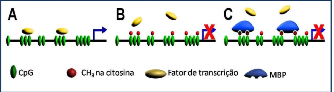
A transcrição gênica pode ser fortemente inibida pela adição de radical metil. A presença de um “capuz” metil sobre uma citosina que precede uma guanina pode inibir a ligação de fatores de transcrição a estas regiões. A não ligação de fatores de transcrição aos seus sítios específicos pode resultar na ausência ou diminuição da transcrição gênica (figura 1). Proteínas chamadas MBPs (Methyl Binding Proteins), com afinidade pelo grupo metil, ligam-se às regiões CpGs localizadas nos promotores e impedem o acesso dos fatores de transcrição aos seus sítios (ATTWOOD et al. 2002).
Figura 1. Mecanismo pelo qual a metilação do DNA inibe a transcrição gênica. A. Região promotora desmetilada permitindo a ligação dos fatores de transcrição. B. Metilação impedindo a ligação dos fatores de transcrição. C. Proteínas que se ligam a metilcitosina em ilhas CpG bloqueiam a ligação dos fatores de transcrição. Fonte: Attwood et al. 2002.
1.2 - RADIAÇÃO SOLAR
A maior parte da energia irradiada pelo sol está na região do espectro Ultravioleta (comprimento de 100-400nm) em comparação ao espectro da luz visível (comprimento de 400-700nm). A radiação UV pode ser subdividida em UVA, UVB e UVC. As frações que atingem a Terra são a UVA e UVB. A atmosfera terrestre bloqueia a passagem da radiação UVC, impedindo que ela chegue à superfície do planeta. Os raios ultravioleta B (UVB) (comprimento de 280-320nm) são absorvidos eficientemente, embora incompletamente pela camada de ozônio, enquanto que os raios ultravioleta A (UVA) (comprimento de 320-400nm) são absorvidos menos eficientemente e assim pode ser mais facilmente transmitido para a atmosfera terrestre (CRUTZEN, 1992). Durante muitos anos a diminuição da camada de ozônio tem sido observada como um resultante da atividade humana, principalmente devido à emissão de compostos contendo halogênio resultando em um aumento da dose de UVB sobre a Terra e consequentemente na pele (CRUTZEN, 1992; DE FABO, 2000).
O efeito da radiação solar tem sido uma das maiores preocupações humanas, pois a exposição aguda da pele à radiação UV causa queimaduras, alteração da pigmentação, inflamação, imunossupressão e danos no tecido conjuntivo da derme (DAMIAN et al., 2011). A exposição crônica por muitos anos altera a arquitetura normal da pele causando o envelhecimento prematuro (foto envelhecimento) (FISHER et al., 2002) e câncer de pele (PFEIFER; BESARATINIA, 2012).
O radiação UVB possui efeitos benéficos a saúde do ser humano, como estimulante para a produção de vitamina D, porém exposição aguda aos raios UVB podem causar efeitos deletérios na pele como eritema e fotodermatoses e nos olhos podem ocorrer queratites e conjutivites. (AMBACH; BLUMTHALER, 1993). Os raios UVB penetram na epiderme e causam as queimaduras solares, e é o principal responsável pelas alterações celulares que predispõem ao câncer da pele. Os raios UVA penetram mais profundamente na pele atingindo a derme, com importante participação nas fotoalergias e também predispõe a pele ao surgimento do câncer (ICHIHASHI, M. et al.,2003).
1.3 PELE
A pele é o maior órgão do corpo humano sendo responsável pela termorregulação, pela defesa, pela percepção e pela proteção. Este órgão se apresenta em duas camadas: a epiderme e a derme. A epiderme é constituída por um epitélio estratificado pavimentoso queratinizado e a derme é um tecido conjuntivo que sustenta a epiderme, sendo constituído por elementos fibrilares, como o colágeno e a elastina (BAUMANN, 2007)
A pele humana representa um órgão com muitas vantagens para o estudo de alterações induzidas pelo envelhecimento e pelos fatores ambientais, pois é o órgão mais exposto (BAUMANN, 2007; GILHAR et al., 2004).
Absorção da radiação solar UV pelas células da pele altera a estrutura química do DNA e causa estresse oxidativo (BIESALSKI et al., 2003; ICHIHASHI et al., 2003; YAAR; GILCHREST, 2007). Essas alterações ativam vias de sinalização celular que regulam múltiplas funções celulares na pele (FISHER et al., 2002; QUAN et al., 2005; RITTIÉ; FISHER, 2002). Células de mamíferos respondem à radiação UV com várias alterações bioquímicas incluindo a expressão de genes distintos chamados de genes de resposta UV, e já foi mostrado que esses genes estão relacionados à transdução de sinal, defesa antioxidante e controle do ciclo celular (SCHARFFETTER–KOCHANEK et al., 2000; SHAULIAN et al., 2000). Ainda, genes que respondem à radiação UVB compreendem muitas proteases, dentre elas as metaloproteases de matriz (MMPs) (QUAN et al., 2009), as quais degradam os componentes da matriz extracelular (HO et al., 2005), bem como reduzem a produção de pro colágeno tipo I (FISHER et al., 2002).
1.4 - QUERATINAS
Queratinas são proteínas pertencentes à família de filamentos intermediários, as quais são predominantemente expressas em células epiteliais. As queratinas representam em torno de trinta proteínas, diferindo pelo peso molecular; sendo o mais baixo (40 KDa), as queratinas 8 e 18, as quais são encontradas no epitélio simples e glandular; queratinas de peso molecular intermediário, encontradas no epitélio estratificado; e as mais pesadas queratinas (aproximadamente 67 KDa) são encontradas no epitélio estratificado queratinizado (MOLL et al., 2008).
A família gênica das queratinas consiste no maior número de membros em humanos com 54 genes funcionais distintos: 31 queratinas epiteliais (citoqueratinas), 15 queratinas específicas do cabelo e 8 queratinas da bainha radicular interna (HESSE et al., 2004). De acordo com a proposta do Comitê de Nomenclatura de Queratina (SCHWEIZER et al., 2006), as queratinas estão divididas em dois grupos: tipo I (K9–K10, K12– K28, e K31–K40 incluindo K33a e K33b) as quais são ácidas e codificadas pelo cromossomo 17, enquanto as do tipo II (K1–K8 (incluindo K6a, K6b e K6c, K71–K86) são básicas e codificadas pelo cromossomo 12 (ROMANO V. et al., 1988). Uma propriedade única das queratinas é que em contraste com outras proteínas dos filamentos intermediários, somente conseguem constituir seus filamentos pela formação de heteropolímeros, formando pares de moléculas do tipo I e do tipo II (1:1) (MOLL et al., 1983).
A principal função das queratinas é manter a integridade e estabilidade mecânicas, através de contatos célula-célula do tecido epitelial. Consequentemente, elas são parte inerente da estabilidade contínua de cada célula na formação do tecido. Como os filamentos de queratina são estabilizadores estruturais das células epiteliais, o interesse por essas moléculas abrange o contexto biológico, embriológico, patológico e dermatológico. Tipicamente, os filamentos de queratinas ficam inseridos aos desmossomos e hemidesmossomos. Dessa forma, contribuem não somente com a estabilidade entre as células epiteliais, mas também com a conexão à membrana basal na ligação entre o epitélio e o tecido conjuntivo (MOLL et al., 2008).
As queratinas também são importantes para a polaridade da membrana das células epiteliais (ORIOLO et al., 2007). Dessa forma, as queratinas apresentam além da sua função mecânica, mas também na sinalização celular.
maturação durante o desenvolvimento. As citoqueratinas apresentam distribuição específica para cada subtipo de epitélio, permitindo que sejam utilizadas como importantes marcadores de sua diferenciação. Todos os epitélios estratificados possuem K5 e K14 nas células basais indiferenciadas, mas diferenças emergem na camada suprabasal com a diferenciação (COULOMBE; LEE, 2012)
Enquanto a troca entre proliferação e diferenciação é normalmente controlada e compartimentalizada dentro da camada basal e suprabasal respectivamente, as células mitóticas podem ocorrer nas camadas diferenciadas durante o processo de reparo e em doenças como o câncer (SINGER; CLARK, 1999). Tumores epiteliais, incluindo metástases, em sua maioria retém o padrão normal de queratinas de seu epitélio de origem (MOLL et al., 1983). De fato, as queratinas são um dos mais potentes marcadores de diferenciação epitelial e marcador tumoral. Técnicas de imunohistoquímica, as quais utilizam anticorpos específicos contra queratinas são usados de forma rotineira em laboratórios para diagnósticos de tumores (BARAK et al., 2004).
A citoqueratina 14 (K14) tipo I e a citoqueratina 5 (K5) tipo II formam o par primário de queratinas do epitlélio estratificado escamoso, incluindo a epiderme como também o epitélio estratificado não queratinizado das mucosas (MOLL et al., 1983). Elas são fortemente expressas nas células indiferenciadas da camada basal contendo células-tronco e são menos expressas na camada suprabasal diferenciada (FUCHS; GREEN, 1980). O par K5/K14 são empacotados como tonofilamentos e se ligam aos desmossomos e hemidesmossomos. Mutações nos genes da K14 e K5 têm sido associadas à Epidermólise Bolhosa Simples, doença de pele caracterizada pela fragilidade da epiderme, mesmo em traumas leves como fricção (ISHIDA-YAMAMOTO; AKEMI ET AL., 1991).
A citoqueratina 19 (K19) tipo I é a menor e diferente das outras queratinas, uma vez que não apresenta cauda alfa-helicoidal (BADER et al., 1986) é expressa em epitélio simples, além de epitélio pseudoestratificado como também na camada basal de células do epitélio estratificado não queratinizado. Já foi observado na mucosa oral que a expressão de K19 pode ser induzida por displasia (LINDBERG; RHEINWALD, 1989) e inflamação (MOLL eta al., 1983). Recentemente, aumento da expressão da K19 foi observado em câncer de tireoide e de fígado (SETHI et al., 2011). Alguns estudos mostram que a K19 é regulada epigeneticamente (PAIVA et al., 2011; PLACHOT; LELIÈVRE, 2004).
Os genes da queratina 14 (KRT14) e19 (KRT19) estão localizados no cromossomo 17
e não possuem ilhas CpG em suas regiões promotora, porém na região proximal do éxon1 da KRT14 há diversos dinucleotídeos CG espalhados, e na região proximal do gene da
1.5 - JUSTIFICATIVA
Estudos de metilação de DNA têm emergido como um importante campo de pesquisa e revelam como o meio ambiente pode afetar nossos genes e alterar drasticamente a expressão gênica, resultando em doenças inflamatórias e tumorais, ou mesmo no envelhecimento.
Já está bem estabelecido como a radiação UV do sol pode causar mutações no DNA e aumentar o risco para o desenvolvimento de câncer de pele (PFEIFER; BESARATINIA, 2009). Entretanto, ainda pouco se sabe sobre a capacidade da radiação UV em causar alterações epigenéticas na pele, e esse pode ser outro mecanismo pelo qual a exposição crônica ao sol leva a tumorigênese.
A maioria das evidências em amostras não tumorais de que a radiação ultravioleta pode causar alterações epigenéticas no DNA são oriundas de
trabalhos experimentais
com ratos, linhagem celulares ou pele reconstruídas in vitro (BERNERD, 2001; MITTALet al., 2003; NANDAKUMAR et al., 2011; CHEN et al., 2012). Raros são os estudos utilizando amostras de pele obtidas de humanos. Um desses estudos mostrou hipermetilação no gene CDH1 (cadherin1) em regiões expostas ao sol
(SATHYANARAYANA et al., 2007). Outro estudo revelou tendência à hipometilação no promotor dos genes KRT75 (queratina 75), SEC31L2 (SEC31 homolog a B (S. cerevisiae), DDAH2 (dimethyl arginine dimethyl aminohydrolase 2) e TET2 (tetmethyl cytosine dioxygenase 2) em amostras de pele expostas ao sol e em adição, mostrou
ainda que, essas alterações eram mais acentuadas em indivíduos mais velhos (GRÖNNIGER et al., 2010).
2. OBJETIVO
O objetivo deste trabalho foi investigar a influência da exposição solar sobre o estado de metilação do DNA no promotor dos genes KRT14 e KRT19 em amostras de células da pele
3. RESULTADOS
O manuscrito a seguir foi submetido para publicação no periódico “Biologicals” e encontra-se
em análise.
3.1 - DNA methylation analysis of cytokeratin 14 (KRT14) and 19 (KRT19) genes of
sun-exposed and sun-protected skin areas
HALINE BARROSO1, ALANNE RAYSSA DA SILVA MELO1, DANIEL UCHÔA DE
ARAÚJO2, FRANCISCO RUIDOMAR PEREIRA3, NAILA FRANCIS PAULO DE
3.2 – Manuscrito
DNA methylation analysis of cytokeratin 14 (
KRT14
) and 19 (
KRT19
) genes of
sun-exposed and sun-protected skin areas
HALINE BARROSO1, ALANNE RAYSSA DA SILVA MELO1, DANIEL UCHÔA DE ARAÚJO2,
FRANCISCO RUIDOMAR PEREIRA3, NAILA FRANCIS PAULO DE OLIVEIRA1,2
1 Programa de Pós Graduação em Biologia Celular e Molecular, Centro de Ciências Exatas e
da Natureza, Universidade Federal da Paraíba, João Pessoa, PB-Brazil
2 Departamento de Biologia Molecular, Centro de Ciências Exatas e da Natureza,
Universidade Federal da Paraíba, João Pessoa, PB-Brazil
3 Departamento de Morfologia, Centro de Ciências da Saúde, Universidade Federal da
Paraíba, João Pessoa, PB-Brazil
Corresponding Author:
Dra. Naila Francis Paulo de Oliveira Universidade Federal da Paraíba
Centro de Ciências Exatas e da Natureza Departamento de Biologia Molecular Cidade Universitária – Campus I João Pessoa-PB
Brazil
CEP 58051-900
ABSTRACT
The ability of environmental factors to induce epigenetic changes has been investigated and many studies have shown a relationship between them. The aim of this study was to investigate the sun exposure influence on DNA methylation status in the cytokeratin 14 (KRT14) and cytokeratin 19 (KRT19) genes of skin cells of subjects without history of skin
disease. Skin biopsies were obtained by punch of exposed (outer forearm) and sun-protected areas (inner arm) from 30 corpses of the Brazilian Service of Death Investigation and genomic DNA was extracted. KRT14 DNA methylation analysis was performed using
Methylation-Specific PCR (MSP) and KRT19 DNA methylation analysis was performed using
Methylation-Sensitive Restriction Enzymes (MSRE) of sun-exposed and sun-protected skin areas. The methylated condition was found to be a common event in skin cells for both
KRT14 and KRT19 genes and no differences were found among areas (p>0.05; McNemar).
We conclude that sun exposure does not induce changes in DNA methylation status in the
KRT14 and KRT19 genes.
1. Introduction
The effect of solar radiation is one of the greatest human concerns. Acute exposure of the skin to UV radiation causes burns, abnormal pigmentation, inflammation, immunosuppression and damage to the connective tissue of the dermis [1,2,3]. Chronic exposure over many years could change the normal architecture of the dermis and epidermis causing premature aging (photoaging) [4] and skin cancer [5]. Data from the National Cancer Institute (INCA-Ministry of Health-Brazil) have shown that skin cancer is what most affects Brazilians, especially in the Northeast, where UV levels are high for most of the year [6].
Absorption of UV radiation by skin cells alters the chemical structure of DNA and causes oxidative stress [7,8]. These changes activate signalling pathways that regulate many cellular functions of the skin [4,9,10]. Mammalian cells respond to UV radiation with various biochemical alterations, including the expression of different genes called UV response genes, and it has been shown that these genes are related to signal transduction, antioxidant defence and cell cycle control [11,12]. It is well established that solar UV radiation can cause mutations in DNA and increase the risk of developing skin cancer [13]. However, little is known about the ability of UV radiation to cause epigenetic changes in the skin, and this may be another mechanism by which chronic exposure to the sun leads to tumorigenesis.
ions [16,17], drugs [18], diet [19], alcohol dependence [20] and smoking [21] are all associated with epigenetic changes.
Surprisingly, the number of studies examining the effects of sun exposure on DNA methylation is limited. Most evidence in non-tumour samples that ultraviolet radiation can cause epigenetic changes is derived from experimental studies with mice or cell culture [22,23,24]. Studies using skin samples obtained from humans suffering from sun exposure are scarce [25,26].
Epidermal architecture is maintained by a family of proteins that compose intermediate filaments of the cytoskeleton, such as keratins. Epithelial keratins are called cytokeratins, which Moll et al. [27] classified into 20 subtypes according to their molecular weight and isoelectric pH in an attempt to clarify the diversity and expression patterns of keratins among epithelial tissue types. The main function of cytokeratins is to maintain the integrity and mechanical stability through cell-cell contacts with epithelial tissue. It has been shown that these proteins are dysregulated in non-tumour samples of skin cells exposed to UV radiation, especially cytokeratins 14 and 19 [28], and also in tumour samples [29].
Based on these facts and in order to contribute more data on the ability of sun exposure causes epigenetic changes, we hypothesised that sun radiation could alter the DNA methylation profile on the cytokeratin 14 (KRT14) and cytokeratin 19 (KRT19) genes of
2. Materials and Methods
2.1. Ethics Statement and Subject Population
Prior to commencement, the study design was approved by the Institutional Review Board of the Federal University of Paraiba (protocol number 430/2011). The sample covered male and female corpses of the Brazilian Service of Death Investigation aged over 30 years. The family of each individual was prompted to sign the consent and agree to participate in the study. The demographic data and general health were obtained via record and individuals with a medical history of skin diseases were not included. Skin samples were collected and ranked according to the criteria of Fitzpatrick [30]. A sample of 30 unrelated subjects was included, and samples were divided into two conditions according to sun exposure: sun-exposed areas (SE) and sun-protected areas (SP).
2.2. Sample Collection and DNA Extraction
Biopsies were collected by punch (5mm diameter) from the outer forearm (sun-exposed area) and inner arm (sun-protected area) up to 10 hours after death. Immediately after removal, biopsies (epidermis and dermis) were stored in a tube containing 800 µL of RNAholder (Bioagency, São Paulo, SP, Brazil) and frozen at -20°C until analyses were performed. After, genomic DNA of skin biopsies was purified using the TRIZOL reagent
(Invitrogen, Carlsband, CA, USA) following the manufacturer’s recommendations. DNA
quantification was performed by NanoDrop spectrophotometer. Samples were then frozen at -20°C until DNA methylation analysis.
2.3. DNA Methylation Analysis
KRT14 - DNA Methylation analysis was performed using Methylation-Specific PCR
DNA Methylation-Gold kit, according to the manufacturer’s instructions. Two fragments were
amplified with specific primers for either methylated or unmethylated targets. Each MSP reaction incorporated 100ng of bisulphite modified DNA, 1µL (10 µM) of each primer and 1x Go Taq Hot Start Green Master Mix (Promega Corporations, Madison, WI, USA) in a final reaction of 20 µL. Methylated and unmethylated cycling conditions were as follows: 95°C x 5 minutes; 40 cycles x (95°C x 1 min, specific for fragment A and B) x 1 min, 72°C x 1 min); and 72°C x 5 min (Table 01). Methylated (Methylated CpG Jurkat Genomic DNA, New England Biolabs) and unmethylated (5-Aza-dc Treated Jurkat Genomic DNA, New England Biolabs) DNA was modified as previously quoted and amplified by PCR as control reactions with primers for the methylated and unmethylated condition, respectively. Amplified PCR samples (6µl) were loaded in 6% polyacrylamide gels and subjected to electrophoresis. DNA bands were detected after silver staining.
KRT19 – Optimal primers for methylation analysis by MSP for the region of interest of
this gene were not found. Therefore, DNA Methylation analysis of KRT19 was performed
using Methylation-Sensitive Restriction Enzymes (MSRE) [32]. Polymerase chain reaction (PCR) analysis was performed, which relied on the inability of restriction enzymes to digest methylated sequences. The examined sites were recognised by one of the following restriction enzymes, whose activity was always blocked by CpG methylation: HhaI and HpaII.
cycles x (95°C for 1min., 58°C for 1 min. and 72°C for 1 min) and 72°C for 1 min. (Table 1). The non-enzyme-treated control DNA sample was always amplified with primers, in parallel with the enzyme-treated samples, and both were subjected to electrophoresis in adjacent lanes. This provided a positive control for the PCR reaction and for DNA loading. Amplified PCR samples (6µl) were loaded in 6% polyacrylamide gels and subjected to electrophoresis. DNA bands were detected after silver staining.
2.4. Statistical Analysis
DNA methylation status among the two conditions was compared by paired non-parametric McNemar Test at a level of 5% using the BIOESTAT 5.0 software (Instituto Mamiraua, Brazil) [33].
3. Results and Discussion
There are many evidences that epigenetic changes are an important mechanism for aging and disease development. Both intrinsic and environmental factors, or a combination of both, may trigger these changes and lead to aging and disease. The most studied environmental factor is cigarette smoke. Little is known about whether sun exposure causes epigenetic changes. It is known that UV radiation is strongly associated with the development of skin cancer and this ability is directly linked to causative mutations; however, the association between UV radiation and epimutations is still limited. In addition, in Brazil, the UV levels are high throughout much of the year, and especially in the Northeast, where the skin cancer rates are also high. Therefore, we were curious to study the effect of solar radiation on the most exposed organ.
aging [35] and photoaging [36]. In particular, KRT14 and KRT19 are located on chromosome
17, and the protein products have a MW of 50 and 40 kDa, respectively [37].
KRT14 - Twenty-eight subjects aged 30-89 years old (60.7±17.6; mean±SD) were
enrolled in the study. Of these, 15 were women and 13 were men. According to skin type, 9 were classified as type III (light brown), 12 as type IV (moderate brown) and 7 as type V (dark brown).
MethPrimer software was used to search CpG islands and sparse CG dinucleotides for KRT14 analysis and we observed that there is no CpG island in the promoter; however, in
the proximal exon 1 there are several CpG sites. The optimal primers were selected for three CpG sites located at position +116, +226 and +272. We observed that most individuals had skin cells in both sun-exposed and sun-protected areas that were positive to the methylated condition (p>0.05) (Figure 1). To date, there have been no studies on the DNA methylation profile in the human KRT14 gene in any context.
KRT19 - Thirty subjects aged 30-89 years old (61.8±18.0; mean±SD) were enrolled in
the study. Of these, 16 were women and 14 were men. According to skin type, 10 were classified as type III (light brown), 13 as type IV (moderate brown) and 7 as type V (dark brown).
Also, in the KRT19 gene promoter, there is no CpG island, as verified by MethPrimer
Software; however, in the proximal exon 1, there is one CpG island with 606 bp and we selected two CpG sites located at position +44, which was recognised by HpaII, and +81,
recognised by HhaI. Our data show that the predominant condition was methylated in both
sun-exposed and sun-protected areas (p>0.05) (Figure 2). KRT19 hypermethylation of
tumour cell lines was shown in renal cancer, but has not been confirmed in samples from patients with renal cancer [38]. However, in the same study, it was observed that KRT19
The data obtained from skin obtained from humans are limited, but it seems that methylation in sun-exposed areas occurs in a specific way. CDH1 methylation was highly
significantly related to sun exposure, and sun-protected specimens had little or no methylation [25]. Another study revealed a tendency to hypomethylation in the promoter of
KRT75 (queratin 75) in skin samples exposed to sunlight. The same study in the promoters
genes SEC31L2 (SEC31 homolog a B (S. cerevisiae), DDAH2 (dimethylarginine
dimethylaminohydrolase 2) and TET2 genes (tet methylcytosine dioxygenase 2) were found
hypermethylated and these changes were more pronounced in older individuals [26]. However, the same study showed no differences between the protected and sun-exposed areas in other CpG sites studied.
Regarding the analysis of gene expression obtained from the skin of sun-exposed and sun-protected areas, the decreased expression of several genes expressed in both the dermis and the epidermis, including cytokeratin family genes, collagen, claudin, aquaporin, and elastin, among others, has been shown [39], along with increased levels of the stratifin protein, which is expressed ubiquitously [40].
Intrinsic factors such as age and gender are also associated with different DNA methylation between individuals [41,42,43]. Although our demographic data show variations in age and gender among individuals, in addition to skin type, we observed that the most commonly found DNA methylation status (methylated condition) reached high frequencies, showing that age, gender and skin type do not influence the methylation profile of the genes studied (Figures 1B, D and 2B, C).
because they can demonstrate how UVA and UVB radiation, which seem to cause different effects [28], can affect the epigenetic profile of the cells. However, the controlled environment of the cell cultures and rodents studies rule out other environmental factors and intrinsic factors of the individuals, which can be protectors or promoters of epigenetic changes.
Epigenetic changes could represent an important pathway by which environmental factors influence aging and disease risks, with a tissue-specific manner. Such comparative analyses could lead to an improved biological understanding of skin aging and photoaging, as well as key biomarkers and new targets for therapeutic intervention of skin diseases.
Our data show that the methylated condition for CpG studied sites for KRT14 and KRT19 genes is a common feature of skin cells. Therefore, we conclude that sun exposure
Funding
This study was supported by the Programa de Pós Graduação em Biologia Celular e Molecular do CCEN da Universidade Federal da Paraíba and the Coordenação de Aperfeiçoamento de Pessoal de Nível Superior (CAPES), Brazil. Haline Barroso and Alanne R S Melo were supported by Coordenação de Aperfeiçoamento de Pessoal de Nível Superior (CAPES), Brazil. Daniel U Araújo was supported by Conselho Nacional de Desenvolvimento Científico e Tecnológico, (CNPq), Brazil.
Acknowledgements
We thank the Brazilian Service of Death Investigation (João Pessoa/PB) and all of the families of the study population for allowing the collection of skin samples.
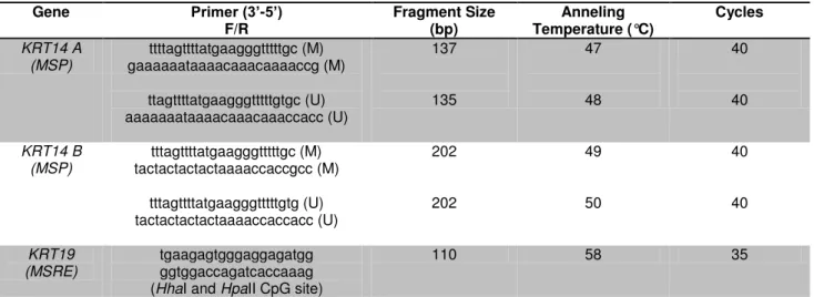
Table 1. Primers sequences for KRT14 (GenBank accession number NG_008624.1) and KTR19 genes (GenBank accession number AF202321.1), and DNA methylation
analysis conditions.
Gene Primer (3’-5’)
F/R Fragment Size (bp) Temperature (°C) Anneling Cycles
KRT14 A (MSP) ttttagttttatgaagggtttttgc (M) gaaaaaataaaacaaacaaaaccg (M) ttagttttatgaagggtttttgtgc (U) aaaaaaataaaacaaacaaaccacc (U) 137 135 47 48 40 40 KRT14 B (MSP) tttagttttatgaagggtttttgc (M) tactactactactaaaaccaccgcc (M) tttagttttatgaagggtttttgtg (U) tactactactactaaaaccaccacc (U) 202 202 49 50 40 40 KRT19 (MSRE) tgaagagtgggaggagatgg ggtggaccagatcaccaaag (HhaI and HpaII CpG site)
110 58 35
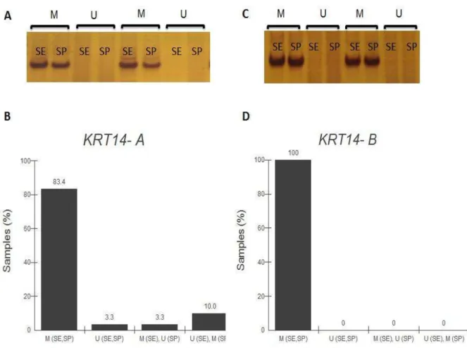
Figure 1 - DNA methylation analysis of KRT14 proximal exon 1 of skin cells. (A) Bands of
two representative samples obtained after polymerase chain reaction of KRT14 fragment A
(135 (M) and 137 (U) bp). (B) Methylation status of the KRT14 gene (p>0.05; McNemar;
n=28). (C) Bands of two representative samples obtained after polymerase chain reaction of
KRT14 fragment B (202 bp). (D) Methylation status of the KRT14 gene (p>0.05; McNemar;
Figure 2 - DNA methylation analysis of KRT19 proximal exon 1 of skin cells. (A) Bands of
representative sample obtained after polymerase chain reaction of KRT19 (110 bp). (B)
Methylation status of the KRT19 gene (p>0.05; McNemar; n=30). ND - non-digested sample,
REFERENCES
[1] Ambach W, Blumthaler M. Biological effectiveness of solar UV radiation in humans. Experientia 1993;49:747–53.
[2] Coelho SG, Choi W, Brenner M, Miyamura Y, Yamaguchi Y, Wolber R, et al. Short- and long-term effects of UV radiation on the pigmentation of human skin. J Investig Dermatol Symp Proc 2009;14:32–5.
[3] Damian DL, Matthews YJ, Phan TA, Halliday GM. An action spectrum for ultraviolet radiation-induced immunosuppression in humans. Br J Dermatol 2011;164:657–9. [4] Fisher GJ, Kang S, Varani J, Bata-Csorgo Z, Wan Y, Datta S, et al. Mechanisms of
photoaging and chronological skin aging. Arch Dermatol 2002;138:1462–70.
[5] Pfeifer GP, Besaratinia A. UV wavelength-dependent DNA damage and human non-melanoma and non-melanoma skin cancer. Photochem Photobiol Sci 2012;11:90–7. [6] Inca. Estimativa Incidência de câncer no Brasil - 2014.
[7] Biesalski HK, Berneburg M, Grune T, Kerscher M, Krutmann J, Raab W, et al. Oxidative and premature skin ageing. Exp Dermatol 2003;12:3–15.
[8] Yaar M, Gilchrest BA. Photoageing: mechanism, prevention and therapy. Br J Dermatol 2007;157:874–87.
[9] Quan T, He T, Voorhees JJ, Fisher GJ. Ultraviolet irradiation induces Smad7 via induction of transcription factor AP-1 in human skin fibroblasts. J Biol Chem 2005;280:8079–85.
[10] Rittié L, Fisher GJ. UV-light-induced signal cascades and skin aging. Ageing Res Rev 2002;1:705–20.
[11] Scharffetter–Kochanek K, Brenneisen P, Wenk J, Herrmann G, Ma W, Kuhr L, et al. Photoaging of the skin from phenotype to mechanisms. Exp Gerontol 2000;35:307–16. [12] Shaulian E, Schreiber M, Piu F, Beeche M, Wagner EF, Karin M. The mammalian UV
response: c-Jun induction is required for exit from p53-imposed growth arrest. Cell 2000;103:897–907.
[13] Pfeifer G, Besaratinia A. Mutational spectra of human cancer. Hum Genet 2009;125:493–506.
[14] Probst AV, Dunleavy E, Almouzni G. Epigenetic inheritance during the cell cycle. Nat Rev Mol Cell Biol 2009;10:192–206.
[15] Anway MD, Leathers C, Skinner MK. Endocrine disruptor vinclozolin induced epigenetic transgenerational adult-onset disease. Endocrinology 2006;147:5515–23. [16] Takiguchi M, Achanzar WE, Qu W, Li G, Waalkes MP. Effects of cadmium on
[17] Shiao Y-H, Crawford EB, Anderson LM, Patel P, Ko K. Allele-specific germ cell epimutation in the spacer promoter of the 45S ribosomal RNA gene after Cr(III) exposure. Toxicol Appl Pharmacol 2005;205:290–6.
[18] Novikova SI, He F, Bai J, Cutrufello NJ, Lidow MS, Undieh AS. Maternal cocaine administration in mice alters DNA methylation and gene expression in hippocampal neurons of neonatal and prepubertal offspring. PLoS One 2008;3:e1919.
[19] Sable P, Randhir K, Kale A, Chavan-Gautam P, Joshi S. Maternal micronutrients and brain global methylation patterns in the offspring. Nutr Neurosci 2013;18.
[20] Zhang R, Miao Q, Wang C, Zhao R, Li W, Haile CN, et al. Genome-wide DNA methylation analysis in alcohol dependence. Addict Biol 2013;18:392–403.
[21] Wan ES, Qiu W, Baccarelli A, Carey VJ, Bacherman H, Rennard SI, et al. Cigarette smoking behaviors and time since quitting are associated with differential DNA methylation across the human genome. Hum Mol Genet 2012;21:3073–82.
[22] Mittal A, Piyathilake C, Hara Y, Katiyar SK. Exceptionally High Protection of Photocarcinogenesis by Topical Application of (-)-Epi gal locatechin-3-Gal late in Hydrophilic Cream in SKH-1 Hairless Mouse Model: Relationship to Inhibition of UVB-Induced Global DNA Hypomethylation. Neoplasia 2003;5:555–65.
[23] Nandakumar V, Vaid M, Tollefsbol TO, Katiyar SK. Aberrant DNA hypermethylation patterns lead to transcriptional silencing of tumor suppressor genes in UVB-exposed skin and UVB-induced skin tumors of mice. Carcinogenesis 2011;32:597–604.
[24] Chen I-P, Henning S, Faust A, Boukamp P, Volkmer B, Greinert R. UVA-induced epigenetic regulation of P16(INK4a) in human epidermal keratinocytes and skin tumor derived cells. Photochem Photobiol Sci 2012;11:180–90.
[25] Sathyanarayana UG, Moore AY, Li L, Padar A, Majmudar K, Stastny V, et al. Sun exposure related methylation in malignant and non-malignant skin lesions. Cancer Lett 2007;245:112–20.
[26] Grönniger E, Weber B, Heil O, Peters N, Stäb F, Wenck H, et al. Aging and chronic sun exposure cause distinct epigenetic changes in human skin. PLoS Genet 2010;6:e1000971.
[27] Moll R, Divo M, Langbein L. The human keratins: biology and pathology. Histochem Cell Biol 2008;129:705–33.
[28] Bernerd F, Del Bino S, Asselineau D. Regulation of keratin expression by ultraviolet radiation: differential and specific effects of ultraviolet B and ultraviolet a exposure. J Invest Dermatol 2001;117:1421–9.
[29] Kurokawa I, Takahashi K, Moll I, Moll R. Expression of keratins in cutaneous epithelial tumors and related disorders - distribution and clinical significance. Exp Dermatol 2011;20:217–28.
[31] Herman JG, Graff JR, Myöhänen S, Nelkin BD, Baylin SB. Methylation-specific PCR: a novel PCR assay for methylation status of CpG islands. Proc Natl Acad Sci U S A 1996;93:9821–6.
[32] Hashimoto K, Kokubun S, Itoi E, Roach HI. Improved quantification of DNA methylation using methylation-sensitive restriction enzymes and real-time PCR. Epigenetics 2007;2:86–91.
[33] Ayres M, Ayres MJ, Ayres DL, Santos ADAS. BioEstat 5.0 Aplicações estatísticas nas áreas das ciências biológicas e médicas. Soc Civ Mamirauá/Imprensa Do Estado Do Pará 2007.
[34] Coulombe P a, Lee C-H. Defining Keratin Protein Function in Skin Epithelia: Epidermolysis Bullosa Simplex and Its Aftermath. J Invest Dermatol 2012;132:763–75. [35] Oender K, Trost A, Lanschuetzer C, Laimer M, Emberger M, Breitenbach M, et al. Cytokeratin-related loss of cellular integrity is not a major driving force of human intrinsic skin aging. Mech Ageing Dev 2008;129:563–71.
[36] Hachiya A, Sriwiriyanont P, Fujimura T, Ohuchi A, Kitahara T, Takema Y, et al. Mechanistic effects of long-term ultraviolet B irradiation induce epidermal and dermal changes in human skin xenografts. Am J Pathol 2009;174:401–13.
[37] Bragulla HH, Homberger DG. Structure and functions of keratin proteins in simple, stratified, keratinized and cornified epithelia. J Anat 2009;214:516–59.
[38] Paiva F, Duarte-Pereira S, Costa VL, Ramalho-Carvalho J, Patrício P, Ribeiro FR, et al. Functional and epigenetic characterization of the KRT19 gene in renal cell
neoplasms. DNA Cell Biol 2011;30:85–90.
[39] McGrath J a., Robinson MK, Binder RL. Skin differences based on age and chronicity of ultraviolet exposure: Results from a gene expression profiling study. Br J Dermatol 2012;166:9–15.
[40] Adachi H, Murakami Y, Tanaka H, Nakata S. Increase of stratifin triggered by ultraviolet irradiation is possibly related to premature aging of human skin Exp Dermatol 2014 Suppl 1:32-6.
[41] El-Maarri O, Becker T, Junen J, Manzoor SS, Diaz-Lacava A, Schwaab R, et al. Gender specific differences in levels of DNA methylation at selected loci from human total blood: A tendency toward higher methylation levels in males. Hum Genet 2007;122:505–14.
[42] Thompson RF, Atzmon G, Gheorghe C, Liang HQ, Lowes C, Greally JM, et al. Tissue-specific dysregulation of DNA methylation in aging. Aging Cell 2010;9:506–18.
[43] Bezerra, SFO, Costa LA, Freitas PAN, de Oliveira NFP. Age-related Changes in DNA Methylation Status of hTERT Gene Promoter of Oral Epithelial Cells. Brazilian Arch
Biol Technol 2015;58:82-89.
[45] Wu J, Zhang J, Qin J. Clinical significance of methylation of E-cadherin and p14ARF gene promoters in skin squamous cell carcinoma tissues. Clin Exp Med 2014;7:1808–
4. CONCLUSÃO
Nossos dados mostram que a condição metilada é um aspecto comum dos genes
KRT14 e KRT19 em células da pele. Assim, concluímos que a exposição solar não
REFERÊNCIAS*
ATTWOOD, J. T.; YUNG, R. L.; RICHARDSON, B. C. Cellular and Molecular Life Sciences. Cell Molecular Life Sciences, v. 59, p. 241– 257, 2002.
AUCLAIR, G.; WEBER, M. Mechanisms of DNA methylation and demethylation in mammals. Biochimie, v. 94, n. 11, p. 2202–11, 2012.
BADER, B. L. et al. Amino acid sequence and gene organization of cytokeratin no. 19, an exceptional tail-less intermediate filament protein. The EMBO journal, v. 5, n. 8, p. 1865–
1875, 1986.
BARAK, V. et al. Clinical utility of cytokeratins as tumor markers. Clinical Biochemistry, v. 37, n. 7, p. 529-40, 2004
BAUMANN, L. Skin ageing and its treatment. The Journal of pathology, v. 211, n. 2, p. 241–51, 2007.
BERGER, S. L. et al. An operational definition of epigenetics. Genes & development, v. 23, n. 7, p. 781–3, 2009.
BERNERD, F. ET AL. Regulation of keratin expression by ultraviolet radiation: differential and specific effects of ultraviolet B and ultraviolet a exposure. The Journal of investigative dermatology, v. 117, n. 6, p. 1421–9, 2001.
BESTOR, T. H. The DNA methyltransferases of mammals. Human molecular genetics, v. 9, n. 16, p. 2395–2402, 2000.
BIESALSKI, H. K. et al. Oxidative and premature skin ageing. Experimental Dermatology, v. 12, p. 3–15, 2003.
CHEN, I. P. et al. UVA-induced epigenetic regulation of P16(INK4a) in human epidermal keratinocytes and skin tumor derived cells. Photochemical & photobiological sciences, v. 11, n. 1, p. 180–90, 2012.
COULOMBE, P. A. ; LEE, C. Defining keratin protein function in skin ephitelia: epidermolysis bullosa simplex and its aftermath. Journal of investigative dermatology, v. 132, n. 3, p. 763-75, 2012.
CRUTZEN, P. J. Ultraviolet on the increase. Nature, v. 356, n. 6365, p. 104–105, 1992. DAMIAN, D. L. et al. An action spectrum for ultraviolet radiation-induced immunosuppression in humans. The British journal of dermatology, v. 164, n. 3, p. 657–9, 2011.
DE BRITO O. C. E.; PACHECO C. Epigenética: regulação da expressão gênica em nível transcricional e suas implicações. Semina: Ciências Biológicas e da Saúde, v. 34, n. 2, p. 125, 24, 2013.
FEINBERG, A. P.; TYCKO, B. The history of cancer epigenetics. Nature reviews Cancer, v. 4, n. February, p. 143–153, 2004.
FISHER, G. J. et al. Mechanisms of photoaging and chronological skin aging. Archives of dermatology, v. 138, p. 1462–1470, 2002.
FUCHS, E.; GREEN, H. Changes in Keratin Gene Expression Differentiation of the Keratinocyte during Terminal. Cell, v. 19, n. April, p. 1033–1042, 1980.
GILHAR, A. et al. Aging of human epidermis: reversal of aging changes correlates with reversal of keratinocyte fas expression and apoptosis. The journals of gerontology, v. 59, n. 5, p. 411–415, 2004.
GRÖNNIGER, E. et al. Aging and chronic sun exposure cause distinct epigenetic changes in human skin. PLoS genetics, v. 6, n. 5, p. e1000971, 2010.
HADLER-OLSEN, E. et al. Regulation of matrix metalloproteinase activity in health and disease. The FEBS journal, v. 278, n. 1, p. 28–45, 2011.
HESSE, M. et al. Comprehensive analysis of keratin gene clusters in humans and rodents. European journal of cell biology, v. 83, p. 19–26, 2004.
HO, J. N. et al. Protective effects of aucubin isolated from Eucommia ulmoides against UVB-induced oxidative stress in human skin fibroblasts. Biological and pharmaceutical bulletin, v. 28, n. 7, p. 1244–1248, 2005.
ICHIHASHI, M. et al. UV-induced skin damage. Toxicology, v. 189, n. 1-2, p. 21–39, 2003.
ISHIDA-YAMAMOTO, Akemi et al. Epidermolysis bullosa simplex (Dowling-Meara type) is a genetic disease characterized by an abnormal keratin-filament network involving keratins K5 and K14. Journal of investigative dermatology, v. 97, n. 6, p. 959-968, 1991.
LI, L.-C.; DAHIYA, R. MethPrimer: designing primers for methylation PCRs. Bioinformatics, v. 18, n. 11, p. 1427–31, 2002.
LINDBERG, K.; RHEINWALD, J. G. Suprabasal 40 kd keratin (K19) expression as an immunohistologic marker of premalignancy in oral epithelium. The American journal of pathology, v. 134, n. 1, p. 89–98, 1989.
MITTAL, A. et al. Exceptionally High Protection of Photocarcinogenesis by Topical Application of (-)-Epi gal locatechin-3-Gal late in Hydrophilic Cream in SKH-1 Hairless Mouse Model: Relationship to Inhibition of UVB-Induced Global DNA Hypomethylation. Neoplasia, v. 5, n. 6, p. 555–565, 2003.
MOLL, R. et al. The human keratins: biology and pathology. Histochemistry and cell biology, v. 129, n. 6, p. 705–33, 2008.
ORIOLO, A. S. et al. GCP6 Binds to Intermediate Filaments : A Novel Function of Keratins in
the Organization of Microtubules in Epithelial Cells. Molecular Biology of the cell, v. 18, n. March, p. 781–794, 2007.
PAIVA, F. et al. Functional and epigenetic characterization of the KRT19 gene in renal cell neoplasms. DNA and cell biology, v. 30, n. 2, p. 85–90, 2011.
PFEIFER, G.; BESARATINIA, A. Mutational spectra of human cancer. Human genetics, v. 125, n. 5-6, p. 493–506, 2009.
PFEIFER, G. P.; BESARATINIA, A. UV wavelength-dependent DNA damage and human non-melanoma and melanoma skin cancer. Photochemical & photobiological sciences, v. 11, n. 1, p. 90–7, 2012.
PLACHOT, C.; LELIÈVRE, S. A. DNA methylation control of tissue polarity and cellular differentiation in the mammary epithelium. Experimental Cell Research, v. 298, p. 122–132, 2004.
POLEFKA, T. G. et al. Effects of solar radiation on the skin. Journal of cosmetic dermatology, v. 11, n. 2, p. 134–43, 2012.
QUAN, T. et al. Ultraviolet irradiation induces Smad7 via induction of transcription factor AP-1 in human skin fibroblasts. The Journal of biological chemistry, v. 280, n. 9, p. 8079–85, 2005.
QUAN, T. et al. Matrix-degrading metalloproteinases in photoaging. In. Journal of
Investigative Dermatology Symposium Proceedings.p. 20-24, 2009.
RITTIÉ, L.; FISHER, G. J. UV-light-induced signal cascades and skin aging. Ageing research reviews, v. 1, n. 4, p. 705-720, 2002.
ROMANO V. ET. AL. Chromosomal assignments of human type I and type II cytokeratin genes to different chromosomes. Cytogenet Cell Genet, v. 48, p. 148–151, 1988.
SATHYANARAYANA, U. G. et al. Sun exposure related methylation in malignant and non-malignant skin lesions. Cancer letters, v. 245, n. 1-2, p. 112–20, 2007.
SCHARFFETTER–KOCHANEK, K. et al. Photoaging of the skin from phenotype to mechanisms. Experimental Gerontology, v. 35, n. 3, p. 307–316, 2000.
SCHWEIZER, J. et al. New consensus nomenclature for mammalian keratins. Journal of Cell Biology, v. 174, n. 2, p. 169–174, 2006.
SETHI, K. et al. Expressions of CK-19, NF-kappaB, E-cadherin, beta-catenin and EGFR as diagnostic and prognostic markers by immunohistochemical analysis in thyroid carcinoma. Journal of experimental therapeutics & oncology, v. 9, p. 187–99, 2011.
SHAULIAN, E. et al. The mammalian UV response: c-Jun induction is required for exit from p53-imposed growth arrest. Cell, v. 245, n. 1-2, p. 112–20, 8 jan. 2000.
USHIJIMA, T.; ASADA, K. Aberrant DNA methylation in contrast with mutations. Cancer science, v. 101, n. 2, p. 300–5, 2010.
WATERLAND, R. A. Assessing the Effects of High Methionine Intake on DNA Methylation. J. Nutr., v. 136, n. 6, p. 1706S–1710, 2006.
WATERLAND, R. A.; JIRTLE, R. L. Transposable Elements : Targets for Early Nutritional
Effects on Epigenetic Gene Regulation. Molecular and Cellular Biology, v. 23, n. 15, p. 5293–5300, 2003.
YAAR, M.; GILCHREST, B. A. Photoageing: mechanism, prevention and therapy. The British journal of dermatology, v. 157, n. 5, p. 874–87, 2007.