Impact of iron overload on interleukin-10 levels, biochemical parameters and oxidative
stress in patients with sickle cell anemia
Maritza Cavalcante Barbosa Talyta Ellen Jesus dos Santos Geane Félix de Souza Lívia Coêlho de Assis Max Victor Carioca Freitas Romélia Pinheiro Gonçalves
Universidade Federal do Ceará – UFC, Fortaleza, CE, Brazil
Conlict-of-interest disclosure:
The authors declare no competing inancial interest
Submitted: 5/31/2012 Accepted: 9/3/2012
Corresponding author: Maritza Cavalcante Barbosa Universidade Federal do Ceará – UFC Rua Capitão Francisco Pedro, nº1210 - Rodolfo Teóilo
60430-370 Fortaleza, CE, Brazil [email protected]
www.rbhh.org or www.scielo.br/rbhh
DOI: 10.5581/1516-8484.20130011
Objective: The aim of this study was to evaluate the impact of iron overload on the proile of interleukin-10
levels, biochemical parameters and oxidative stress in sickle cell anemia patients.
Methods: A cross-sectional study was performed of 30 patients with molecular diagnosis of sickle cell anemia.
Patients were stratiied into two groups, according to the presence of iron overload: Iron overload (n = 15) and Non-iron overload (n = 15). Biochemical analyses were performed utilizing the Wiener CM 200 automatic analyzer. The interleukin-10 level was measured by capture ELISA using the BD OptEIAT commercial kit. Oxidative stress parameters were determined by spectrophotometry. Statistical analysis was performed using GraphPad Prism software (version 5.0) and statistical signiicance was established for p-values < 0.05 in all analyses.
Results: Biochemical analysis revealed signiicant elevations in the levels of uric acid, triglycerides, very
low-density lipoprotein (VLDL), alanine aminotransferase (ALT), lactate dehydrogenase (LDH), urea and creatinine in the Iron overload Group compared to the Non-iron overload Group and signiicant decreases in the high-density lipoprotein (HDL) and low-density lipoprotein (LDL). Ferritin levels correlated positively with uric acid concentrations (p-value < 0.05). The Iron overload Group showed lower interleukin-10 levels and catalase activity and higher nitrite and malondialdehyde levels compared with the Non-iron overload Group.
Conclusion: The results of this study are important to develop further consistent studies that evaluate the effect of iron overload on the inlammatory proile and oxidative stress of patients with sickle cell anemia.
Keywords: Anemia, sickle cell; Iron overload; Interleukin-10; Oxidative stress; Nitric oxide/metabolism
Introduction
Sickle cell anemia (SCA) is a chronic hemolytic anemia resulting from a mutation (GAG → GTG) in the sixth codon of the β globin gene, resulting in the substitution of valine for glutamic acid and the formation of hemoglobin S (Hb S)(1,2). It is characterized by a
chronic inlammatory state with elevated levels of proinlammatory cytokines and constant cell activation, which contributes signiicantly to the pathogenesis of SCA(3).
Cytokines may participate in several mechanisms that promote a vaso-occlusive process, such as vascular endothelial activation, adhesion of erythrocytes and leukocytes to the vascular endothelium and platelet activation(4). Although changes in levels of proinlammatory cytokines and anti-inlammatory properties have been previously demonstrated in SCA, the role of cytokines in SCA has not been established yet(5).
Interleukin-10 (IL-10), primarily produced by monocytes but also secreted by T and B lymphocytes when activated, is a cytokine with anti-inlammatory activity(6,7). IL-10 suppresses
inlammation by several mechanisms, including by decreasing the production of inlammatory
cytokines and the toxic effects of radicals such as nitrites (NO2-)(8). Few studies have evaluated the levels of IL-10 in patients with SCA and the results are still conlicting(5,9,10). However,
hydroxyurea (HU) therapy appears to increase the levels of the anti-inlammatory cytokine but
the mechanism remains unclear(5).
Red blood cell transfusions have an important role in the treatment of acute and chronic complications of SCA by reducing the anemia, the concentration of Hb S and hemolysis(11). The clinical use of this treatment has often demonstrated a signiicant reduction in the incidence of acute chest syndrome, painful events, stroke and the number of hospital
admissions(12,13). However, clinical and laboratory signs resulting from iron overload (IO)
are observed after ten to twenty transfusions as there is no effective route of excreting iron
from the organism(14).
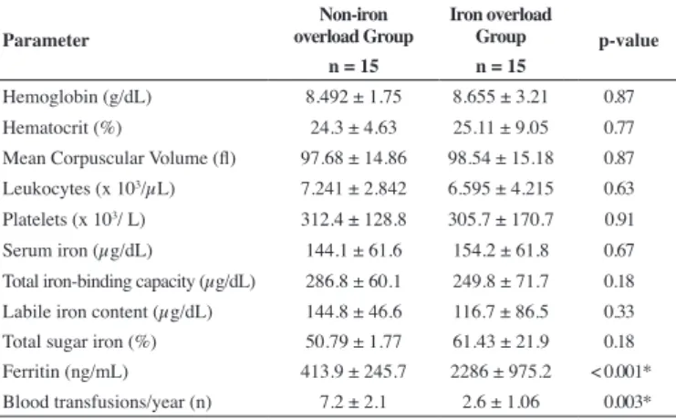
Table 1 - Iron proile and hematological parameters
Parameter
Non-iron overload Group
Iron overload
Group p-value n = 15 n = 15
Hemoglobin (g/dL) 8.492 ± 1.75 8.655 ± 3.21 0.87
Hematocrit (%) 24.3 ± 4.63 25.11 ± 9.05 0.77
Mean Corpuscular Volume (l) 97.68 ± 14.86 98.54 ± 15.18 0.87
Leukocytes (x 103/µL) 7.241 ± 2.842 6.595 ± 4.215 0.63
Platelets (x 103/ L) 312.4 ± 128.8 305.7 ± 170.7 0.91
Serum iron (µg/dL) 144.1 ± 61.6 154.2 ± 61.8 0.67
Total iron-binding capacity (µg/dL) 286.8 ± 60.1 249.8 ± 71.7 0.18 Labile iron content (µg/dL) 144.8 ± 46.6 116.7 ± 86.5 0.33
Total sugar iron (%) 50.79 ± 1.77 61.43 ± 21.9 0.18
Ferritin (ng/mL) 413.9 ± 245.7 2286 ± 975.2 < 0.001* Blood transfusions/year (n) 7.2 ± 2.1 2.6 ± 1.06 0.003* Results expressed as mean ± standard deviation. Student t-test: *p-value < 0.05
Methods
Patients
A cross-sectional study was carried out of 30 adult patients of both genders with the molecular diagnosis of SCA. All were treated with hydroxyurea and followed up in the hematology service of a referral hospital in Fortaleza, Brazil. The exclusion criteria were positive serology for viral infections (hepatitis B virus, hepatitis C virus and Human immunodeiciency virus) and evidence of clinical manifestations, such as vaso-occlusive and infectious events during the three months prior to blood collection. Patients were stratiied into two groups according to the presence of iron overload. One group consisted of patients with sickle cell anemia with iron overload (Iron overload Group; n = 15) and the other, patients with sickle cell anemia but without iron overload (Non-iron overload Group; n = 15). The criterion for diagnosis of the IO was serum ferritin, with two consecutive determinations of not less than 1000 ng/mL(17). All the patients with IO were undergoing treatment with
iron chelation. An informed consent form was signed by all participants and this study was approved by the Ethics Research Committee of the Universidade Federal do Ceará in accordance with Resolution 196/96 of the National Health Institute.
Laboratory methods
Molecular diagnosis of patients was based on the polymerase chain reaction (PCR) followed by digestion using the restriction
enzyme DdeI. The IL-10 levels were determined by enzyme-linked
immunoabsorbent assay (ELISA) using the OptEIA BD™ commercial kit (BD Biosciences, San Diego, CA). The biochemical proile and iron levels were determined in a Modular P800 automatic analyzer Hitachi. Plasma levels of malondialdehyde (MDA) were quantiied by the reaction with thiobarbituric acid. In this reaction, two molecules of the acid react with one of MDA to form a chromophore which has maximal absorbance in the range 532-535 nm(18). Nitrite, a metabolite of nitric oxide, was determined in plasma using Green’s method, based on the diazotization reaction, which forms a chromophore with peak absorbance at 560 nm(19). The catalase activity in erythrocytes was determined by the spectrophotometric method, whereby the decomposition of hydrogen peroxide was monitored at 240 nm in hemolysate samples collected in ethylenediaminetetraacetic acid (EDTA) anticoagulant and diluted in a Tris-EDTA buffer(20).
Statistical Analysis
The GraphPrism (version 5.01) program was used for statistical analysis. The Kolmogorov-Smirnov test was used to check for normal distribution of the data. The results are expressed as mean ± standard deviation. Statistical differences between groups were identiied using Fisher’s exact and the Mann-Whitney tests. Correlation analysis was performed using the Pearson correlation test. The level of signiicance was set for p-values < 0.05 in all analyzes.
Results
Of the 30 patients who participated in the study, 19 (63.3%)
were female and 11 (36.7%) were male. The mean age of the study population was 38.5 ± 15.6 years. Ethnicity was predominantly Mulattos (70%), followed by Blacks (30%). There were no statistically signiicant differences between the studied groups in respect to the demographic parameters (p-value > 0.05). The iron proile and hematological parameters are shown in Table 1. The ferritin concentration and the number of transfusions per year were signiicantly different between the two groups (p-value < 0.05).
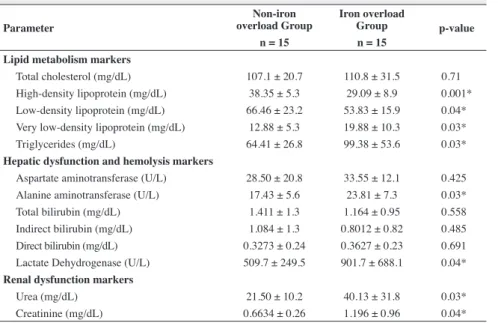
An analysis of biochemical parameters revealed elevated alanine aminotransferase (ALT), lactate dehydrogenase (LDH), triglycerides, very low-density lipoprotein (VLDL), urea and creatinine levels and lower high-density lipoprotein (HDL) and low-density lipoprotein (LDL) levels in the Iron overload Group when compared to Non-iron overload Group (p-value < 0.05). There was no statistically signiicant difference for the parameters of total cholesterol, AST and total bilirubin or its fractions (Table 2).
The mean IL-10 value in the Iron overload Group was 41.0 ± 19.0 pg/mL and in the Non-iron overload Group it was 145.4 ± 97.3 pg/mL; this difference is statistically signiicant (p-value < 0.05 - Figure 1). There was a negative correlation between the IL-10 levels and ferritin levels (p-value = 0.003; r = -0.48) in the participants of this study (Figure 2).
The mean uric acid level in the Iron overload Group was 6.368 ± 1.9 mg/dL, while in the Non-iron overload Group it was 4.512 ± 1.5 mg/dL (p-value = 0.0038 - Figure 3). There was a negative correlation between the IL-10 levels and uric acid levels in the study participants (p-value = 0.001; r = 0.56 - Figure 4).
The oxidative stress analysis showed that the Iron overload Group presented higher levels of nitrites and MDA and lower catalase activity compared to the Non-iron overload Group (p-value < 0.05 - Table 3). The levels of IL-10 correlated negatively with MDA levels (p-value = 0.0075; r = -0.4631) and nitrite levels (p-value = 0.0396; r = -0.3735) and positively with catalase activity (p-value = 0.0059; r = 0.4691 - Figure 5).
Table 2 - Evaluation of biochemical parameters
Parameter
Non-iron overload Group
Iron overload
Group p-value n = 15 n = 15
Lipid metabolism markers
Total cholesterol (mg/dL) 107.1 ± 20.7 110.8 ± 31.5 0.71
High-density lipoprotein (mg/dL) 38.35 ± 5.3 29.09 ± 8.9 0.001*
Low-density lipoprotein (mg/dL) 66.46 ± 23.2 53.83 ± 15.9 0.04*
Very low-density lipoprotein (mg/dL) 12.88 ± 5.3 19.88 ± 10.3 0.03*
Triglycerides (mg/dL) 64.41 ± 26.8 99.38 ± 53.6 0.03*
Hepatic dysfunction and hemolysis markers
Aspartate aminotransferase (U/L) 28.50 ± 20.8 33.55 ± 12.1 0.425
Alanine aminotransferase (U/L) 17.43 ± 5.6 23.81 ± 7.3 0.03*
Total bilirubin (mg/dL) 1.411 ± 1.3 1.164 ± 0.95 0.558
Indirect bilirubin (mg/dL) 1.084 ± 1.3 0.8012 ± 0.82 0.485
Direct bilirubin (mg/dL) 0.3273 ± 0.24 0.3627 ± 0.23 0.691
Lactate Dehydrogenase (U/L) 509.7 ± 249.5 901.7 ± 688.1 0.04*
Renal dysfunction markers
Urea (mg/dL) 21.50 ± 10.2 40.13 ± 31.8 0.03*
Creatinine (mg/dL) 0.6634 ± 0.26 1.196 ± 0.96 0.04*
Results expressed as mean ± standard deviation. Student t-test: *p-value < 0.05
Figure 2 - Correlation between IL-10 levels and ferritin levels (n = 30) Pearson correlation test; *p-value < 0.05
Figure 3 - Evaluation of uric acid (n=30) Mann-Whitney test; *p-value < 0.05
Discussion
Excess intracellular iron is potentially toxic due to catalytic reactions that favor the formation of free radicals that promote lipid peroxidation of cellular components, progressing to necrosis and/or apoptosis(21). The results of this study revealed a signiicant increase in ALT, urea and creatinine levels in the Iron overload Group and is thus in accordance with the literature, which reports that an excess of iron can trigger and exacerbate liver and kidney damage in patients with SCA(22-24).
LDH has been identiied as an important prognostic marker in patients with SCA at risk of developing leg ulcers, pulmonary hypertension and priapism(25,26). In this study, the elevation of
serum LDH in patients with IO suggests more severe hemolysis in these patients compared to patients without IO. The excess of free iron in the organism favors the formation of free radicals and with oxidative stress can damage the membranes of erythrocytes, exacerbating the hemolysis.
In general, SCA patients have alterations in their lipid metabolism. Hypocholesterolemia and reductions in the HDL and LDL levels are the most documented indings in these patients, however their clinical signiicance is not yet fully known(27). Our
data revealed signiicant decreases in total cholesterol, HDL and LDL levels and increases in VLDL and triglyceride levels in patients with IO, which is in agreement with literature data. This shows that oxidative stress generated by chronic hemolysis and IO may be associated with reduced plasma lipids except triglycerides. Moreover, hepatic dysfunction, which is normally present in these situations, reduces the endogenous cholesterol and ampliies the changes in the lipid proile in SCA patients(28,29).
Some studies suggest that high uric acid levels can promote organ damage and may have a deleterious effect on endothelial
cell function(10). Furthermore, uric acid can act as pro-oxidant,
particularly at high concentrations(11). These observations illustrate a potentially negative contribution of high uric acid levels in SCA. Our results conirm this statement as shown by the negative correlation between uric acid and IL-10 levels.
IL-10 is a cytokine with potent anti-inlammatory activity which reduces the production of various cytokines including IL-1, IL-6, IL-8, IL-12, TNF-α and GM-CSF to promote uptake and
retention of iron in the reticuloendothelial system(7,30,31). There are
no reports in the literature on the inluence of IO on the level of this cytokine in SCA. The result of the correlation between IL-10 levels and MDA, nitrite and catalase activity in our study suggest that signiicantly reduced IL-10 levels in the group with IO can contribute to a hyperoxidative state due to lower uptake of free iron leading to organ damage by ROS. Furthermore, the reduced IL-10 levels may be associated with elevated proinlammatory cytokine levels that are, in part, responsible for the vaso-occlusive phenomenon. The negative correlation between the IL-10 levels and ferritin levels reinforces the existence of a clear inlammatory state in patients with IO as a result of iron excess.
Several studies have demonstrated a signiicant increase in oxidative stress biomarkers in SCA patients compared with healthy subjects(32-35). In SCA, oxidative stress occurs by
mechanisms intrinsic to the pathophysiology of the disease, such as the increased rate of auto-oxidation associated to Hb S and reduced bioavailability of nitric oxide (NO)(36,37). The oxidative
stress analysis in patients of this study revealed a hyperoxidative status exacerbated in the group with IO by high MDA and nitrite
Table 3 - Oxidative stress biomarkers
Biomarker
Non-iron overload Group
Iron overload
Group p-value n = 15 n = 15
Malondialdehyde (µM) 5.33 ± 2.34 8.32 ± 2.25 0.0008*
Nitrite (µM) 1.019 ± 1.13 2.513 ± 1.78 0.0003*
Catalase (U/mL) 1391 ± 322.3 982.0 ± 216 0.0002*
Results expressed as mean ± standard deviation. Student t-test: *p-value < 0.05
levels and by a reduction in the antioxidant enzyme catalase activity when compared with the group without IO. Our results are consistent with the literature, which indicates that transfusional IO is a enhancing factor of oxidative stress in SCA(38,39).
MDA is a biomarker of damage caused by ROS derived from lipid peroxidation of membranes; its accumulation changes the organization of membrane phospholipids, contributing to the process
of cellular degeneration(34). Excess iron plays an important role in the increased production of ROS, including the hydroxyl radical (OH). In addition to damage to the cell membrane and cellular components, the radical OH- reacts with NO, thereby reducing its bioavailability(33). NO is known for its antioxidant activity. One of the mechanisms involved is to induce heme-oxidase 1 (HO1). HO-1 removes the heme in the plasma, thereby preventing its participation in reactions to form the toxic species of the organism and peroxidation, such as OH-(40). With the increased consumption
of NO in patients with IO, its antioxidant activity is compromised, thereby increasing the hyperoxidative state and inlammation present in SCA individuals. The correlations in this study between the levels of IL-10 and oxidative stress parameters support this theory.
Conclusion
The results of this study are important to develop more consistent studies that evaluate the effect of IO on the inlammatory proile and oxidative stress in SCA patients, as well as to investigate new markers able to predict organ damage with greater speciicity and thus contribute to the treatment and prognosis of these patients.
References
1. Kato GJ, Gladwin MT, Steinberg MH. Deconstructing sickle cell disease: reappraisal of the role of hemolysis in the development of clinical subphenotypes. Blood Rev. 2007;21(1):37-47.
2. Bonini-Domingos CR. Metodologias laboratoriais para o diagnóstico de hemoglobinopatias e talassemias. Brasil: HN; 2006. 121 p.
3. Conran N, Franco-Penteado CF, Costa FF. Newer aspects of the pathophysiology of sickle cell disease vaso-occlusion. Hemoglobin. 2009;33(1):1-16.
4. Makis AC, Hatzimichael EC, Bourantas KL. The role of cytokines in sickle cell disease. Ann Hematol. 2000;79(8):407-13.
5. Lanaro C, Franco-Penteado CF, Albuqueque DM, Saad ST, Conran N, Costa FF. Altered levels of cytokines and inlammatory mediators in plasma and leukocytes of sickle cell anemia patients and effects of hydroxyurea therapy. J Leukoc Biol. 2009;85(2):235-42.
6. Moore KM, Vieira P, Fiorentino DF, Tronustine ML, Khan TA, Mosmann TR. Homology of cytokine synthesis inhibitory factor (IL-10) to the Epstein-Barr virus gene BCRFI. Science. 1990;248(4960):1230-4. Erratum in: Science 1990;250(4980):494.
7. Yssel H, De Waal Malefyt R, Roncarolo MG, Abrams JS, Lahesmaa R, Spits H, et al. IL-10 is produced by subsets of human CD4+ T cell clones and peripheral blood T cells. J Immunol. 1992;149(7):2378-84. 8. Moore KW, O’Garra A, de Waal Malefyt R, Vieira P, Mosmann TR.
Interleukin-10. Annu Rev Immunol. 1993;11:165-90.
9. Bourantas KL, Dalekos GN, Makis A, Chaidos A, Tsiara S, Mavridis A. Acute phase proteins and interleukins in steady state sickle cell disease. Eur J Haematol. 1998;61(1):49-54.
10. Graido-Gonzalez E, Doherty JC, Bergreen EW, Organ G, Telfer M, McMillen MA. Plasma endothelin-1, cytokine, and prostaglandin E2 levels in sickle cell disease and acute vaso-occlusive sickle crisis. Blood. 1998;92(7):2551-5.
11. Rees DC, Williams TN, Gladwin NT. Sickle-cell disease. Lancet. 2010;376(9757):2018-31.
12. Inati A, Khoriaty E, Musallam KM. Iron in sickle-cell disease: what have we learned over the years? Pediatr Blood Cancer. 2011;56(2):182-90. 13. Fung EB, Harmatz P, Milet M, Ballas SK, De Castro L, Hagar W, Owen
W, Olivieri N, Smith-Whitley K, Darbari D, Wang W, Vichinsky E; Multi-Center Study of Iron Overload Research Group. Morbidity and mortality in chronically transfused subjects with thalassemia and sickle cell disease: a report from the multi-center study of iron overload. Am J Hematol. 2007;82(4):255-65.
14. Porter JB. Practical management of iron overload. Br J Haematol. 2001;115(2):239-52.
15. Ward R. An update on disordered iron metabolism and iron overload. Hematology. 2010;15(5):311-7.
16. Ferreira AL, Matsubara LS. Radicais livres: conceitos, doenças relacionadas, sistema de defesa e estresse oxidativo. Rev Assoc Med Bras. 1997;43(1):61-8.
17. Cançado RD. Sobrecarga e quelação de ferro na anemia falciforme. Rev Bras Hematol Hemoter. 2007;29(3):316-26.
18. Draper HH, Hadley M. Malondialdehyde determination as index of lipid pero-xidation. Methods Enzymol. 1990;186:421-31.
19. Green LC, Wagner DA, Glogowski J, Skipper PL, Wishnok JS, Tannenbaum SR. Analysis of nitrate, nitrite and (15N) nitrate in biological luids. Anal Biochem. 1982;126(1):131-8.
20. Aebi H. Catalase in vitro. Methods Enzymol. 1984;105:121-6.
21. Fillet G, Beguin Y, Baldelli L. Model of reticuloendothelial iron metabolism in humans: Abnormal behavior in idiopathic hemochromatosis and in inlammation. Blood. 1989;74(2):844-51.
22. Olynyk JK, Trinder D, Ramm GA, Britton RS, Bacon BR. Hereditary hemochromatosis in the post HFE era. Hepatology. 2008;48(3):991-1001. 23. Gianini EG, Testa R, Savarino V. Liver enzyme alteration: a guide for
clinicians. CMAJ. 2005;172(3):367-79.
24. Núñez-Quintana A, Hondal-Álvarez NI, Ayllón-Valdés L. Alteraciones renales en la drepanocitosis. Rev Cub Hematol Inmunol Hemot. 2011;27(2):168-78.
25. Cumming V, King L, Fraser R, Serjeant G, Reid M. Venous incompetence, poverty and lactate dehydrogenase in Jamaica are important predictors of leg ulceration in sickle cell anaemia. Br J Haematol. 2008;142(1):119-25. 26. Kato GJ, McGowan V, Machado RF, Little JA, Taylor J 6th, Morris CR,
et al. Lactate dehydrogenase as a biomarker of hemolysis-associated nitric oxide resistance, priapism, leg ulceration, pulmonary hypertension, and death in patients with sickle cell disease. Blood. 2006;107(6):2279-85. 27. Naoum FA. Alterações do peril lipídico nas anemias. Rev Bras Hematol
Hemoter. 2005;27(3):223-6.
28. Rahimi Z, Merat A, Haghshenass M, Madani H, Rezaei M, Nagel RL. Plasma lipids in Iranians with sickle cell disease: hypocholesterolemia in sickle cell anemia and increase of HDL-cholesterol in sickle cell trait. Clin Chim Acta. 2006;365(1-2):217-20.
29. Oztas Y, Durukan I, Unal S, Ozgunes N. Plasma protein oxidation is correlated positively with plasma iron levels and negatively with hemolysate zinc levels in sickle-cell anemia patients. Int J Lab Hematol. 2012;34(2):129-35.
xxx
31. Jison ML, Munson PJ, Barb JJ, Suffredini AF, Talwar S, Logun C, et al. Blood mononuclear cell gene expression proiles characterize the oxidant, hemolytic, and inlammatory stress of sickle cell disease. Blood. 2004;104(1):270-80.
32. Manfredini V, Lazzaretti LL, Griebeler IH, Santin AP, Brandao VD, Wagner S, et al Blood antioxidant parameters in sickle cell anemia patients in steady state. J Natl Med Assoc. 2008;100(8):897-902. 33. Wood KC, Granger DN. Sickle cell disease: role of reactive
oxygen and nitrogen metabolites. Clin Exp Pharmacol Physiol. 2007;34(9):926-32.
34. Hundekar P, Suryakar A, Karnik A, Ghone R, Vasaikar M. Antioxidant status and lipid peroxidation in sickle cell anaemia. Biomed Res. 2010;21(4):461-4.
35. Elias DB, Freitas RM, Gonçalves RP, Magalhães HY, Sousa JH, Magalhães SM. Evaluation of the concentration of malondialdehyde and nitrite in patients with sickle cell anemia treated or not with hydroxyurea einstein (São Paulo). 2010;8(4 pt 1):414-8.
36. Hebbel RP, Eaton JW, Balasingam M, Steinberg MH. Spontaneous oxygen radical generation by sickle erythrocytes. J Clin Invest. 1982;70(6):1253-9. 37. Morris CR, Kato GJ, Poljakovic M, Wang X, Blackwelder WC, Sachdev
V, et al. Dysregulated arginine metabolism, hemolysis-associated pulmonary hypertension, and mortality in sickle cell disease. Comment in: JAMA. 2005;294(19):2432-3; author reply 2433-4; JAMA. 2005;294(19):2433; author reply 2433-4.
38. Gonçalves RP, Elias DB, Magalhães HI, de Souza JH. Study of correlation of nitrite levels with malonaldehyde and the prognosis of patients with sickle cell disease on hydroxyurea, Ceará-Brazil. J Clin Lab Anal. 2011;25(5):369-73.
39. Alsultan AL, Seif MA, Aminn TT, Naboli M, Alsuliman AM. Relationship between oxidative stress, ferritin and insulin resistance in sickle cell disease. Eur Rev Med Pharmacol Sci. 2010;14(6):527-38.