Original Article
35 Rev Bras Hematol Hemoter. 2013;35(1):35-8
Molecular matching of red blood cells is superior to serological matching in sickle cell
disease patients
Daiane Cobianchi da Costa Jordão Pellegrino Jr
Gláucia Andréia Soares Guelsin Karina Antero Rosa Ribeiro Simone Cristina Olenscki Gilli Lilian Castilho
Universidade Estadual de Campinas - UNICAMP, Campinas, SP, Brazil
Conlict-of-interest disclosure:
The authors declare no competing inancial interest
Submitted: 6/17/2012 Accepted: 9/3/2012
Corresponding author: Daiane Cobianchi da Costa
Instituto Nacional de Ciência e Tecnologia do Sangue - Hemocentro – UNICAMP
Rua Carlos Chagas, 480, Caixa Postal 6198 CEP 13081-970 Barão Geraldo, Campinas, SP, Brazil
Phone: 55 19 3521-8749 [email protected]
This study was supported by the Fundação de Amparo à Pesquisa do Estado de São Paulo (FAPESP) grant numbers 2008/07544-8 (DCC) and 2009/05924-0 (LC)
www.rbhh.org or www.scielo.br/rbhh DOI: 10.5581/1516-8484.20130012
Objective: To evaluate the usefulness of DNA methods to provide a means to precisely genotypically match donor blood units for the antigen-negative type of 35 sickle cell disease patients.
Methods: Red blood cell units were investigated for ABO, D, C, c, E, e, K, Fya, Fyb, Jka, Jkb, S, s, Dia and
RH variants by performing a molecular array (Human Erythrocyte Antigen BeadChipTM, BioArray Solutions),
polymerase chain reaction followed by restriction fragment length polymorphism analysis and sequencing of patient samples and donor units that had been serologically matched based on the ABO, Rh and K phenotypes and the presence of antibodies.
Results: Matches for 21 of 35 sickle cell disease patients presented discrepancies or mismatches for multiple
antigens between the genotype proile and the antigen proile of their serologically-matched blood units. The
main discrepancies or mismatches occurred in the RH, FY, JK and MNS systems. Eight Rh alloimmunized
patients presented RHD and RHCE variants that had not been serologically identiied. According to these results better matches were found for the patients with genotyped units and the patients beneited as shown by
better in vivo red blood cell survival.
Conclusion: Molecular matching is superior to serological matching in sickle cell disease patients, decreasing the risk of transfusion reactions, especially delayed transfusion reactions to existing alloantibodies and preventing alloimmunization.
Keywords: Anemia, sickle cell; Molecular typing; Blood group antigens; Isoantibodies/blood
Introduction
Red blood cell (RBC) transfusions have played an important role in allowing sickle cell disease (SCD) patients to live longer. However, their use is complicated by the high incidence of RBC alloimmunization(1-7), making the identiication of compatible RBC products dificult,
and is associated with delayed hemolytic transfusion reactions (DHTRs). For it to be possible to transfuse SCD patients effectively, more effective ways to reduce risk of transfusion reactions, transfusion-associated hyperhemolysis syndrome and alloimmunization must be found.
Although transfusion services establish protocols to reduce alloimmunization, there is still no consensus on the best practical approach even though the obvious goal is to provide blood that will survive for the maximum period of time(8-10). Three common approaches used
to supply RBC products (ABO and D compatible) to SCD patients are (i) to give speciic
antigen-negative RBCs after the patient has made the alloantibody, (ii) to match for C, E, c, e and K antigens and (iii) to match for C, E, c, e, K, Fya, Fyb, Jka and Jkb antigens(11-14).
In recent years, molecular DNA-based genetic methods have greatly improved transfusion therapy for SCD patients because they can be used to genotype patients and donors and maintain an inventory of DNA-typed units to identify compatible donors. Previous studies have demonstrated the relevance of genotyping blood groups to manage multiply transfused SCD patients by allowing
the determination of the true blood group genotype and by assisting the identiication of suspected
alloantibodies and the selection of antigen-negative RBCs for transfusion(15,16).
By testing the patient and donors it is possible to provide more extensively matched blood for patients thereby preventing additional alloimmunization. A study by Klapper et al.(17) using
the human erythrocyte antigen (HEA) BeadChipTM DNA analysis and a web-based inventory
management system to model donor-recipient matching showed that even with a limited donor pool, matching for Rh, Kell, Duffy, Kidd and MNS could be achieved at least 50% of the time. Herein the use of DNA methods to precisely match donor blood units for the antigen-negative genotypes of SCD patients is reported.
Methods
Samples
36
da Costa DC, Pellegrino J Jr, Guelsin GA, Ribeiro KA, Gilli SC, Castilho L
Rev Bras Hematol Hemoter. 2013;35(1):35-8
serologically matched for patients based on their ABO, Rh and K phenotypes and the presence of antibodies. Antigen-matched RBC units were investigated for recipients using blood group genotypes of the ABO, D, C, c, E, e, K, Fya, Fyb, Jka, Jkb, S, Lua,
Dia, Jsb, Doa and Dob systems. All the patients and donors agreed
to participate in this study by signing an Institute Review Board approved informed consent form.
Data processing
To perform data management, a web-hosted inventory management system was designed for this study. An electronic
link using speciic software to compare the blood donor units
and the patients’ needs was established allowing automatic
identiication of the most compatible blood.
DNA preparation
The genomic DNA was extracted from 200-µL aliquots of whole blood by manual spin column separation (QIAmp, Qiagen, Valencia, CA), according to the instructions of the manufacturer and eluted into 100 µL of buffer. The DNA concentration of each sample was calculated by the measurement of optical density at 260 and 280 nm and an 8-µL aliquot, containing ~10 ng of gDNA, was transferred for the polymerase chain reaction (PCR).
Human erythrocyte antigen BeadChipTM DNA analysis
The DNA array analysis was performed using RHD and RHCE BeadChipTM analysis which uses probes directed
to polymorphic sites for RHD and RHCE variants and HEA BeadChipTM containing probes directed to polymorphic
sites in the RHCE, FY (including FY-GATA and FY265), DO (including HY and JO), CO, DI, SC, GYPA, GYPB (including
markers permitting the identiication of U-negative and
U-variant types), LU, KEL, JK and LW genes and one mutation
associated with hemoglobinopathies (Hb S) (BioArray
Solutions, Warren, NJ, USA) for all donor and patient samples.
The HEA BeadChipTM assay was performed in accordance
with a previously described protocol(16,18,19).
Polymerase chain reaction-restriction fragment length polymorphism
PCR followed by restriction fragment length polymorphism (RFLP) was used to identify the RHCE*ceBI and RHD*DOL variants prevalent in African descendents. Screening of samples was carried out by analyzing RHCE*818 and RHCE*1132 in standard PCR products generated from genomic DNA using
RHCE-speciic-primers, followed by RFLP using the MwoI and
Tsp45I restriction enzymes, respectively(20). Genomic DNA was
ampliied using RHD-speciic primers lanking exon 4 and exon
8, with the products being submitted to sequencing to determine the presence of the RHD*DOL (nt 509T>C in exon 4) and RHD*DOL-2 alleles (nt 509T>C in exon 4 and nt 1132C>G in exon 8). PCR products were sequenced in both directions.
Results
Molecular matching
Of the 35 SCD patients studied, 21 presented discrepancies or mismatches for multiple antigens between their extended
HEA (xHEA) proile and the antigen proiles of their
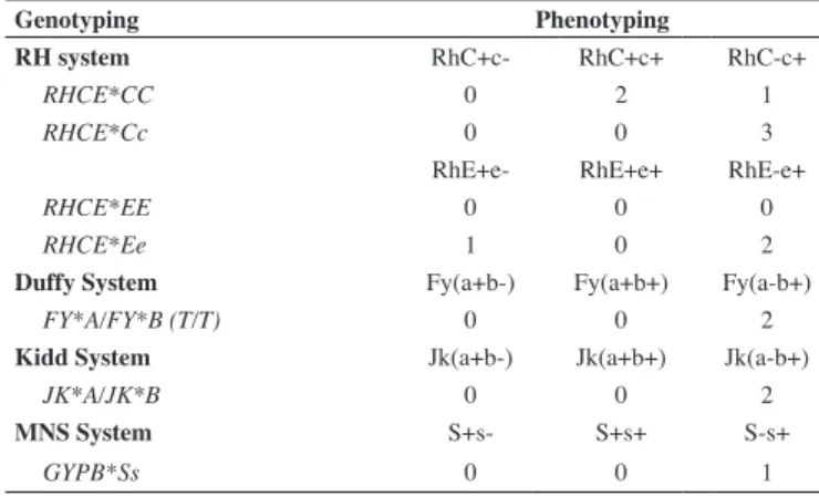
serologically-matched blood units. The main discrepancies or mismatches occurred in the RH, FY, JK and MNS systems. Discrepancies between the previous phenotype and genotype-derived phenotype were found in 14 alloimmunized chronically transfused patients (Table 1) who were not having good in vivo RBC survival and mismatches for multiple antigens were found in 17 patients receiving blood units matched for ABO, Rh and K. Eight Rh alloimmunized patients presented RHD and RHCE variants that had not been serologically identiied or that had been misinterpreted as autoantibodies. Of these eight patients, two had the RHCE*ceAR associated with the RHD*DAR variant, two had the RHCE*ceBI associated with the RHD*DOL variant, two had RHCE*ce48C,733G and two had RHCE*ce48C,733G, 1006T, both associated with the RHD*DIIIa variant (Table 2). According to these results, better matches were found for the patients in the institution’s DNA-typed units, and in the majority of cases, the degree of matching was enhanced and the patients
beneited by receiving transfusions that provided better in vivo RBC survival. The transfusion timeline before genotyping was about 1 week, and with extended genotyped-matched blood this changed to 30 days. These patients were followed up for one year and have not developed other antibodies.
Table 1 - Phenotyping and genotyping discrepancies found in the samples of 14 sickle cell disease patients
Genotyping Phenotyping
RH system RhC+c- RhC+c+ RhC-c+
RHCE*CC 0 2 1
RHCE*Cc 0 0 3
RhE+e- RhE+e+ RhE-e+
RHCE*EE 0 0 0
RHCE*Ee 1 0 2
Duffy System Fy(a+b-) Fy(a+b+) Fy(a-b+)
FY*A/FY*B (T/T) 0 0 2
Kidd System Jk(a+b-) Jk(a+b+) Jk(a-b+)
JK*A/JK*B 0 0 2
MNS System S+s- S+s+ S-s+
GYPB*Ss 0 0 1
Table 2 - RHD and RHCE variants found in eight sickle cell disease patients
n RHD RHCE
2 RHD*DAR RHCE*ceAR
2 RHD*DOL RHCE*ceBI
2 - RHCE*ce48C, 733G
37 Molecular matching of red blood cells is superior to serological matching in sickle cell disease patients
Rev Bras Hematol Hemoter. 2013;35(1):35-8
extensive DNA-based blood group typing of donors and patients has shown to be a cost-effective procedure and now chronically SCD patients can be screened for at least the lack of the C, c, E, e, K, S, Fya, Jka and Jkb antigens.
The reliable prediction of xHEA phenotypes offers a potential alternative to the current serologic methods of donor unit screening and transfusion recipient typing. DNA array technology can contribute to the management of transfusions in SCD patients by facilitating transfusion support with antigen-matched blood. It has the potential to replace the routine blood group phenotyping with a reduction in costs as well as the workload involved in donor and patient antigen typing.
Conclusion
In summary, the implementation of molecular matching can decrease the risk of transfusion reactions, especially delayed transfusion reactions to existing alloantibodies, and prevents alloimmunization. Additionally, the degree of enhanced matching was higher than that which occurred by random based matching of only the ABO, Rh and K systems. Matching at the DNA level
may provide an added level of safety and eficacy by reducing
transfusion requirements, decreasing the risk of transfusion-related acute lung injury and potential exposure to infectious diseases.
Acknowledgments
We thank Débora Castilho Credidio and Maria Zelma Machado Perdigão for their technical support.
References
1. Giblett ER. A critique of the theoretical hazard of inter- vs. intra-racial transfusion. Transfusion. 1961;1:233-8
2. Davies SC, McWilliam AC, Hewitt PE, Devenish A, Brozovic M. Red cell alloimmunization in sickle cell disease. Br J Haematol. 1986;63(2):241-5.
3. Reisner EG, Kostyu DD, Philips G, Walker C, Dawson DV. Alloantibody response in multiply transfused sickle cell patients. Tissue Antigens. 1986;30(4):161-6
4. Cox JV, Steane E, Cunningham G, Frenkel EP. Risk of alloimmunization and delayed hemolytic transfusion reactions in patients with sickle cell disease. Arch Intern Med. 1988;148(11):2485-9.
5. Rosse WF, Gallagher D, Kinney TR, Castro O, Dosik H, Moohr J, Wang W, Levy PS. The cooperative study of sickle cell disease. Transfusion and alloimmunization in sickle cell disease. Blood. 1990;76(7):1431-7.
6. Vichinsky EP, Earles A, Johnson RA, Hoag MS, William A, Lubin B. Alloimmunization in sickle cell anemia and transfusion of racially unmatched blood. N Engl J Med. 1990;322(23):1617-21. Comment in: N Engl J Med. 1990; 323(20):1420-2; N Engl J Med. 1990; 322(23):1666-8.
7. Orlina AR, Sosler SD, Koshy M. Problems of chronic transfusion in sickle cell disease. J Clin Apher. 1991;6(4):234-40.
8. Sosler SD, Jilly BJ, Saporito C, Koshy M. A simple, practical model for reducing alloimmunization in patients with sickle cell disease. Am J Hematol. 1993;43(2):103-6.
9. Castro O, Sandler SG, Houstoun-Yu P, Rana S. Predicting the effect of transfusing only phenotype-matched RBCs to patients with sickle cell disease: theoretical and practical implications. Transfusion. 2002;42(6):684-90.
These results were also very important to clarify and conirm the speciicities of alloantibodies and were essential to distinguish
between alloantibodies and autoantibodies, particularly in patients with RH variants.
Discussion
The provision of antigen-negative blood forms the basis for safe blood transfusions by decreasing the risk of hemolytic transfusion reactions and preventing new instances of alloimmunization(21).
High-throughput genotyping based on DNA arrays is a very feasible method to obtain a large pool of well-typed blood donors and can contribute to the management of transfusions in SCD patients by allowing a more accurate selection of donor units to reduce transfusion requirements. The ability to test patients and a large number of donors simultaneously for several antigens, together with computer analysis and interpretation of data(16,17), facilitates the matching of RBC
components to the recipients’ blood type making the process feasible and easily increasing the inventory of donor units for SCD patients. Klapper et al.(17) concluded that if patients and
donors are extensively DNA typed, it is feasible to provide units for transfusion that are more extensively matched than in the current standard of practice.
In the Blood Center of UNICAMP, SCD patients who
require multiple transfusions are placed on chronic prophylactic transfusion protocols. For the effective application of genotyping of these patients it is important that suitably phenotyped/ genotyped donors are available.
When molecular-matching was applied to 35 SCD patients, discrepancies or mismatches were found for multiple antigens
between their xHEA proile and the antigen proile of their
serologically-matched blood units. Additionally, RH variants that
would never be identiied by serology were found in eight patients
and alloantibodies were distinguished from autoantibodies. It was
possible to ind a better match in our xHEA-typed units for these
patients, enhancing the degree of matching and reducing the risk of alloimmunization and delayed transfusion reactions.
The patients who beneited from receiving antigen-matched
RBCs based on genotyping (as shown by better in vivo RBCs survival) had increased hemoglobin levels and diminished frequency of transfusions. These patients presented autoantibodies
that masked clinically signiicant alloantibodies and the use
of this DNA approach was essential to distinguish between autoantibodies and alloantibodies.
Alloimmunization may cause several problems ranging from inconvenience due to delay in obtaining matched blood to
DHTRs. The beneits that donor-recipient blood group genotype
matching would have on reducing the incidence of delayed transfusion reactions were demonstrated in this study. Extended matching of donors and SCD patients for the ABO, D, C, c, E, e, K, Fya, Fyb, Jka, Jkb, S, s and Dia antigens and RH variants would
also reduce the development of the majority of alloantibodies that currently exist in more than 90% of the immunized SCD patients.
38
da Costa DC, Pellegrino J Jr, Guelsin GA, Ribeiro KA, Gilli SC, Castilho L
Rev Bras Hematol Hemoter. 2013;35(1):35-8 10. Chou ST, Westhoff CM. The role of molecular immunohematology in
sickle cell disease. Transfus Apher Sci. 2011;44(1):73-9.
11. Vichinsky EP, Luban NL, Wright E, Olivieri N, Driscoll C, Pegelow CH, Adams RJ; Stroke Prevention Trail in Sickle Cell Anemia. Prospective RBC phenotype matching in a stroke-prevention trial in sickle cell anemia: a multicenter transfusion trial. Transfusion. 2001;41(9):1086-92. Comment in: Transfusion. 2002;42(5):658-9; author reply 659-60.
12. Beiboer SH, Wieringa-Jelsma T, Maaskant-von Wijk PA, van der Schoot CE, Van Zwieten R, Roos D, et al. Rapid genotyping of blood group antigens by multiplex polymerase chain reaction and DNA microarray hybridization. Transfusion. 2005;45(5):667-79. Comment in: Transfusion. 2005;45(5):652-3.
13. Bugert P, McBride S, Smith G, Dugrillon A, Klüter H, Ouwehand WH, et al. Microarray-based genotyping for blood groups: comparison of gene array and 5’-nuclease assay techniques with human platelet antigen as a model. Transfusion. 2005;45(5):654-9. Comment in: Transfusion. 2005;45(5):652-3.
14. Flickinger C. In search of red blood cells for alloimmunized patients with sickle cell disease. Immunohematology. 2006;22(3):136-42.
15. Castilho L, Rios M, Pellegrino Jr J, Saad ST, Costa FF. Blood group genotyping facilitates transfusion of b thalassemia patients. J Clin Lab Anal. 2002;16(5):216-20.
16. Ribeiro KR, Guarnieri MH, da Costa DC, Costa FF, Pellegrino J Jr, Castilho L. DNA array analysis for red blood cell antigens facilitates the transfusion support with antigen-matched blood in patients with sickle cell disease. Vox Sang. 2009;97(2):147-52.
17. Klapper E, Zgang Y, Figueroa P, Ness P, Stubbs J, Abumuhor I, et al. Toward extended phenotype matching: a new operational paradigm for the transfusion service. Transfusion. 2010;50(3):536-46.
18. Hashmi G, Shariff T, Seul M, Vissavajjhala P, Hue-Roye K,
Charles-Pierre D, et al. A lexible array format for large-scale, rapid
blood group DNA typing. Transfusion. 2005;45(5):680-8. Comment in: Transfusion. 2005;45(5):652-3.
19. Hashmi G, Shariff T, Zhang Yi, Cristobal J, Chau C, l Seul M, et al. Determination of 24 minor red blood cell antigens for more than 2000 blood donors by high-throughput DNA analysis. Transfusion. 2007;47(4):736-47. Erratum in: Transfusion. 2007;47(5):952.
20. Halter Hipsky C, Costa DC, Omoto R, Zanete A, Castilho L, Reid ME. Prevalence of RHD*DOL and RHCE*ce(818T) in two populations. Immunohematology. 2011;27(2):66-7.
21. Schonewille H, van de Watering LM, Brand A. Additional red blood cell alloantibodies after blood transfusions in a nonhematologic alloimmunized patient cohort: is it time to take precautionary measures? Transfusion. 2006;46(4):630-5.