STUDY OF THE EFFECT OF USING A COLLIMATOR ON A GAMMA
PROBE IN RADIOGUIDED SURGERY*
Iran José Oliveira da Silva1
, Helen Jamil Khoury2
, Márcia Rosana Leite de Lemos3
, Paulo José de Almeida Filho3, Maria Inês Calil Cury Guimarães4
OBJECTIVE: The aim of the present study is to evaluate the performance of the Europrobe gamma probe spatial resolution in radioguided surgery. MATERIALS AND METHODS: In the radioguided surgery technique, after a radiotracer injection into the primary tumor, a gamma detector probe is utilized to determine the lo-calization of the sentinel lymph node. In order to simulate the region of the radiotracer injection the sentinel lymph node, two Tc-99m sources, with 20.42 MBq and 0.70 MBq, were positioned in a water-filled tank. With a collimator cap attached to the probe, scans over the water surface were made. So, the count rate was measured by means of lateral displacement of the probe in relation to the both sources with distances ranging between 30 mm and 60 mm. RESULTS: Results showed that the use of the collimator contributes to improve the spatial resolution of the probe allowing the identification of a sentinel lymph node within a distance up to 30 mm from the radiotracer injection point. CONCLUSION: The utilization of a collimator cap with a 3.5 mm diameter central orifice on a Europrobe gamma probe, allows the identification of a sentinel lymph node within a distance up to 30 mm from the radiotracer injection point.
Keywords: Intraoperative probe; Radioguided surgery; Sentinel lymph node localization; Spatial resolution.
Estudo do efeito do uso de colimador na sonda gama utilizada em cirurgia radioguiada.
OBJETIVO: O objetivo deste trabalho consiste em estudar a influência da resolução espacial da sonda gama Europrobe que é utilizada em cirurgia radioguiada. MATERIAIS E MÉTODOS: Na técnica de cirurgia radio-guiada, após a injeção de um radiotraçador no tumor primário, é utilizada uma sonda detectora de radiação gama a fim de determinar a localização do linfonodo sentinela. Para simular a região dos pontos de injeção do radiotraçador e o linfonodo sentinela, duas fontes de Tc-99m, com 20,42 MBq e 0,70 MBq, foram posi-cionadas no interior de um recipiente preenchido com água. Em seguida, com a janela de entrada da sonda coberta com um colimador, realizou-se varredura sobre a superfície da água. Assim, foi possível registrar a taxa de contagens variando-se a distância lateral da sonda em relação às duas fontes, as quais foram sepa-radas por uma distância variando entre 30 mm e 60 mm. RESULTADOS: Os resultados mostraram que o uso do colimador contribui para melhorar a resolução espacial da sonda, permitindo a identificação do linfonodo sentinela distante até 30 mm do ponto de injeção. CONCLUSÃO: Esse estudo permite concluir que a sonda Europrobe, quando utilizada com capa colimadora com orifício central de 3,5 mm de diâmetro, é capaz de identificar o linfonodo sentinela posicionado a até 30 mm de distância em relação à região do ponto de in-jeção do radiotraçador.
Unitermos: Sonda intra-operativa; Cirurgia radioguiada; Localização de linfonodo sentinela; Resolução espacial.
Abstract
Resumo
* Study developed at Real Hospital Português de Pernambuco, Recife, PE, Brazil.
1. Master Degree in Energetic and Nuclear Technologies. 2. Doctorate in Dosimetry and Nuclear Instrumentation. 3. Specialists in Nuclear Medicine.
4. Doctorate in Dosimetry and Nuclear Medicine. Mailing address: Dr. Iran José Oliveira da Silva. Universidade Federal de Pernambuco, Departamento de Energia Nuclear. Ave-nida Professor Luiz Freire, 1000, Recife, PE, Brazil 50740-540. E-mail: [email protected]
Received January 11, 2006. Accepted after revision March 15, 2006.
INTRODUCTION
Neoplasms or cancers may disseminate throughout the body, basically by two ways: by the blood, through veins and arteries, or by the lymphatic system. This allows the
cancer, from a determined site (primary tumor), to generate tumors of the same type in other distant organs (metastasis).
Usually, when a cancer is detected, an evaluation is performed for the presence, or not, of tumor cells dissemination to some lymphatic basin proximal to the primary tumor. If this has occurred, it is necessary to evaluate the degree of involvement of this lymph node basin, since the tumor cells may migrate to other lymph nodes and other parts of the body. The traditional ap-proach involves the removal for analysis of all (10 to 20) lymph nodes in the tumor-draining area(1). However, a technique al-lowing a higher accuracy in the evaluation
of the suspected lymphatic basin involve-ment degree is based on the identification and removal for analysis of the key lymph node, also known as sentinel lymph node (SLN), which may include cells from the primary tumor.
count-ing-rate in relation to the background radia-tion counting rate. Studies have shown that, in more than 90% of cases, this region is where the SLN is located(2).
This procedure, known as radioguided surgery, requires the best of a surgeon’s ability, since it is necessary to handle the probe in search of regions where there is a high enhancement in relation to the mea-sured counting rates, i.e., regions for which a little change in the probe positioning or inclination may cause abrupt variations in counting rates.
Since in the radioguided surgery tech-nique the major difficulty consists in local-izing the SLN in sites near the primary tu-mor, the objective of the present study is to analyze the effect in the probe spatial reso-lution with the use of a collimator, as well as the variation of the distance between the SLN and the radiotracer injection site.
GAMMA PROBE OPERATING PRINCIPLES
Gamma probes are compact devices ba-sically constituted of two components: a
gamma radiation detection system, usually conceived as the probe itself that may be scintillator crystal-based or semiconductor-based, and an electronic system, where the signal is amplified, processed and visual-ized by means of an analogical or digital display(3).
Scintillator crystal-based probe
This type of probe operates basically in the same way of conventional scintillation-detectors, and in this case, for a detection system to acquire small dimensions, a light guide or a optical fiber beam is coupled between the scintillator crystal and the photomultiplier tube, so that the light pro-duced in the crystal due to the interaction with the radiation is transmitted through the light guide to the photomultiplier tube where it is converted into an electrical sig-nal.
Some scintillator crystal-based probes utilize the crystal coupled with a Si photo-diode replacing the photomultiplier tube. A critical point in this type of probe is the choice of a high input impedance, low-noise preamplifier, since photodiodes have not an internal signal amplifier device.
Usually, gamma probes scintillator crys-tals are NaI(Tl), CsI(Tl), Bi4Ge3O12 or CdWO4-based, the two first elements be-ing the most frequently utilized. These crystals present the advantage of an in-creased atomic number and high specific mass, which contributes to the increase of the gamma radiation absorption coeffi-cient.
Semiconductor crystal-based probe
In the semiconductor crystal, the radia-tion-sensitive volume corresponds to the depletion region formed by the junction of two types of semiconductor materials: one of P-type, and another of N-type(4).
The majority of gamma probes utilize HgI2, CdTe or CdZnTe-based semiconduc-tor crystals. These materials have not only a high gamma radiation absorption coeffi-cient, but also a prohibited wideband range. This latest feature allows the probe to be utilized at the human body temperature (37°C), with less interference of noise re-sulting from thermal generation by pairs of electron-vacancies in the crystal.
However, differently from Ge or Si semiconductors, in these crystals, because of the low electrons-vacancies mobility, the charge carriers collection is strongly af-fected by the presence of defects or impu-rities (Cd, Au, Zn atoms, etc.) in the crys-talline net. This contributes to limit the construction of probes utilizing highly thick semiconductor detectors necessary for the detection of high and intermediate energy gamma rays.
PARAMETERS AFFECTING GAMMA PROBES PERFORMANCE
Detection sensitivity and efficiency
The sensitivity is defined as the relation between number of photons depositing all the energy into the probe detector sensitive volume and the amount of photons emitted by the source. Therefore, the sensitivity is heavily affected by the source-detector geometry, besides being directly propor-tional to the sensitive area of the detector, and inversely proportional to the squared distance between source and detector, if the source is punctual.
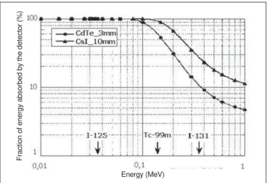
The total radiation energy absorption by the detector is dependent on its physical characteristics such as dimensions, effec-tive atomic number, and mass density. One may observe that, for example, in the graphic on Figure 1, for photons with en-ergy higher than 150 keV, the enen-ergy frac-tion absorbed, in the case of a CdTe (3 mm thick) semiconductor detector, decreases considerably in relation to the absorbed energy fraction in the CsI (10 mm thick) scintillator detector(5). Therefore, the scin-tillator crystal-based probes present higher sensitivity for high energies than semicon-ductor-based probes.
Resolution in energy
Since the pulse height produced in the probe output is proportional to the incident radiation energy deposited in the crystal, the radiation energies spectrum is
trans-Figure 1. Variation of the fraction of absorbed energy in a CdTe semiconductor and in a CsI scintillator
crystal as a function of photons energy (5).
Energy (MeV)
formed into a pulse-height spectrum which is recorded by the electronic system. How-ever, it is important to note that a little fluc-tuation may occur in the pulse-height, cor-responding to the same energy deposited, basically because of two factors: the statis-tical fluctuation of the number of the semi-conductor detector charge carriers, or the number of the scintillator detector photons produced by radiation, as well as the influ-ence from noise of the electronic detection system components (detector, preamplifier, filters, etc.). As a result, the energies dis-tribution spectrum obtained with the probe from a monoenergetic source, acquires a Gaussian shape(4). So, the resolution in energy of the detection system is defined as the photopeak FWHM — full width at half maximum.
Therefore, when the probe is utilized to localize a small radiation source, it is nec-essary to select a window in the pulse dis-crimination system, so that the probe de-tects just the pulses corresponding to the photons emitted directly from the source, and rejects the photons scattered by the source region.
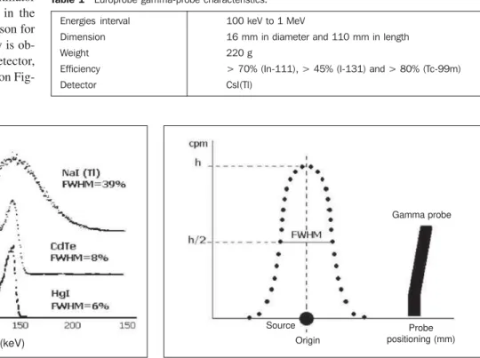
As the radiation incident on the semi-conductor detector produces a number of charge carriers interaction about ten times superior to that produced in the scintillator crystal, the statistical fluctuation in the number of carriers is lower, the reason for which a better resolution in energy is ob-tained with the semiconductor detector, according to the scheme presented on Fig-ure 2(6).
Spatial resolution
The probe spatial resolution is ex-pressed as the FWHM of the point spread function (PSF), correspondent to the graphic relating the counting rate detected in a punctual source as a function of the lateral distance from the detector central axis, as shown in Figure 3.
So, when the probe is utilized for sur-veying the region of interest, the spatial resolution reflects the ability of the probe to separately identify the radiation emitted by two punctual sources very close to each other, i.e., in the case of a radioguided sur-gery, this parameter reflects the ability of the probe to differentiate the SLN activity from the activity in the region of the ra-diotracer injection site, as per Figure 4. Consequently, amongst all the mentioned parameters, the spatial resolution is a criti-cal parameter for the gamma probe in the task of localizing the SLN.
For determining the differentiation be-tween measures and countings obtained, i.e., the node identification, Britten(7), based on the probe response as per Figure 4, has determined that the percent value of the difference between the PSF peak value
corresponding to the node P, and a point in this curve base, M, which is known as peak reduction value (PRV), where (P–M) / P × 100%, is a value reflecting the probe abil-ity to localize the SLN. Based on labora-tory data, Britten has determined that the smallest distance between the injection site and SLN allowing differentiation from countings corresponds to a 25% peak re-duction.
MATERIALS AND METHODS
In the present study we have utilized an Europrobe gamma probe, whose character-istics are shown in Table 1. In this probe, the light generated in the CsI(Tl) crystal, due to the interaction with radiation, is converted into electrical pulses in the Si photodiode coupled with the electronic system by means of a low-noise and high input impedance preamplifier.
For studying the probe spatial resolu-tion, we have utilized an experimental ar-rangement, as per Figure 5. The gamma probe was connected to an also Europrobe electronic system which allows the visual-ization of the counting rates on a digital dis-play. For studying the sources in an
envi-Figure 2. Comparison of the resolution in energy obtained for the 140 keV
photopeak measured with three types of gamma probes(6).
Figure 3. Scheme for determination of the probe spatial resolution.
Table 1 Europrobe gamma-probe characteristics.
Energies interval Dimension Weight Efficiency Detector
100 keV to 1 MeV
16 mm in diameter and 110 mm in length 220 g
> 70% (In-111), > 45% (I-131) and > 80% (Tc-99m) CsI(Tl)
Countings
Energy (keV)
Gamma probe
Probe positioning (mm) Source
ronment similar to that of the human body, we have utilized a phantom constituted of a water thank measuring 250 mm in diam-eter, and 110 mm in height. On the borders of this thank an acrylic guide was posi-tioned, with a millimetric scale on it, allow-ing the probe to scan the water surface, identifying the probe central axis lateral distance in relation to the source.
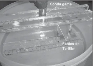
According to the Figure 6, the source of Tc-99m (the radioisotope usually utilized for SLN localization) was placed inside two small acrylic cylinders measuring 7 mm in diameter × 5 mm in height, fixed on acrylic supports which allowed the variation of the source depth in relation to the water surface. Therefore, for studying the probe ability to separately identify the SLN and the injection site region activities, one of the sources (20.42 MBq activity) was utilized to represent the injection sites on the primary tumor, and the other (0.70 MBq activity), to represent the SLN.
Initially, we have studied the variation of the probe spatial resolution as a function of the type of environment (air or water) and distance between the source and the probe window. For this purpose, setting the electronic system of the probe for the rater meter mode, we have measured the count-ing rates with the probe scanncount-ing the wa-ter surface, the lawa-teral distance varying at each 5 mm from a Tc-99m (20.42 MBq) source positioned at 15 mm from the probe window. These measurements were per-formed with the source in the air, and fol-lowing, with the source in the water. Then, with the source still submerged,
measure-ments were performed with the source at 25 mm from the probe window.
Latter, for studying the variation of the probe spatial resolution power as a result of the use of a collimator on the detector window, we have utilized a lead cylinder with a 1.5 mm-thick wall and a central hole. In this case, besides performing the mea-surements with the probe without the col-limator, we have also performed measure-ments with the probe utilizing both a col-limator with a 2.5 mm-hole, and another with a 3.5 mm-hole. These measurements of counting rates were performed with the probe scanning the water surface, the lat-eral distance varying at each 5 mm between the probe central axis and the radiotracer injection site, whose positioning was con-sidered as the landmark on the guide, as per Figure 7. In all of the measurements per-formed, the radiotracer injection site and
Figure 4. PSF for two punctual sources, representing the injection site and the
SLN separated by a d distance.
Figure 5. Arrangement utilized for studying the gamma probe spatial reso-lution.
Figure 6. Positioning of Tc-99m sources, representing the injection site and the SLN.
the SLN, respectively, were placed at 10 mm and 20 mm from the water surface, with a 60 mm gap between them.
Finally, aiming at studying the probe spatial resolution variation as a function of the distance between the radiotracer injec-tion site and the SLN, measurements were performed with the same experimental ar-rangement previously described, for the injection site and the SLN separated by 30 mm, 40 mm, 50 mm and 60 mm distances, and with the probe utilizing a 3.5 mm-thick collimator.
RESULTS AND DISCUSSION
The results of the study of the probe spatial resolution variation as a function of the type of environment (air or water), as well as the distance between the source and the probe window, are shown in the
graph-Gamma probe Electronic system
Guide
Phantom
PFS (injection site) PFS (node)
PFS (injection site + node)
Figure 7. Scheme of the arrangement utilized to measure PSF for two
punc-tual sources, representing the injection site and the SLN, separated by a d
distance.
Gamma probe
O (origin)
SLN Injection site
Figure 8. Spatial resolution variation as a function of the type of environment and distance between the source and the probe window.
Table 2 FWHM measured for the source in different types of environments and distances from the pro-be window.
Type of environment
Air Water Water
Distance between the source and the probe window (mm)
15 15 25
FWHM (mm)
16.4 24.2 26.6
ics on Figure 8. This Figure presents the PSF obtained for different environments and distances between the source and the probe window. Based on each curve, the FWHM was determined, as per Table 2. The results analysis demonstrates that a change in the type of environment where the source is localized, i.e., from air to water, implies a strong deterioration of the FWHM, that is to say, the resolution wors-ens, increasing about 48%. This increase in the FWHM value is a result of the radiation photons scattering in the water. On the other hand, the results also demonstrate that a 10 mm increase in the distance be-tween the source and the probe window, even in the water, causes little deterioration in the FWHM value (about 1% increase) when compared with the result obtained with the change in the source environment.
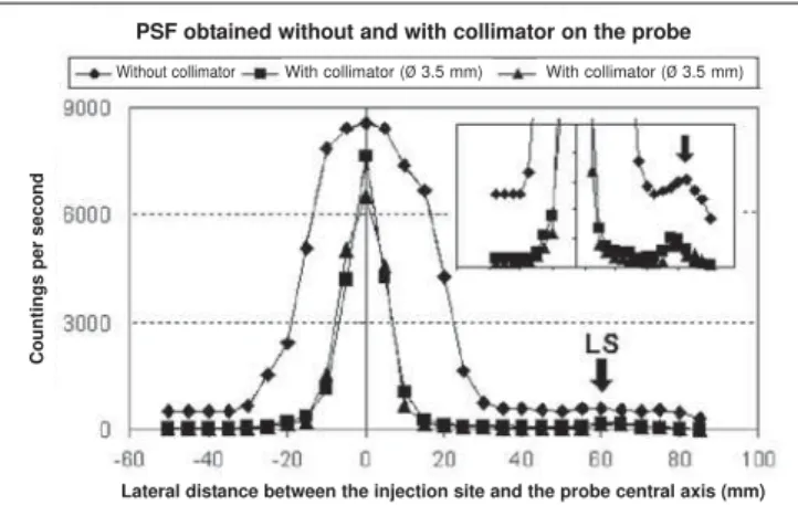
The results of the study of the probe spatial resolution power variation as a func-tion of the collimafunc-tion on the detector win-dow are shown in the graphics included on Figure 9 and Table 3. The results analysis demonstrates that the utilization of collima-tors with 3.5 mm- and 2.5 mm-holes results in an increase in PRP of respectively 283% and 376%, in comparison with the PRP determined with the probe without collima-tor. This demonstrates that the use of a
col-limator on the probe window contributes to an accurate SLN identification.
The results of the study of the probe spatial resolution variation as a function of the distance between the radiotracer injec-tion site and the SLN, with a 3.5 mm-hole collimator on the probe, are shown in the graphics included on Figure 10 and Table 4. The analysis of these results demon-strates that as the distance between the in-jection site and the SLN is reduced from
Source in water at 2.5 cm from the detector
Source in the air at 1.5 cm from the detector Source in water at 1.5 cm from the detector
Lateral distance between the source and the probe central axis (mm)
Countings per second
Figure 9. PFS for the injection site and the SLN at respectively 10 mm and 30 mm from the water surface, and separated by 60 mm.
Without collimator With collimator (Ø 3.5 mm) With collimator (Ø 3.5 mm)
Lateral distance between the injection site and the probe central axis (mm)
Countings per second
PSF obtained without and with collimator on the probe
Figure 10. PSF for different distances between the injection site and the SLN. The arrows indicate the position with higher counting rate measured in the SLN region.
Lateral distance between the injection site and the probe central axis (mm)
Countings per second
60 mm to 30 mm, there is a 45% decrease in the spatial resolution power as a result of the counting rates superposition. How-ever, it is important to note that, even for a 30 mm distance between the injection site and the SLN, a precise localization of the SLN is feasible, according to Britten(7).
An accurate localization of the SLN in the radioguided surgery is essential for a precise determination of the degree of in-volvement of the lymphatic basin near the primary tumor. Therefore, the study of the gamma probe spatial resolution allows the
Table 3 PRV for the injection site and SLN sepa-rated by a 60 mm-distance and positioned, respec-tively, at 10 and 20 mm from the probe window.
Probe without collimator
Probe with collimator (Ø3.5 mm)
Probe with collimator (Ø 2.5 mm)
PRV (%)
16.4 62.8 78.0
Distance between the injection site and the SLN (mm)
PRV (%)
30
40.8 40
62.8 50
72.0 60
74.4 Table 4 PRV variation as a result of the distance between the radiotracer injection site and the SLN.
evaluation of the actual probe potential in the task of localizing the SLN.
The present study demonstrates that the radiation scattering in the environment has a great influence on the probe spatial reso-lution power, difficulting the localization of the SLN in deeper regions. Additionally, one may also conclude that the utilization of a collimator on the probe window in-creases its resolution power, contributing to an accurate identification of the SLN, in spite of causing a decrease in the probe detection efficiency. Finally, it was shown that the Europrobe probe can identify a SLN localized at up to 30 mm from the radiotracer injection sites.
REFERENCES
1. Halkar RK, Aarsvol JN. Intraoperative probes. J Nucl Med Technol 1999;27:188–193. 2. Lima MV, Tavares JM, Silveira RA, Filho MT,
Silva FA, Silva LF. Intraoperative use of gamma probe for identification of sentinel node in penile cancer. Braz J Urol 2002;28:123–129. 3. Zanzonico P, Heller S. The intraoperative gamma
probe: basic principles and choices available. Semin Nucl Med 2000;30:33–48.
4. Knoll GF. Radiation detection and measurement. 2nd ed. New York: John Wiley & Sons, 1989. 5. Ricard M. Intraoperative detection: probes and
radiation protection. Revue de I’Acomen 2000;3: 127–133.
6. Zanzonico P, Heller S. The intraoperative gamma probe: basic principles and choices available. Semin Nucl Med 2000;30:33–48.
7. Britten AJ. A method to evaluate intra-operative gamma probes for sentinel lymphatic node local-ization. Eur J Nucl Med 1999;26:76–83.