Article
ISSN 0102-695X
http://dx.doi.org/10.1590/S0102-695X2011005000161 Received 21 Nov 2010 Accepted 3 Jun 2011 Available online 2 Sep 2011
litwinowii
roots are apoptotic for HeLa cells
Zahra Tayarani-Najarani,
1Javad Asili,
2Heydar Parsaee,
1Seyed
Hadi Mousavi,
1Naser Vadati Mashhadian,
3Alireza Mirzaee,
2Seyed Ahmad Emami
*,21Pharmacological Research Centre of Medicinal Plants, School of Medicine,
Mashhad, University of Medical Sciences, Mashhad, Iran,
2Department of Pharmacognosy, School of Pharmacy, Mashhad, University of
Medical Sciences, Mashhad, Iran,
3Medical Toxicology Research Center, Mashhad, University of Medical Sciences,
Mashhad, Iran.
Abstract: Chemical investigation on the CH2Cl2 fraction of the Scutellaria litwinowii Bornm. & Sint., Lamiaceace, root extract for the i rst time resulted in the isolation of
wogonin, and neobaicalein. These compounds were evaluated for their cytotoxicity towards HeLa cell lines and lymphocytes. Meanwhile, the role of apoptosis was explored in this toxicity. The cells were cultured in RPMI medium and incubated with different
concentrations of isolated l avonoids. Cell viability was quantii ed by MTS assay.
Apoptotic cells were determined using propidium iodide staining of DNA fragmentation
by l ow cytometry (sub-G1peak). Wogonin, and neobaicalein inhibited the growth of
malignant cells in a dose-dependent manner. The IC50 values of 46.62 and 79.34 µM were, respectively, found for neobaicalein and wogonin against HeLa cells after 48 h
of treatment. Neobaicalein induced a sub-G1 peak in the l ow cytometry histogram of
treated cells compared to control cells indicating that apoptotic cell death is involved in neobaicalein toxicity. Neobaicalein exerts cytotoxic and pro-apoptotic effects in HeLa cell lines and could be considered as a potential chemotherapeutic agent in cancer treatment. Keywords:
apoptosis Lamiaceae neobaicalein
Scutellaria litwinowii
wogonin
Introduction
Scutellaria, Lamiaceae, is a genus of about 300 species of plants (Mabberley, 1993) and has been used as traditional medicine in China, India, Korea, Japan, Europe and North America (Shang et al., 2010). Scutellaria spp. possesses effective anticancer activity and this property
is due to some l avones such as baicalin, baicalein and wogonin (Li-Weber, 2009). Scutellaria extracts are not only cytostatic but also cytotoxic in vitro and in vivo
(Scheck et al., 2006; Shang et al., 2010).
Recent pharmacologic researches have coni rmed that total extracts or l avonoids of the genus Scutellaria possess anti-lipoperoxidation, anti-platelet,
anti-inl ammatory, antitumor, hepatoprotective, antioxidant, antibacterial and antiviral activities (Ye et al., 2004; Parajuli et al., 2009; Zhang et al., 2006; Burnett et al., 2007; Sonoda et al., 2004). In general, l avonoids are the
most crucial class of compounds that have been isolated from several species of Scutellaria (Malikov & Yuldashev, 2002). However, only a handful of the Scutellaria species have been studied for potential anticancer activity in the new literature.
Scutellaria litwinowii Bornm. & Sint., Lamiaceace, is one of the Iranian species of Scutellaria. In our previous study we reported that the CH2Cl2 fraction of the S. litwinowii extract has cytotoxic effects against
human gastric adenocarcinoma (AGS), human cervix
carcinoma cell line (HeLa), human breast cancer cell line (MCF-7), and rat pheochromocytoma cell line (PC12) (Tayarani-Najaran et al., 2009).
In the course of our search for anti-tumor compounds, analysis of the CH2Cl2 fraction obtained from S. litwinowii extract led to the isolation of two
l avones: wogonin and neobaicalein that are cytotoxic
toward HeLa cell. Details of the isolation, structural elucidation, and cytotoxic activities are described herein. The growth-inhibitory activities of neobaicalein (1) and wogonin (2) toward HeLa cells were compared with
6-hydroxyl avone (3) and baicalein (4).
Materials and Methods
Chemicals
sodium citrate, 6-hydroxylavone (3), baicalein (4) and triton X-100 were purchased from Sigma (St
Louis, MO, USA); tetrazolium compound,
3-(4,5- dimethylthiazol-2-yl)-5-(3-carboxymethoxyphenyl)-2-(4-sulphophenyl)-2H-tetrazolium (MTS) from Promega
(Madison, WI, USA); RPMI 1640 and FCS from Gibco (Grand Island, USA); silica gel (230-400 mesh) for column chromatography and precoated silica gel GF254
sheets from Merck (Darmstadt, Germany); methanol and distilled water as HPLC grade from Duksan (South Korea); the other solvents as analytical grade from Dr.
Mojallali lab. (Tehran, Iran).
Plant material
The roots of Scutellaria litwinowii Bornm. & Sint., Lamiaceace, were collected from Hosseinabad
valley (2100 m height) in Pivejan, a village at 65 km
south-west of Mashhad, Razavi Khorasan province, northeast of Iran. The plant was authenticated by Mr. M.R. Joharchi, Ferdowsi University of Mashhad Herbarium (FUMH). Voucher specimen (No: 14175) was deposited in herbarium of School of Pharmacy, Mashhad University
of Medical Sciences (Ghahreman & Attar, 1999).
Instrumentation
Melting points were taken in a single capillary
tube on a Toshniwal melting point apparatus (Mumbai, India) and are uncorrected. NMR spectra were recorded
on a Bruker Avance 500 at 25 ºC using CDCl3 as solvent
and TMS as internal standard (proton and carbon
frequencies were 500.13 and 125.75 MHz respectively).
Reversed-phase semi-preparative HPLC separations
were carried out on a Wellchrom Knauer system (Herbert Knauer GmbH, Berlin, Germany), that consisted of a
Knauer K-1001 pump, using RP18 (250 mm×16 mm, 5 µm) column, eluted isocratically with MeOH/H2O (9:1) mixed with 0.05% phosphoric acid v/v (adjusted by triethylamine to pH 3) at 2 mL/min. Knauer K-2600 UV-Vis detector was set at 270 nm. The pure compounds were resuspended in DMSO and were then screened for growth inhibition of HeLa cells by MTS assay.
Extraction, isolation and puriication of neobaicalein
and wogonin
Dry powdered roots of S. litwinowii (400 g) were extracted three times with 2 L of methanol 90% for
24 h at room temperature. The combined iltered extracts
were evaporated to dryness under vacuum at 50 °C to afford 25 g of crude methanol extract (yield 6.25%). The residue was dissolved in MeOH/H2O (9:1) and the
solution was extracted subsequently with n-hexane, CH2Cl2, ethylacetate, n-butanol and inally water using the
partitioning method described previously (Otsuka, 2006).
Then, based on the superior cytotoxic and apoptogenic properties of the CH2Cl2 fraction among other fractions, mentioned in our previous report (Tayarani-Najaran et al., 2009), the CH2Cl2 fraction (3.7 g) was selected and fractionated by silica gel column chromatography using a mixture of petroleum ether and ethyl acetate with increasing polarity to yield twelve fractions. The
fractions were pooled according to TLC proiles and
evaporated. Then the fractions (petroleum ether/ethyl acetate, 78:22, and 70:30) were repeatedly subjected to reversed-phase semiparaperative HPLC. This yielded 9.5 and 6.4 mg of compounds neobaicalein (1) and wogonin (2) respectively. The structure of these compounds was determined using 1D and 2D NMR methods (Table 1).
O OH H3CO
H3CO
OCH3
O
HO
OCH3 O
O OCH3
HO OH
2 1
O
O HO
O
O HO
OH HO
4 3
HeLa cells culture
HeLa cells were obtained from Pasteur Institute
(Tehran, Iran) and maintained at 37 °C in a humidiied
atmosphere (90%) containing 5% CO2. Cells were
cultured in Roswell Park Memorial Institute medium
(RPMI 1640 medium) with 10% (v/v) fetal bovine serum, 100 U/mL penicillin and 100 mg/mL streptomycin. HeLa cells were seeded overnight and then incubated with various concentrations of neobaicalein (1), wogonin (2),
6-hydroxylavone (3), and baicalein (4) for 48 h.
For MTS assay, cells were seeded at 5×10³ cells
per well onto 96-well culture plates. For assay of apoptosis, cells were seeded at 104 cells per well onto a 24-well plate.
For each concentration and time course study, there was a control sample that remained untreated and received an
equal volume of medium.
Leukocyte culture
Human umbilical cord blood samples (50 mL) were collected from a fresh umbilical cord attached to
containing citrate buffer as an anticoagulant. The
sample was diluted with an equal volume of Phosphate Buffered Saline (PBS), then layered over Ficoll-Hypaque
density gradient separation solution (1.077 g/mL), and centrifuged at 800 × g for 20 min at room temperature. The mononuclear cell layer was removed, washed twice in PBS and resuspended in RPMI 1640 medium supplemented with 2 mM glutamine (Sigma Chemical Co.), antibiotics
and 10% FCS. Leukocytes (5×10⁴ cells per well) were
incubated with various concentrations of neobaicalein (1) (1.5-100 µM) in 96-well microtiter plates, for 48 h. This study protocol was approved by the ethical committee of Mashhad University of Medical Sciences.
Cell proliferation and viability assays
The MTS assay (Malich et al., 1997), is based on the reduction, by mitochondrial dehydrogenase in metabolically active cells, of the MTS to the colored, water-soluble formazan that absorbs at 490 nm. The cells were seeded in each well of a 96-microwell plate and treated with various concentrations of neobaicalein (1), wogonin (2), 6-hydroxylavone (3), and baicalein (4). After 48 h of
incubation, CellTiter 96® AQ
ueous Assay reagent (Promega,
Madison, WI, USA), which is composed of the solutions
of a novel tetrazolium compound MTS and an electron coupling reagent phenazine methosulfate (PMS, a redox intermediary), was added to each well according to the manufacturer’s instructions. After 1 h, cell viability was determined by measuring the absorbance at 490 nm using an ELISA microplate reader (Awareness, Palm City, FL, USA).
The cytotoxicity was expressed as IC50, which
was calculated using Graph Pad Software (Graph Pad
prism 5 software) and presented as mean±SEM of three independent experiments with three replicates for each concentration.
Cell morphology
The HeLa cells were plated in 96-well plates at a density of 5×10³ cells/well and grown for 24 h in order to
attach to the surface of the plates completely. Neobaicalein (1), wogonin (2), 6-hydroxylavone (3), and baicalein (4) were added in different concentrations (0, 25, 50 and 100 µM) to the cells and then the cells were grown at 37 oC in
Table 1. 1HNMR (500.13 MHz) and 13CNMR (125.75 MHz) data for neobaicalein (1) (Takido et al., 1975; Iinuma et al., 1979) and
wogonin (2) (Li et al., 2004) in CDCl3.
Position neobaicalein (1) wogonin (2)
δ-13C (ppm) δ-1H (ppm), J(Hz) δ-13C (ppm) δ-1H (ppm), J(Hz)
1 _ _ _ _
2 162 _ 163.5 _
3 112.2 6.62 s 105.9 6.68 s
4 183.3 _ 182.4 _
5 152.9 _ 157.7 _
6 136.3 _ 98.8 6.44 s
7 146.4 _ 148.8 _
8 132.8 _ 126.8 _
9 149.7 _ 155.2 _
10 106.8 _ 105.27 _
1′ 108.7 _ 131.1 _
2′ 158.8 _ 126.2 7.92 d ( J=6.87)
3′ 102.9 6.66 d (J=8.5 Hz) 129.2 7.56 m
4′ 132.8 7.28 t (J=8.5 Hz) 131.9 7.56 m
5′ 110.1 6.51 d (J=8.5 Hz) 129.2 7.56 m
6′ 156.4 _ 126.2 7.92 d (J=6.87)
6′-OCH3 55.8 3.80 s _ _
6-OCH3 61.1 3.92 s _ _
7-OCH3 61.2 4.10 s _ _
8-OCH3 62.1 3.91 s 62.0 4.05 s
2′-OH _ 8.23 s _ _
a humidii ed atmosphere with 5% CO2 for 48 h. For cell
morphology experiments, the culture plates were examined and photographed by the inverted light microscope.
Apoptosis
Apoptotic cells were detected using PI staining of
treated cells followed by l ow cytometry to detect the so-called sub-G1 peak (Nicoletti et al., 1991; Tayarani-Najaran et al., 2010). Briel y, HeLa cells were cultured overnight
in a 24-well plate and treated with various concentrations of neobaicalein (1) for 48 h. Floating and adherent cells were then harvested and incubated at 4 °C overnight in
the dark with 750 mL of a hypotonic buffer (50 mg/mL PI in 0.1% sodium citrate+0.1% Triton X-100) before l ow cytometric analysis using a Partec l ow cytometer (GmbH, Münster, Germany) was conducted. Ten thousand events were acquired.
Statistical analysis
One-way analysis of variance (ANOVA) and Bonferroni’s post hoc were used for data analysis. All results were expressed as mean±SEM. p<0.05 was
considered statistically signii cant. The IC50 values and their 95% coni dence intervals (CI 95%) were obtained by nonlinear regression using the Graphpad program
(Intuitive Software for Science, San Diego, CA, USA).
Results
Identii cation of individual chemical constituents in S. litwinowii with growth-inhibitory activity
In our previous study we analyzed different fractions of Scutellaria litwinowii Bornm. & Sint., Lamiaceace, root extract for their possible cytotoxic activity. Among them, the CH2Cl2 fraction was found to be more effective than the other fractions on human cancer cells (Tayarani-Najaran et al., 2009). To identify individual biologically active compounds within the chemically complex CH2Cl2 fraction of S. litwinowii, we separated the CH2Cl2 fraction of S. litwinowii by CC and HPLC and screened the isolated compounds for the ability to inhibit HeLa cells growth by MTS assay.
Normal-phase column chromatography of the CH2Cl2 fraction of this root extract, followed by semi-preparative reversed-phase HPLC afforded two compounds. Compound 1 (neobaicalein or
skullcapflavone II) was a yellow powder (9.5 mg),
with a melting point of 181-183 oC (Djerassi, 1994),
that was obtained from petroleum ether/ethyl acetate (78:22) fraction and compound 2 was a yellow powder (6.4 mg), with a melting point of 203 oC (Djerassi,
1994), that was obtained from petroleum ether/ethyl
acetate (70:30) fraction. The 1H-NMR and 13CNMR of
the isolated compounds are shown in Table 1. Based on these results and literature reports of compounds
known to be in other Scutellaia spp. (Shang 2010), it was confirmed that the compounds are wogonin (mw 284, 2), and neobaicalein (mw 374, 1).
Effects of neobaicalein on cell proliferation and viability
of tumor cells and lymphocytes
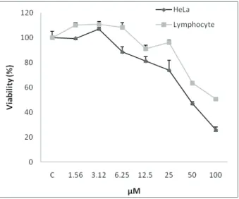
The cytotoxicity of neobaicalein (1) to HeLa cells was investigated using the colorimetric MTS assay. Firstly, HeLa cells were incubated with various concentrations of neobaicalein (1.5-100 µM) for 48 h. As shown in Figure 1 neobaicalein decreased cell viability of HeLa cells in a concentration-dependent manner. This toxicity was associated with morphological changes including reduction of cell volume and rounding of the cells. The substantial morphological changes observed in neobaicalein-treated HeLa cells were examined and photographed by the inverted light microscope.
Damaged cells became round and shrunken, while the
untreated cells retained their normal size and shape (Figure 2).
According to the MTS assay, incubation of neobaicalein in the same range of concentrations produced less toxicity to lymphocytes than HeLa cells (Figure 1).
Figure 1. A. Comparison of the cytotoxic effects of neobaicalein on HeLa cells and normal proliferating lymphocytes from human umbilical cord cells. Cells were treated with different concentrations of neobaicalein for 48 h. Neobaicalein, was less cytotoxic on normal proliferating lymphocytes. Data are representative of three independent experiments and express the mean±SEM.
Comparative analysis of cell growth inhibition by neobaicalein (1), wogonin (2), 6-hydroxyfl avone (3), and
Figure 3. Effect of neobaicalein (1), wogonin (2),
6-hydroxyl avone (3), and baicalein (4) on cell viability of HeLa cells. (A) HeLa cells were treated with different concentrations of
neobaicalein, wogonin, 6-hydroxyl avone, and baicalein for 48 h. Viability was quantitated by MTS assay. Data are representative
of three independent experiments and express the mean±SEM. Cytotoxicity of neobaicalein (1), wogonin (2),
6-hydroxyl avone (3), and baicalein (4) was examined on HeLa cells. In order to compare the cytotoxicity of
isolated l avonoides with 6-hydroxyl avone and baicalein
against malignant cells, another MTS assay was carried out for different concentrations (1.5-100 µM). Among them, neobaicalein was found to be more effective than the other compounds (Figure 2, 3) and its toxicity started at a concentration as little as 6 µM. All the compounds showed inhibitory effects on the HeLa cells in a concentration-dependent manner. The IC50 values of all the compounds against HeLa cells after 48 h of treatment are shown in Table 2.
Role of apoptosis in HeLa cells treated with neobaicalein
Apoptosis following treatment with neobaicalein (1) (6-100 µM) was measured with PI staining and l ow
cytometry, aiming to detect the sub-G1 peak resulting
from DNA fragmentation. Flow cytometry histogram of
the neobaicalein-treated cells exhibited a sub-G1 peak in
HeLa cells which indicates the involvement of an apoptotic process in neobaicalein-induced cell death (Figure 4).
Table 2. IC50 values of neobaicalein (1), wogonin (2), 6-hydroxyl avone (3), and baicalein (4) against HeLa cell line and lymphocytes.
Data are presented as IC50 values and 95% coni dence interval (CI 95%) from three independent experiments, performed in
triplicate.
IC50 µM (CI 95%)
neobaicalein (1) wogonin (2) 6-hydroxyl avone (3) baicalein (4)
HeLa cell line 46.62
(39.98 to 54.36)
79.34
(58.77 to 107.1) >100 >100
Lymphocytes >100 --- ---
---Figure 4. Role of apoptosis in neobaicalein-induced toxicity in HeLa cells. Flow cytometry histograms of PI-stained HeLa cells
treated with neobaicalein for 48 h. Sub-G1 peak as an indicative of apoptotic cells, was induced in neobaicalein-treated but not in
Discussion
The present study was conducted to evaluate the cytotoxic activity of putative bioactive compounds found in the CH2Cl2 fraction of Scutellaria litwinowii Bornm. & Sint., Lamiaceace, and to explore the potential bioactive components responsible for this cytotoxic activity.
The HPLC fractionation of S. litwinowii
identiied two lavones that were cytotoxic toward HeLa
cell line: neobaicalein (1), and wogonin (2). In this study, we evaluated the active compound neobaicalein as an
anticancer agent on HeLa cells for the irst time and most important is that our indings revealed that apoptosis
is involved in neobaicalein mediated cancer cell death. Based on the result of our previous research, the inhibition of tumor growth by the CH2Cl2 fraction of S. litwinowii may be through apoptosis. In this study, the cytotoxic and proapoptotic effects of neobaicalein on HeLa cells were investigated.
A major complication of chemotherapy is toxicity to normal cells, which is due to the inability of drugs to differentiate between normal and malignant cells. This
often impacts the eficacy of treatment and even makes it impossible to cure the patients. One of the requisites of a
cancer chemo-preventive agent is elimination of damaged or malignant cells through cell cycle inhibition or induction of apoptosis without or with less toxicity in normal cells
(Srivastava & Gupta, 2006). The growth inhibition assays
we carried out on HeLa cells and lymphocytes treated with neobaicalein revealed differences in potency among normal and human tumor cells. A balance between therapeutic vs toxic effects is important while assessing the therapeutic potential of a given compound. In regard to chemotherapy
side effects, it is essential to know whether the compound
exerts a harmful effect on normally dividing cells such as proliferating lymphocytes (Anazetti et al., 2003). In this context, neobaicalein seems to be less cytotoxic toward normal cells (lymphocytes), since an increased IC50 was calculated at similar concentrations against normal cells (lymphocytes).
The growth inhibition assays we carried out on HeLa cells by neobaicalein (1), wogonin (2),
6-hydroxylavone (3), and baicalein (4) revealed differences in potency among the four compounds. The IC50 values of 46.62 and 79.34 µM were, respectively, found for neobaicalein and wogonin against HeLa cells after 48 h
of treatment. While the IC50 values for 6-hydroxylavone,
and baicalein were more than 100 µM.
The apoptotic process comprises a series of events
including cell shrinkage, increased cytoplasmic density,
and chromatin condensation and separation into sharply bounded masses that abut the nuclear membrane and can
form blister-like budding. The latter then segregate to
produce membrane-bound apoptotic bodies. The integrity of the plasma membrane is preserved to the late stages of
apoptosis. In contrast, necrotic cells demonstrate chromatin
clumping into ill-deined masses and gross swelling of
organelles. Rupture of the plasma membrane occurs later and the contents of the cytoplasm are released from the cell.
It should be notiied that cell death is not always
accompanied by the typical features of either apoptosis or necrosis. Examples of cell death have been described in which the pattern of morphological and/or biochemical changes neither resembled typical apoptosis nor necrosis but often had features of both. In some cases, the integrity of the plasma membrane was preserved, but DNA degradation was random, without evidence of internucleosomal cleavage. In other situations, DNA degradation was typical of apoptosis, but nuclear fragmentation and other features
of apoptosis were not apparent. Generally, while most
hematopoietic lineage cell types are primed to apoptosis and their death has typical features of apoptosis, the death of epithelial type cells is more complex and sometimes
dificult to classify. Furthermore, some drugs which cause
apoptosis may also mystify the pattern of cell death due to the drug-induced secondary effects on the cell. For example, when apoptosis is triggered by drugs affecting cell structure and function or by drugs affecting one or more pathways of the apoptotic cascade, particular features
of apoptosis may not be apparent. Likewise, prolonged
cell arrest in the cell cycle induced by some drugs leads
to growth imbalance which may signiicantly alter cell biochemistry and morphology (Darzynkiewicz et al.,
1997).
All four compounds share the same lavone backbone. The major differences between the chemical
structures are the presence and position of hydroxyl and methoxy groups. Previous studies involving baicalein and
wogonin showed that these compounds inluence multiple cellular processes (Shang et al., 2010; Wakabayashi & Yasui, 2000; Hui et al., 2002). In contrast, little is known about the activities of 6-hydroxylavone and neobaicalein.
The only growth inhibition assay that has been carried out with neobaicalein was done on the LNCaP and PC-3 cells (Bonham et al., 2005). Neobaicalein-mediated inhibition of HeLa cells growth has not been described before, although wogonin has been reported to reduce the proliferation of
several cell types (Ikemoto et al., 2000; Sonoda et al., 2004) including HeLa cells (Yang et al., 2009; Yu et al.,
2007).
It is considered important to screen apoptotic inducers from plants, either in the form of crude extracts or as components isolated from them. In the present study neobaicalein-induced apoptosis was involved in the induction of cell death. Apoptotic cells exhibit several
biochemical modiications such as protein cleavage, protein cross-linking, DNA fragmentation, and phagocytic
No previous literature has reported the possible apoptotic activity of neobaicalein in the inhibition of cancer cell growth. According to Figure 1, a concentration-dependent induction of apoptosis was detected in the neobaicalein-treated HeLa cells. It has been reported that DNA fragmentation creates small fragments of DNA that can be eluted following incubation in a hypotonic
phosphate–citrate buffer. When cells are stained with a quantitative DNA-binding dye such as PI, aiming to detect the sub-G1 peak resulting from DNA fragmentation, cells that have lost DNA will take up less stain and will appear to the left of the G1 peak (Brohem et al., 2009).
Neobaicalein is a lavone with two phenolic
hydroxyl and four methoxyl groups. QSAR studies
of lavone have indicated that methylated lavones in
general possess antiproliferative properties. It has been
demonstrated that fully methylated lavones appear
to have great potential as cancer chemopreventive/ chemotherapeutic agents, in particular in oral cancer
(Walle et al., 2007).
Administering individual pure compounds is advantageous because it eliminates the inconsistencies involved in plant cultivation and extraction procedures and reduces the side effects that may be attributed to undesirable chemicals within the plant (Nelson & Montgomery 2003). The results reported here indicate that most of the activities of S. litwinowii toward the HeLa cells that we evaluated can be recapitulated with
two puriied isolavones.
Neobaicalein might be one of the potential compounds in the crude extract of S. litwinowii that could be effective in the prevention and/or treatment of cancer.
Acknowledgments
The authors would like to thank Dr. H. Nasirli for her assistance in low cytometry. This work was supported
by grants from Research Affairs of Mashhad University of Medical Sciences, the Specialized Research Fund (No.
87887) for the Pharmacy Doctoral Program. We are also
grateful for the editorial assistance of Dr. N. Tayarani-Najaran.
References
Anazetti MC, Melo PS, Dura´n N, Haun M 2003. Comparative cytotoxicity of dimethylamide-crotonin in the
promyelocytic leukemia cell line (HL60) and human
peripheral blood mononuclear cells. Toxicology 188: 261-274
Bonham M, Posakony J, Coleman I, Montgomery B, Simon
J, Nelson PS 2005. Characterization of Chemical Constituents in Scutellaria baicalensis with
Antiandrogenic and Growth-Inhibitory Activities toward
Prostate Carcinoma. Clin Cancer Res 15: 3905-3914.
Brohem CA, Sawada TCH, Massaro RR, Almeida RL, Rivelli
DP, Ropke CD, da Silva VV, de Lima TM, Curi R, Barros
SBM, Maria-Engler SS 2009. Apoptosis induction by 4-nerolidylcatechol in melanoma cell lines. Toxicol In vitro 23: 111-119.
Burnett BP, Jia Q, Zhao Y, Levy RM 2007. A medicinal extract
of Scutellaria baicalensis and Acacia catechu acts as a dual inhibitor of cyclooxygenase and 5- lipoxygenase to
reduce inlammation. J Med Food 10: 442-451.
Darzynkiewicz Z, Juan G, Li X, Gorczyca W, Murakami T,
Traganos F 1997. Cytometry in Cell Necrobiology: Analysis of Apoptosis and Accidental Cell Death (Necrosis). Cytometry 27: 1-20
Djerassi C 1994. Dictionary of natural products, 1sted , Great Britain; Cambridge: Chapman and Hall, 2: 1702 and 3:
2789.
Ghahreman A, Attar F 1999. Biodiversity of plant species in Iran
Tehran, Vol. 1. Iran: Tehran University Publication, 384.
Hui KM, Huen MS, Wang HY 2002. Anxiolytic effect of
wogonin, a benzodiazepine receptor ligand isolated from
Scutellaria baicalensis Georgi. Biochem Pharmacol 64:
1415-1424.
Ikemoto S, Sugimura K, Yoshida N, Yasumotoa R, Wadaa S,
Yamamotoa K, Kishimoto T 2000. Antitumor effects of
Scutellariae Radix and its components baicalein, baicalin, and wogonin on bladder cancer cell lines. Urology 55: 951-955.
Iinuma M, Matsuura S, Kusuda K 1979. C-NMR spectral studies
on poly substituted lavonoids. Chem Pharm Bull 28: 708-716.
Li BH, Jiang Y, Chen F 2004. Separation methods used for
Scutellaria baicalensis active components. J Chromatogr
812: 277-290
Li-Weber M 2009. New therapeutic aspects of lavones: The
anticancer properties of Scutellaria and its main active
constituents Wogonin, Baicalein and Baicalin. Cancer Treat Rev 35: 57-68.
Mabberley DJ 1993, The Plant-Book, 2nd edn. UK: Cambridge
University Press, 532.
Malich G, Markovic B, Winder C 1997. The sensitivity and speciicity of the MTS tetrazolium assay for detecting the in vitro cytotoxicity of 20 chemicals using human cell lines. Toxicology 124: 179-192.
Malikov VM, Yuldashev MP 2002. Phenolic compounds of
plants of the Scutellaria L. genus. Distribution, structure, and properties. Chem Nat Compd 38: 358-406.
Mousavi SH, Tayarani-Najaran Z, Hersey P 2008. Apoptosis,
from signaling pathways to therapeutic tools. Iran J Basic Med Sci 11: 121-142.
Nelson PS, Montgomery B 2003. Unconventional therapy for
prostate cancer: good, bad or questionable? Nat Rev Cancer 3: 845-858.
Nicoletti I, Migliorati G, Pagliacci MC, Grignani F, Riccardi
low cytometry. J Immunol Methods 139: 271-279.
Otsuka H 2006. Puriication by solvent extraction using partition coeficient. In: Sarker SD, Latif Z, Gray AL (eds). Natural
Products Isolation, 2nd edn. Totowa: New Jersey, Humana
Press, 269-273.
Parajuli P, Joshee N, Rimando AM, Mittal S, Yadav AK 2009.
In vitro antitumor mechanisms of various Scutellaria extracts and constituent lavonoids. Planta Med 75: 41-48.
Scheck AC, Perry K, Hank NC, Clark WD 2006. Anticancer
activity of extracts derived from the mature roots of Scutellaria baicalensis on human malignant brain tumor cells. BMC Complem Altern M 6: 27-36.
Shang X, He X, He X, Li M, Zhang R, Fan P, Zhang Q, Jia Z
2010. The genus Scutellaria an ethnopharmacological and phytochemical review. J Ethnopharmacol 128: 279-313.
Sonoda M, Nishiyama T, MatsukawaY, Moriyasu M 2004. Cytotoxic activities of lavonoids from two Scutellaria
plants in Chinese medicine. J Ethnopharmacol 91: 65-68.
Srivastava JK, Gupta S 2006. Tocotrienol-rich fraction of palm
oil induces cell cycle arrest and apoptosis selectively in human prostate cancer cells. Biochem Bioph Res Co 346: 447-453.
Takido M, Aimi M, Takahashi S, Yamanouchi S, Torii H 1975.
[Studies on the constituents in the water extracts of crude drugs. I. On the roots of Scutellaria baicalensis Georgi (Wōgon) (1) (author's transl)]. Yakugaku Zasshi 95: 108-13.
Tayarani-Najaran Z, Emami SA, Asili J, Mirzaei A, Mousavi SH
2009. Analyzing cytotoxic and apoptogenic properties of
Scutellaria litwinowii root extract on cancer cell lines.
Evid Based Complement Alternat Med doi:10.1093/
ecam/nep214. [Epub ahead of print]
Tayarani-Najaran Z, Mousavi SH, Asili J, Emami SA 2010. Growth-inhibitory effect of S. lindbergii in human cancer cell lines. Food Chem Toxicol 48: 599-604.
Walle T, Ta N, Kawamori T, Wen X, Tsuji PA, Walle UK
2007. Cancer chemopreventative properties of orally
bioavailable lavonoids-Methylated versus unmethylated lavones. Biochem Pharmacol 73: 1288-1296.
Wakabayashi I, Yasui K 2000. Wogonin inhibits inducible
prostaglandin E2: production in macrophages. Eur J Pharmacol 406: 477-481.
Yang L, Zhang HW, Hu R, Yang Y, Qi Q, Lu N, Liu W, Chu YY, You QD, Guo QL 2009. Wogonin induces G1 phase arrest through inhibiting Cdk4 and cyclin D1 concomitant with
an elevation in p21Cip1 in human cervical carcinoma HeLa cells. Biochem Cell Biol 87: 933-942
Ye F, Wang H, Jiang S, Wu J, Shao J, Cheng X, Tu Y, Zhang
DY 2004. Quality evaluation of commercial extracts of Scutellaria baicalensis. Nutr Cancer 49: 217-222.
Yu J, Liu H, Lei J, Tan W, Hu X, Zou G 2007. Antitumor activity
of chloroform fraction of Scutellaria barbata and its active constituents. Phytother Res 21: 817-822.
Zhang Y, Wang X, Xu Z, Liu Z, Ni Q, Chu X, Qiu M, Zhao A, Jia W 2006. Protective effect of lavonoids from Scutellaria
baicalensis Georgi oncerebral ischemia injury. J
Ethnopharmacol 108: 355-360.
*Correspondence
Seyed Ahmad Emami
Department of Pharmacognosy, School of Pharmacy, Mashhad University of Medical Science
P.O. Box 9177948564, Mashhad, Iran [email protected]