Pedro Freire de Sousa Martins
Licenciado em Biologia
Biological Evaluation of Chlorogold Complexes
as Potential Anti-Tumoural Compounds:
Unraveling Mechanisms of Action
Dissertação para obtenção do Grau de Mestre em Genética Molecular e Biomedicina
Orientadora: Maria Alexandra Núncio de Carvalho Ramos
Fernandes, Professora Doutora, FCT/UNL
Presidente: Professora Doutora Ilda Santos Sanches
Arguente: Professora Doutora Luísa M.D.R.S. Martins
Vogal: Professora Doutora Maria Alexandra Núncio de
Carvalho Ramos Fernandes
iii
UNIVERSIDADE NOVA DE LISBOA
FACULDADE DE CIÊNCIAS E TECNOLOGIA
DEPARTAMENTO DE CIÊNCIAS DA VIDA
Pedro Freire de Sousa Martins
Biological Evaluation of Chlorogold Complexes as Potential Anti-tumoural
Compounds: Unraveling Mechanisms of Action
Dissertação apresentada para a obtenção do Grau de Mestre em Genética Molecular e Biomedicina, pela Universidade Nova de Lisboa, Faculdade de Ciências e Tecnologia
Orientadora:
Professora Doutora Alexandra Fernandes (FCT/UNL)
v My master studies have resulted in the following publications:
Martins, P., Rosa, D., Fernandes, A.R., Baptista, P. V 2014b. Nanoparticle Drug Delivery Systems : Recent Patents and Applications in Nanomedicine. Recent Patents on Nanomedicine 3: 1–14.
vii Biological Evaluation of Chlorogold Complexes as Potential Anti-tumoural Compounds: Unraveling
mechanisms of action
Copyright Pedro Freire Martins, FCT/UNL, UNL
ix
Acknowledgements
Probably the best way to describe the last few months of my live is as being a bumpy ride, with some good ―ups‖ but definitely with some ―downs‖ as well. Nonetheless the path I took could not have been accomplished without the help of some important people.
First and foremost I would like thank my supervisor Prof. Alexandra Fernandes for accepting me in her laboratory. The influence, the constant support and the challenges thrown at me were undoubtedly paramount for the fulfillment of this thesis as well as for my upbringing from an academic point of view. I am deeply grateful for the opportunity.
From the CIGMH, the research group next door, first would like to thank Prof. Pedro V. Baptista for the good sense of humor as well as for all the valuable inputs in my research, and secondly to all the remaining members that were helpful enough to share their insights, and expertise.
Also, a big thanks to Dr. Guadalupe Cabral and to all CEDOC personnel for the collaboration and availability which allowed for the accomplishment of flow cytometry assays, and to the Centro de Química Estrutural do Instituto Superior Técnico, for providing the tested compounds.
For all the advice, orientation and support in the lab I would like to express my appreciation to Joana Silva. Her expertise and experience in the field as well as her willingness to teach others are of great value to this laboratory, playing an important role in my current knowledge. Every cell culture in the future will certainly remind me of her. Not only gifted by her intellect, she is a fun and amusing person to a hang around contributing to an enjoyable environment in the lab and in that sense she is a true cornerstone in the research group. For all the precious help and fun moments thank you!
To Lidia and Soraia, fantastic members of this lab, I would like to thank the companionship, the laughs and all the support given when needed. The countless shared coffees in the patio, and good moments surely won’t be forgotten!!
Even though I consider her as having a rather complicated temperament, Mara is probably one of the persons that most surprised me in a good way. I also would like to thank her for the good company and shared knowledge.
x Some good old friends could not have been more important as well. For the company, the laughs, the nights out, the countless coffees and the patience throughout this moment of my life I also would like to thank to João Carneiro, Raimundo Diz, Rodolfo Marques, David Lopes, Sónia Castro, Bruna Pereira to Diana Bordalo and to Nádia Albuquerque.
To my entire family I would like to express my gratitude for the concern and support during the elaboration of this thesis. However there aren’t enough words to describe how grateful I am to my grandfather. His incredible joy and interest in my career inspired me and gave me strength to complete my thesis and even though he is not around anymore I am sure he would be proud of my accomplishments.
Finally, for all the unconditional love and continuous support my next thank you notes are unquestionably directed towards my mom and dad. For being ―there‖ through thick and thin situations, for the way they raised me to be a better person, and for the way they always presented themselves available for any problem I had, THANK YOU for being who you are!
xi
Resumo
A quimioterapia citotóxica encontra-se actualmente definida como sendo característica tradicional do tratamento oncológico; no entanto efeitos secundários adversos e a aquisição de ressistência a medicamentos quimioterapeuticos permanecem grandes desafios a ultrapassar. Nos últimos anos tem sido desenvolvido um grande esforço no sentido de encontrar novos compostos quimioterapeuticos com propriedades farmacodinâmicas e farmacocinéticas de forma a atingir maior especificidade tumoral e efeitos secundários reduzidos. Este trabalho teve como objectivo caracterizar o efeito anti-tumoral de compostos de cloro-ouro e explorar os mecanismos pels quais estes exercem a sua actividade antiproliferativa, como parte deste esforço. Ensaios de citotoxicidade in vitro envolvendo os compostos cloro(trimetilfosfina)ouro(I) e cloro(trifenilfosfina)ouro(I) na linha A549 revelaram valores de IC50 de 44.4 e 30.0 μM respectivamente, e 3.3 e 5.4 μM na linha H1975 respectivamente. A citoselectividade dos mesmos compostos para linhas não tumorais, avaliada na cultura normal de fibroblastos revelou valores de IC50 de 7.7 e 19.1 μM respectivamente. Características morfológicas de apoptose foram igualmente confirmadas. Marcação por Hoechst 33258 nas linhas A549 e H1975 quando expostas aos compostos ao seu valor de IC50 revelaram indícios de fragmentação nuclear e condensação da cromatina. Estes resultados foram ainda comprovados por citometria de fluxo. A dupla marcação com Anexina V-FITC e IP das células A549 após exposição ao composto B, demonstrou a capacidade de indução de mecanismos de morte celular por apoptose de uma forma dependente de dose. O composto cloro(trimetilfosfina)ouro(I) demonstrou ainda capacidade de induzir atrasos no ciclo celular sendo este efeito mais evidente na fase S. Interação dos compostos com a molécula de DNA revelou ser relativamente fraca ou inexistente revelada através da incapacidade dos compostos de comprometerem a conformação de DNA plasmidíco. Os estudos proteómicos apesar de pouco conclusivos quanto ao mecanismo de acção dos compostos, permitiram identificar potenciais novos biomarcadores para o prógnostico de adenocarcinoma pulmonar.
Palavras Chave: Cancro, Complexos de Cloro-ouro, Citotoxicidade, Apoptose, Ciclo Celular,
xiii
Abstract
Cytotoxic chemotherapy at the present state is set as the traditional hallmark of oncological treatment; however host toxicity and drug resistance acquisition remain as main challenges to overcome. Over the past few years there has been a great effort towards finding new chemotherapeutic compounds with improved pharmacodynamic and pharmacokinetic properties in order to achieve higher cancer specificity and reduced undesirable side effects. The present work intended to characterize and elucidate the anti-tumoural effect of chlorogold complexes bearing phosphine or N,O-donor ligands, and explore the mechanisms by which they exert their antiproliferative properties as a part of this effort. Chloro(trimethylphosphine)gold(I) and Chloro(triphenylphosphine)gold(I) in vitro cell viability assays in A549 tumour cell line exhibited IC50values of 44.4 and 30.0 μM respectively, plus 3.3 and, 5.4 μM in H1975 respectively. Predisposition of the chlorogold complexes to target non-tumoural cells, evaluated in fibroblasts normal cell line, revealed an IC50value of 7.7 and 19.1 μM respectively. Apoptosis morphological features were also confirmed. A549 and H1975 cell line exposure to both chlorogold complexes at their respective IC50 values, revealed nuclear fragmentation and chromatin condensation, observed by Hoechst 33258 staining. These results were further proven for by flow cytometry analysis. Double staining with Annexin V-FITC and Propidium Iodide in A549 after compound exposure revealed the ability to induce mechanisms of cell death by apoptosis in a dose-dependent fashion. Chloro(triphenylphosphine)gold(I) was further proven to be able to induce cell cycle delay, this effect being especially evident for S-phase. Moreover compound interaction with the DNA molecule was proven to be either weak or inexistent through electrophoretic mobility assay revealed by the compounds’ inability to compromise plasmid DNA conformation. Proteomic studies even though not conclusive regarding chlorogold compounds’ mechanism of action, allowed the identification of new potential biomarkers for prediction of prognosis in non-small-cell lung carcinoma.
xv
General Contents
Figure Index ... xvii
Table Index ... xxi
Abbreviation List ... xxiii
Units List ... xxv
Symbol List ... xxv
1. Introduction ... 1
1.1. Cancer Etiology ... 1
1.1.1. From Carcinogens and Lifestyle Habits to Genetic Influence ... 1
1.2. Incidence and Mortality Rates ... 3
1.2.1. Lung Cancer Incidence ... 4
1.3. Cancer Biology: Cellular and Molecular Basis ... 5
1.3.1. Genomic instability and other Contributing Factors for Neoplasic Transformation ... 5
1.3.1.1 p53 Tumour Suppressor ... 7
1.3.2. Mechanisms of Carcinogenesis: A Multi-step Process ... 9
1.3.3. Mechanisms of Cell Death: Apoptosis, Autophagy & Necrosis ... 12
1.3.3.1. Apoptosis... 13
1.3.4. Cell Division and Carcinogenesis Interplay ... 16
1.3.4.1. Cell Cycle Regulation and Deregulation in Cancer ... 17
1.4. Principles of Cancer Therapy ... 19
1.4.1. Chemotherapy: Metal Based and other Anti-Tumoural Compounds ... 21
1.4.4.1. Gold(I) and Gold(III) Based Chemotherapeutic Compounds ... 24
1.4.4.2. Doxorubicin ... 25
1.4.4.3. Erlotinib ... 26
1.5. Combination Chemotherapy ... 26
1.6. Aims and Objectives ... 27
2. Materials and Methods ... 29
2.1. Compounds ... 29
2.2. Human Cell Lines ... 29
2.3. Cell Line Handling and Maintenance ... 30
2.4. Quality Control: Mycoplasma Analysis of Cell Lines ... 31
2.5. Growth Inhibition Assays ... 32
2.5.1. Combination Chemotherapy: Cell Viability Assessment ... 33
2.6. Apoptotic Potential Evaluation ... 34
xvi
2.6.2. AnnexinV-FITC and Propidium Iodide Staining ... 34
2.7. DNA Interaction Studies ... 35
2.7.1. UV/Vis Spectrophotometric Analysis... 35
2.7.2. DNA cleavage assay ... 36
2.8. Cell Cycle Progression Assay ... 37
2.9. Proteomic Studies: Two-dimensional (2-D) Gel Electrophoresis ... 38
2.9.1. Sample Preparation, and Compound Exposure ... 38
2.9.2. Whole Protein Extraction: Ultrasonication ... 38
2.9.3. Whole Protein Precipitation and Purification: 2-D Clean-Up Kit ... 39
2.9.4. Whole Protein Quantification: Pierce Reagent 660 nm ... 39
2.9.5. 2-D Gel Electrophoresis: Isoelectric Focusing ... 40
2.9.6. 2-D Gel Electrophoresis: SDS-PAGE ... 40
2.9.7. Detection and Digital Imaging ... 41
2.10. Multidrug Resistance Induction ... 41
3. Results and Discusion ... 43
3.1. Cytotoxic Potential Evaluation ... 43
3.2. Combination Chemotherapy and Implications in NSCLC Treatment ... 47
3.3. Evaluation of the Apoptotic Potential ... 50
3.3.1. Hoechst 33258 Labeling: Nuclear Morphology Alterations ... 50
3.3.2. Annexin-FITC and Propidium Iodide Double Labeling: Necrotic vs Apoptotic Cells... 53
3.4. Cell Cycle Evaluation: Cell Cycle Arrest ... 57
3.5. Compound-DNA Interactions ... 61
3.5.1. UV-Vis Spectroscopy Analysis ... 61
3.5.2. DNA cleavage assay ... 61
3.6. Proteome Evaluation: Comparative Proteomics ... 64
3.7. Morphological Characterization of Human Colorectal and Lung Adenocarcinoma Cell Lines with Multidrug Resistance ... 71
4. Conclusions and Future Perspectives ... 72
5. References ... 76
Appendix A ... a Appendix B ... b Appendix C ... d
xvii
Figure Index
Figure 1.1 – Incidence and mortality rates worldwide for most common cancers (Adapted from
(―GLOBOCAN,‖ 2012). 3
Figure 1.2 – Lung and bronchus cancer incidence and mortality rates in the United States (―Cancer of the
Lung and Bronchus - SEER Stat Fact Sheets,‖ 2010). 4
Figure 1.3 – p53 as a barrier against tumour development. Oncogene activation or dysregulated cell cycle progression leads to stalled DNA replication forks and activation of the DNA damage response (DDR)
(Adapted from Farnebo, et al., 2010). 6
Figure 1.4 – p53 intricate circuit board of biological processes and their respective regulation, with
discriminated transcriptional and cytoplasmic roles (Adapted from Vousden and Prives, 2009). 8
Figure 1.5 – Schematics illustrating the multistage facet of carcinogenesis. Premalignant cells are the result of a initiating event such as a compromised tissue repair response, conferring an enhanced survival.
Sequentially early tumour nodules would be the result of the accumulation of successive mutational events, like immune evasion or epigenetic modifications. In the end angiogenesis factors and loss of cell adherence would lead to metastasis and to the formation of an advanced tumour (Adapted from Grivennikov et al.,
2010) . 9
Figure 1.6 – Illustration of the six hallmarks that characterize and influence the process of carcinogenesis
(Adapted from Hanahan and Weinberg, 2011). 11
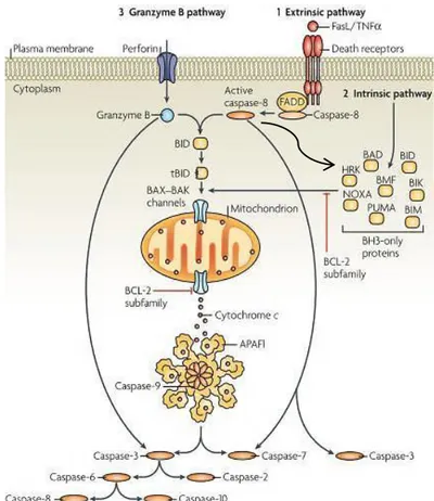
Figure 1.7 – Illustration of the different activation pathways of apoptosis. Extrinsic pathway (1) as well as intrinsic (2) and granzime B pathway (3) are depicted alongside with their main intervenients. Irrespective of
the actual route to caspase activation, all pathways lead to the activation of the major effector caspases, caspase-3, caspase-6 and caspase-7, and these carry out much of the proteolysis that is seen during apoptosis
(Adapted from Taylor et al., 2008). 14
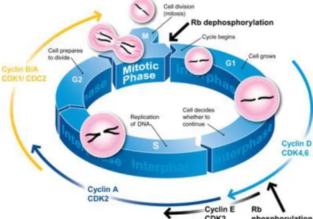
Figure 1.8 – Illustration of the regulation of the mammalian cell cycle. Key CDK/cyclin complexes role in the distinct phases of the cell cycle is also displayed. Cdk4/cyclin D complexes phosphorylate Rb in mid G1, while Cdk2/cyclin A and Cdk2/cyclin E phosphorylate Rb at the G1 to S transition. Dephosphorylation of Rb allows binding to E2F family of transcription factors, forming a silencing complex restriction the progression
of cell cycle, by transcription prevention of cell cycle control genes. Cdk2/cyclin A and Cdk1/cyclin B/A complexes and their kinase activity is essential for progression through S phase and entry in M phase
(Adapted from (―Propidium iodide staining of cells to assess DNA cell cycle,‖ 2014). 17
Figure 1.9 –(Left) Doxorubicin chemical structure and (right)doxorubicin’s DNA intercalation mechanism
through computer modeling tools (Adapted from Hannon, 2007). 25
Figure 3.1 –Dose dependent cytotoxicity of chlorogold compounds, B (left) and D (right), on non-small cell lung adenocarcinoma cell line (A549). The respective relative IC50 of each compound is displayed in the
upper right corner of each chart. The data are represented as means ± SEM of at least three independent experiments; *p < 0.05, as compared with the control group. Cell viability values were normalized in relation
xviii
Figure 3.2 –Dose dependent cytotoxicity of chlorogold compounds, B (left) and D (right), on non-small cell lung adenocarcinoma cell line (H1975). The respective relative IC50 of each compound is displayed in the
upper right corner of each chart. The data are represented as means ± SEM of at least three independent experiments; *p < 0.05, as compared with the control group. Cell viability values were normalized in relation
to the control group without compounds (only DMSO). 45
Figure 3.3 –Dose dependent cytotoxicity of chlorogold compounds, B (left) and D (right), on fibroblast cell line. The respective relative IC50 of each compound is displayed in the upper right corner of each chart. The
data are represented as means ± SEM of at least two independent experiments; *p < 0.05, as compared with the control group. Cell viability values were normalized in relation to the control group without compounds
(only DMSO). 46
Figure 3.4 – Cell viability in NSCLC A549 cell line in response to compound B and doxorubicin in combination therapy. Combination strategies’ (1º Dox_B; 1º B_Dox; B&Dox) effectiveness was compared to
single agent (SA) treatments (Dox_SA; B_SA). All strategies resorted to the IC50 values of each compound.
The data are represented as means ± SEM of at least three independent experiments; *p < 0.05, as compared with the control group. Cell viability values were normalized in relation to the control group without
compounds (only DMSO). 47
Figure 3.5 –Cell viability in NSCLC A549 cell line in response to erlotinib and compound B (Left) and erlotinib plus doxorubicin (Right) in combination therapy. Combination strategies’ (1º Erlo_B; 1º B_Erlo; Erlo&B; Erlo_Dox; Dox_Erlo; Erlo&Dox) effectiveness was compared to single agent (SA) treatments
(Dox_SA; Erlo_SA; B_SA). All strategies resorted to the IC50 values of each compound. The data are
represented as means ± SEM of at least three independent experiments; *p < 0.05, as compared with the control group. Cell viability values were normalized in relation to the control group without compounds (only
DMSO). 48
Figure 3.6 – NSCLC A549 cell line nuclear staining with Hoechst 33258 in response to 44.4 μM of compound B (B) and 30.0 μM of compound D (D) bringing to evidence nuclear morphological alterations
indicative of apoptosis. Qualitative results were compared with the respective solvent of the compounds: 0.1
% (v/v) DMSO (A;C). White arrows point out evidences of initial apoptosis hallmarks such as chromatin
condensation or aberrant nuclear morphology. White circles indicate nuclear fragmentation. 51
Figure 3.7 – NSCLC H1975 cell line nuclear staining with Hoechst 33258 in response to 3.36 μM of compound B (B) and 5.43 μM of compound D (D) bringing to evidence nuclear morphological alterations
indicative of apoptosis. Qualitative results were compared with the respective solvent of the compounds: 0.1 % (v/v) DMSO (A;C). White arrows point out evidences of initial apoptosis hallmarks such as chromatin
condensation or aberrant nuclear morphology. White circles indicate nuclear fragmentation. 52
Figure 3.8 – Proportion of viable, apoptotic and necrotic cells in NSCLC A549 cell line, when exposed to different concentrations of compound B and doxorubicin or when exposed to both in combination at their respective IC50 values. Cells treated with 0.1% (v/v) DMSO were used as control. Data was analysed by flow
cytometry after Annexin-V/fluorescein isothiocyanate (FITC) and propidium iodide (PI) double staining. The
xix
Figure 3.9 – Effect of compound B in single agent treatment and of compound B and doxorubicin in combination therapy on NSCLC A549 cell cycle. Cells were treated with 0.1% (v/v) DMSO as control of the
experiment or with 44.4 μM compound B and compound B & Dox at their respective IC50 during different
time points (3, 6, 9 h). DNA was stained with propidium iodide, and DNA content was analysed by flow
cytometry. The data are represented as means ± SEM of at least two independent experiments. 58
Figure 3.10 – Exposure effect of 200 ng of pUC18 plasmid DNA to either 0.62 % (v/v) DMSO or to increasing concentrations of compound B (0, 10, 30, 60 80, 100, 150, 200 μM). Resulting products of a 24 h exposure period at 37 ºC were submitted to electrophoresis in agarose gel 0.7 % (w/v) (upper panel). λ/HindIII – molecular weight marker; C – control with plasmidic DNA pUC18; DMSO – control with DMSO at 0.62 % (v/v) without compound; L – Linearized pUC18 with EcoRI. I – Supercoiled isoform; II – Nicked isoform (not showed); III – Linear isoform. pUC18 plasmidic DNA isoform distribution, illustrated in the bar
chart (lower panel) was obtained through software analysis tool GelAnalyzer. 62
Figure 3.11 – Exposure effect of 200 ng of pUC18 plasmid DNA to either 2.23 % (v/v) DMSO or to increasing concentrations of compound D (0, 60, 80, 100, 150, 200, 250, 300 μM). Resulting products of a 24
h exposure period at 37 ºC were submitted to electrophoresis in agarose gel 0.7 % (w/v) (upper panel). λ/HindIII – molecular weight marker; C – control with plasmidic DNA pUC18; DMSO – control with DMSO at 0.62 % (v/v) without compound; L – Linearized pUC18 with EcoRI. I – Supercoiled isoform; II – Nicked
isoform (not showed); III – Linear isoform. pUC18 plasmidic DNA isoform distribution, illustrated in the bar
chart (lower panel) was obtained through software analysis tool GelAnalyzer. 63
Figure 3.12 –Comparative proteome profiling of HCT-116 (left) and A549 (right) cell lines when subjected to 0.1 % (v/v) DMSO for an exposure period of 48 h. 2-DE gels were obtained from at least 200 μg of whole
protein extract and resulting spots were stained with Comassie Blue. Spots whose abundance variance levels
were considered significantly altered were marked and number tagged. 65
Figure 3.13 –Categorization of the proteome profiling of HCT-116 and A549 into functional categories such as, chaperone/stress response, metabolism cytoskeleton mobility, protein turnover/detoxification, signal transduction and other. Protein functional properties were based on STRING 9.1 database. The data is
xxi
Table Index
Table 1.1 – Main groups of chemotherapeutic compounds discriminating their main representatives and
modes of action (Adapted from Baba and Câtoi, 2007). 22
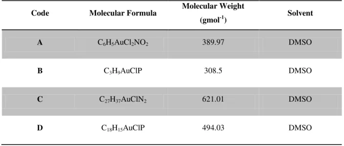
Table 2.1 – Molecular and structural characteristics of the studied compounds, and necessary information for
preparation of the respective stock solutions. 29
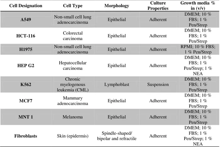
Table 2.2– Details and characteristics of human cell lines used in this work. Cell lines are discriminated by cell designation, type; morphology, culture properties and growth media. (DMEM - Dulbecco’s Modified Eagle Medium; FBS - Fetal Bovine Serum; NEA - Non-essential amino acids; Pen/Strep -
Penicilin-Streptomicin). 30
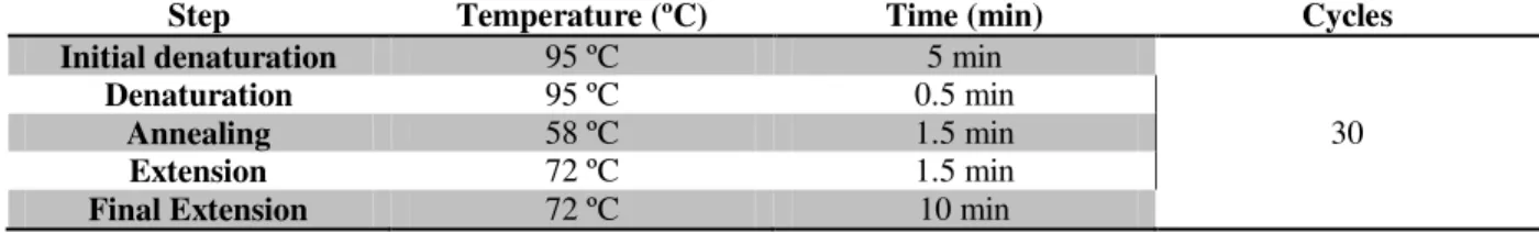
Table 2.3– Primer set used in Mycoplasma analysis, detailing forward and reverse primer sequences, as well
as the expected size of the specific amplicon for the 16S rRNA gene. 31
Table 2.4– PCR amplification program for the 16S rRNA gene in mycoplasma detection protocol. 32
Table 2.5– Ultrasonication protein extraction protocol. 39
Table 2.6– 2-D gel electrophoresis IEF five step program. 40
Table 3.1–IC50values for chlorogold compounds B and D, in non-small cell lung (A549; H197), human
colorectal (HCT116), hepatocellular (HepG2), breast (MCF-7) and melonotic (MNT-1) carcinoma cell lines
as well as on chronic myelogenous leukemia (K562) and human fibroblasts. 44
Table 3.2– Total percentage of viable, early apoptotic, late apoptotic and necrotic cells, in NSCLC A549 cell line, when exposed to different concentrations of compound B and doxorubicin or when exposed to both in combination at their respective IC50 values. Cells treated with 0.1% (v/v) DMSO were used as control. Data
was analysed by flow cytometry after Annexin-V/fluorescein isothiocyanate (FITC) and propidium iodide (PI) double staining. Data values are referent to those reported in Figure 3.8, and are represented as means ±
SEM of at least three independent experiments. 54
Table 3.3– Total percentage of NSCLC A549 cells at different stages of the cell cycle, when exposed to compound B and doxorubicin at their respective IC50 values either in single agent treatment or in combination
therapy. Data was analysed by flow cytometry after propidium iodide (PI) staining of DNA. Cells treated with 0.1% (v/v) DMSO were used as control. Data values are referent to those reported in Figure 3.9, and are
represented as means ± SEM of at least two independent experiments. 57
Table 3.4 – Proteome evaluation: Total number of proteins identified in 2-D gel electrophoresis, with the indication of the spot ID referent to figure 3.7, UniProt ID, protein identification, isoelectric point, molecular
weight, function and abundance variance levels between HCT-116 and A549 cell lines. Proteins whose abundance variance levels were considered significantly altered were highlighted. Values under 0.7 (red) and
xxiii
Abbreviation List
Abs Absorbance
Abs260/Abs230 Ratio between 260 nm and 230 nm absorbance Abs260/Abs280 Ratio between 260 nm and 280 nm absorbance Apaf-1 Apoptosis protease activating factor-1
APS Ammonium Persulfate
ATP Adenosine-5'-triphosphate
BAX Encoding gene for pro-apoptotic protein Bax, of the protein family Bcl-2
Bax Bcl-2-associated X protein
BCL-2 Encoding gene for pro-apoptotic protein Bcl-2, of the protein family Bcl-2 Bcl-2 B-cell lymphoma protein 2
Bid BH3 interacting domain death agonist
Bis N,N'-Methylenebisacrylamide
BRCA1 Breast cancer 1 susceptibility gene BRCA2 Breast cancer 2 susceptibility gene
CASP3 Encoding gene for Cysteine-aspartic protease 3 Caspase Cysteine-aspartic protease
Cdk Cyclin-dependent kinases
cDNA Complementar DNA
CHAPS (3-[(3-Cholamidopropyl)dimethylammonio]-1 propanesulfonate)
CT-DNA Calf Thymus-DNA
DISC Death inducing signaling complex
DMEM Dulbecco’s Modified Eagle Medium
DMSO Dimethyl Sulfoxide
Dnase Desoxirribonuclease
DOX Doxorrubicin
DTT Dithiothreitol
E2F E2 transcription factor
EGF Epidermal growth factor
EMSA Electrophoretic Mobility Shift Assay
FADD Fas-associated death domain
FasL Fatty acid synthetase ligand
FBS Fetal Bovine Serum
FITC Fluorescein Isothiocyanate
HCT116 Colorectal carcinoma cell line HepG2 Hepatocellular carcinoma cell line
HER Human epidermal growth factor receptor-2 HER2 Codifying gene for membrane receptor HER
xxiv IC50 50 % growth inhibition concentration
IFNγ Interferon gamma
IKKβ Serine/threonine protein kinase that phosphorylates the I-kappa-B protein JNK1 Protein kinase of the MAPK family
Kb Affinity binding constant
MTS 3-(4,5-dimetiltiazol-2-il)-5-(3-carboximetoxifenil)-2-(4-sulfofenil)-2H-tetrazólio
PBS Phosphate Buffered Saline
PI Propidium Iodide
PMS Phenazine Methosulphate
PMSF Phenylmethylsulfonyl fluride
pUC18 Plasmid DNA
Rb Retinoblastoma tumour suppressor protein
RIPK Receptor interacting protein kinases
ROS Reactive Oxygen Species
RPMI-1640 Rosswell Park memorial institute medium 1640
SDS Sodium dodecyl sulfate
SDS-PAGE Sodium dodecyl sulfate - Polyacrylamide gel electrophoresis
SGTA Small glutamine-rich tetratricopeptide repeat-containing protein alpha SMAC/DIABLO Second mitochondria-derived activator of caspase/Direct IAP-binding protein
with low pI
TAE Tris base, acetic acid and EDTA buffer
TEMED Tetramethylethylenediamine
TNF Tumour necrosis factor
TNFR1 Tumour necrosis factor receptor 1
TOPO II Topoisomerase II
TP53 p53 protein encoding gene
TRADD TNF receptor-associated death domain
TrxR Thioredoxin reductase
Tris-HCl Tris-Hidroclorite
xxv
Units List
% (w/v) Weight/volume percentage % (v/v) Volume/volume percentage A; mA Amperes; miliamperes
AU Absorbance units
bp Base pairs
°C Celsius degrees
H; min; s Hours; minutes; seconds kDa; Da KiloDalton; Dalton (10-3kg)
Kg; g; mg; μg; ng Quilogramas; grama (10-3kg); miligrama (10-6 kg); micrograma (10-9 kg); ng
– nanograma (10-12 kg)
L; mL; μL Liter; mililiter(10-3L); μL – microliter (10-6 L)
m; cm; mm; nm Meter; centimeter (10-2 m); milimeter (10-3 m); nanometer (10-9 m) M; mM; μM Molar (mol/L); milimolar (10-3 M); micromolar (10-6 M)
mol; pmol Mole; picomole
rpm Rotations per minute
U Unit; mU – miliunit
V Volts
W Watts
Symbol List
[Complex] Complex concentration
[DNA] DNA concentration
Σ Summation
ε Molar extinction coefficient
εa Apparent molar extinction coefficient
εb Molar extinction coefficient when bound to DNA
εf Molar extinction coefficient when unbound
λ Wavelength
1
1.
Introduction
1.1. Cancer Etiology
At any given moment an adult human is composed of approximately 1015 cells endowed with grand versatility and plasticity, paramount for different morphogenesis mechanisms and maintenance of adult tissues through cell turnover processes (Bertram, 2001). These numerous processes are usually tightly regulated by a network of overlapping molecular mechanisms which govern, among other factors, both cell proliferation and programmed cell death in a perfect balance. At the same time the genomic sequences’ susceptibility to corruption, either by intrinsic or extrinsic factors may tip this intricate homeostatic balance, leading to changes that are incompatible with an organismic structure. In other words cells that undergo this process, typically become unresponsive to normal cell signaling factors, and ―behave‖ in a organismic-independent fashion, often leading to altered cell proliferation programs and consequently to large populations of renegade cells that ultimately lead to cancer formation (Bertram, 2001; Weinberg, 2013).
An important concept to grasp when considering cancer is that it is generally accepted as a genetic disease or as a group of genetic diseases, although not always heritable. Even though hereditary cancer predisposition syndromes have become more and more easily identified and studied, only 5 to 10 % of all cancer cases can actually be attributed to such genetic defects (Garber and Offit, 2005; Anand et
al., 2008). In fact since the review by Doll and Peto in 1981 (Doll and Peto, 1981) it was concluded that the main causes of cancer are mainly related to environmental and lifestyle factors, accounting for the remaining 90 to 95 % of incidence cases (Anand et al., 2008).
1.1.1.
From Carcinogens and Lifestyle Habits to Genetic Influence
2 and meta-analysis studies, to increase to variable extent, the risk of contracting colorectal, breast and/ or prostate cancer (World Cancer Report, 2008). Furthermore the relationship between diet and cancer becomes more obvious when observing incidence rates among populations in migratory studies (McCracken et al., 2007). For instance Asians have been shown to have 25 times lower incidence of prostate cancer and 10 times lower incidence of breast cancer (Anand et al., 2008) however the rates of these types of cancer tend to increase in migrating Asian-American populations indicating that they have been subjected to a more Western lifestyle, becoming simultaneously more prone to the same risk factors associated for these specific cancers (McCracken et al., 2007). Smoking and related mutagens on the other hand, can almost be considered as particular class due to its importance and influence on the tinkering of specific molecular networks and mechanisms and consequently on the emergence of cancer. In particular cigarette smoke is known to contain approximately 60 mutagens (Takahashi et al., 2011) ranging from polycyclic aromatic hydrocarbons to specific nitrosamines. Exposure to such mutagens to airway epithelial cells through continuous smoking leads to severe molecular lesions and diminished repair capability, being accountable as the major cause of cancer in humans, and correlated to 13 different types such as lung, oral cavity, nasal cavity and nasal sinuses, pharynx, larynx, oesophagus, stomach, pancreas, liver, urinary bladder, kidney, uterine cervix and myeloid leukaemia (World Cancer Report, 2008). Benzopyrenediol epoxide is one example of a cigarette smoke metabolite that as actually been etiologically linked to lung cancer, which is known to form adducts on guanine residues with a particular incidence on the tumour suppressor gene TP53 (Lemjabbar et al., 2003). Similarly DNA adduction and mutagenesis were also reported for aromatic amines derived from cigarette smoking (Besaratinia and Tommasi, 2013). Recent studies have also connected the capacity of tobacco smoke to trigger chronic inflammation, through IKKβ and JNK1 pathway activation (Takahashi et al., 2011), which has been extensively demonstrated to play decisive roles at different stages of tumour development (Grivennikov et al., 2010).
3 On the other hand DNA replication itself can be considered as a source of mutational events. DNA replication enzymes inherent mutation rate is as little as 1 in 109 (Loeb et al., 1974) however error prone phenotypes in DNA replication enzymes or in translesion DNA inherently increase this error rate, leading to genomic instability and to cancer susceptibility (Suzuki and Takahashi, 2013). Genetic disturbances can range from chromosome alterations to point mutations which ultimately can disable tumour suppressor genes or activate proto-oncogenes that respectively can alter cell proliferation and survival pathways (Suzuki and Takahashi, 2013).
1.2. Incidence and Mortality Rates
Cancer burden worldwide is known to be increasing over the years, in part as a reflection of westernized habits, but also as consequence of the continuous growth and aging of the world’s population (Jemal et al., 2011). Accordingly to the most recent data from the World Health Organization (WHO), in 2012 alone it was estimated 14.1 million new cancer cases, alongside with 8.2 million related deaths, turning cancer into a major leading cause of death worldwide (―World cancer factsheet,‖ 2014). Of particular importance 44 % of all cancer cases and 53 % of related deaths occurred in low or medium developing countries (Rise, 2013). Globally, the most commonly diagnosed cancers were those of the lung (1.8 million, 13.0 % of the total), breast (1.7 million, 11.9 %), and colorectal (1.4 million, 9.7 %) whereas the most common causes of cancer death were cancers of the lung (1.6 million, 19.4 % of the total), liver (0.8 million, 9.1 %), and stomach (0.7 million, 8.8 %) (Figure 1.1) (―GLOBOCAN,‖ 2012).
Figure 1.1 – Incidence and mortality rates worldwide for most common cancers (Adapted from ―GLOBOCAN,‖ 2012). The close relationship evidenced between incidence and mortality rates for some types of cancer such as lung, colorectal and stomach, allows not only to infer about social and cultural habits, as well as draws attention to a more pressing issue that is the inefficiency of current therapeutic strategies applied for these same cancers. In the end with current trends it is predicted that globally, cancer burden will increase significantly, up to 19.3 million new cancer cases per year by 2025
(―GLOBOCAN,‖ 2012; ―World cancer factsheet,‖ 2014) stressing out ever more the importance of a
4 and of the establishment of priorities and control strategies to fray down the overwhelming spread of cancer globally.
1.2.1. Lung Cancer Incidence
In 2012 1.8 million new cases of lung cancer were estimated (―GLOBOCAN,‖ 2012), compared with 1.6 million in 2008 and is considered the most common cause of death in developing and developed countries (Jemal et al., 2011). Notably in Europe and in Eastern Asia the incidence rates for men surpasses the 50 cases per 100000 persons while in women these rates are slightly lower due to historic variants. The highest estimate rates for women are evidenced for North America and Northern Europe with values of 32.8 and 23.7 cases per 100000 persons respectively. In contrast low developing countries in the Western and Middle Africa area report the lowest incidence rates in an order of magnitude of 0.8 to 1.1 cases per 100000 persons (―GLOBOCAN,‖ 2012).
Figure 1.2 – Lung and bronchus cancer incidence and mortality rates in the United States (―Cancer of the Lung and Bronchus - SEER Stat Fact Sheets,‖ 2010).
Despite these statistical numbers the fact is that the incidence and mortality rates for lung cancer is actually decreasing for the majority of western countries including many European countries (Jemal et
5 Stat Fact Sheets,‖ 2010), emphasizing the continuous need for better control programs and particularly treatment strategies.
1.3. Cancer Biology: Cellular and Molecular Basis
1.3.1. Genomic instability and other Contributing Factors for Neoplasic
Transformation
The occurrence of aberrational events within the genome, either of temporary or permanent nature, commonly referred to as genomic instability in the literature, are often associated and characteristic of most cancers (Negrini et al., 2010). Both numerical aberrations and structural rearrangements of chromosomes or even the accelerated rate of chromosomal alterations, which result in gains or losses of whole chromosomes as well as inversions, deletions, duplications and translocations of large chromosomal segments, are the most frequent form of genomic instability. Although expansion and contraction of the number of oligonucleotides repeats in microsatellite sequences have also been described (Negrini et al., 2010; Gordon et al., 2012).
Even though the genomic instability in hereditary cancers is well understood, and strongly linked to mutations in DNA repair genes such as those involved in the nucleotide and base excision repair processes (BRAC1, BRAC2, MSH2, MYH), the molecular basis is not so clear for sporadic cancers. Recent reports have however shed some light over this subject mentioning that mutations in caretaker genes (like DNA repair genes) are infrequent in early cancer development (69 to 97 % of cancers did not present mutations in caretaker genes) and propose oncogene-induced DNA replication stress as the most likely responsible for the processes involved in genomic instability for these cancers (Negrini et
6
Figure 1.3 – p53 as a barrier against tumour development. Oncogene activation or deregulated cell cycle progression leads to stalled DNA replication forks and activation of the DNA damage response (DDR) (Adapted from Farnebo, et al., 2010)
In addition a recent cohort study involving 3281 tumours across 12 tumour types as part of the Cancer Genome Atlas effort, has demonstrated that TP53 is in fact the most frequently mutated gene (Kandoth
et al., 2013). Other reports have actually linked TP53 mutations to over 50 % of all human cancers including lung carcinoma where it represents an extremely common event (Vincenzi et al., 2006; Farnebo et al., 2010; Wong, 2011). Furthermore PIK3CA was found to be second most frequently mutated gene representing a frequency of over 10 % of most cancer types (Kandoth et al., 2013), and the remaining frequently deregulated genes mainly encoded classical oncoproteins like epidermal growth factor receptor (EGFR), KRAS and NRAS family, phosphatase and tensin homologue (PTEN) (Negrini et al., 2010; Kandoth et al., 2013).
Apart from the genetic mechanisms described above, epigenetic studies are gaining increasing relevance as more and more evidences point out a more significant role for DNA alterations and associated chromatin changes in gene expression and in tumourigenesis (Waldmann and Schneider, 2013). Hallmarks of epigenetic changes like DNA methylation and post-translational modifications of histones may contribute to oncogenesis through transcriptional silencing of tumour suppressor genes (Waldmann and Schneider, 2013). Global hypomethylation, possibly the first epigenetic modification to actually be linked with tumourigenesis is commonly observed in malignant cells being associated with chromosomal instability in colorectal cancer (Berman et al., 2012). However methylation changes occurring at CpG islands equally play an important role in modelling transcriptional responses. Hypermethylation frequency of such regions was found to be often high. In fact typically unmethylated CpG islands in promoter genes, around 5 to 10 %, have actually been confirmed to be methylated in various cancer genomes (Dawson and Kouzarides, 2012; Waldmann and Schneider, 2013; Esteller et al., 2001), but the relevance of this alterations is still to be deciphered.
7 to be linked to some specific cancers such as gliomas. Nevertheless all in all, the consequences of such mutations remain to be unravelled and further studied (Waldmann and Schneider, 2013).
Additionally specific mutations in numerous epigenetic gene regulators have also been found in several cancers which in part might explain differences in methylation patterns during oncogenesis. For example histone-lysineN-methyltransferase gene, MLL2, has been recurrently found mutated in several cancers (Kandoth et al., 2013), but in particular high incidence in follicular lymphoma. Plus
UTX, a histone demethylase has been found to be mutated in up to 12 histological distinct cancers (Dawson and Kouzarides, 2012). Mutations were still found in other chromatin remodelling genes such as MLL3, MLL4 for bladder, lung and endometrial cancers, and demethylase gene KDM5C in kidney carcinoma (Kandoth et al., 2013). By understanding the mechanisms involved in epigenetic control and their associated mutations, the foundations are laid for the production of new drugs and treatments to override such alterations that may cause cancer and many other diseases.
Other factors such as proteotoxic, metabolic and oxidative stress have reportedly influence the process of neoplasic transformation and tumourigenesis. In regards to metabolic stress, for instance, it is known that tumoural cells produce ATP through glycolysis which in turn grants tumoural cells an adaption to hypoxia conditions, and allows for the acidification of the surrounding environment. In turn it facilitates tumour invasion and immune suppression. More detailed information is presented elsewhere (Luo et al., 2010).
1.3.1.1 p53 Tumour Suppressor
8
Figure 1.4 – p53 intricate circuit board of biological processes and their respective regulation, with discriminated transcriptional and cytoplasmic roles (Adapted from Vousden and Prives, 2009).
In turn it is the ―impairment‖ of the auto-regulatory loop between Mdm2 and p53 (which regulates the overall stability and activity of p53 through post-translational modifications) through these cellular stress inputs that lead to the accumulation of p53 (Alarcon-Vargas and Ronai, 2002). This then triggers the activation of p53 target genes, such as p21, GADD45, Bax, Puma, Noxa, DR5, p53AIP, PIDD, and others, triggering apoptosis, autophagy, cell cycle arrest or senescence (Farnebo et al., 2010; Alarcon-Vargas and Ronai, 2002). It is then easy to understand that activation of such pathways by p53, in normal conditions, leads to tumour suppression (Figure1.4).
For instance cell cycle arrest can be achieved through p53 mediated activation of the cyclin-dependent kinase inhibitor p21, alongside with target genes such as 14-3-3 sigma and GADD45 (Vousden and Prives, 2009). Particularly p21 inhibits cell cycle progression mainly through the inhibition of CDK2 and CDK1, which are involved in a myriad of phosphorylation cascades (such as the phosphorylation of RB; key for cell cycle entry), leading to a temporary growth arrest in G1/S stage of the cell cycle. Plus p21 binding to proliferating cell nuclear antigen hinders DNA polymerase activity, inhibiting replication and allowing for the activation of DDR processes (Manuscript, 2010). Together these processes concede time for DNA repair.
9
Figure 1.5 – Schematics illustrating the multistage facet of carcinogenesis. Premalignant cells are the result of an initiating event such as a compromised tissue repair response, conferring an enhanced survival. Sequentially early tumour nodules would be the result of the accumulation of successive mutational events, like immune evasion or epigenetic modifications. In the end angiogenesis factors and loss of cell adherence would lead to metastasis and to the formation of an advanced tumour (Adapted from (Grivennikov et al., 2010).
or Bcl-xL family can either lead the promotion or inactivation of pro-apoptotic Bcl-2 multi-domain proteins and consequently to cell death or cell survival (Chipuk and Green, 2006).
In short p53 specific DNA binding and transcriptional regulation of target genes can be hindered by a myriad of mutations which in turn compromise the ability of a controlled response to tumour suppression, and the wide number of other biological processes in which it is involved. Plus contrary to general believe, recent reports state that some of the p53 mutations, besides abrogating tumour suppression and general transcriptional regulation circuit board, actually endow the protein a set of characteristics that have been found to be fundamental for oncogenesis (Oren and Rotter, 2010).
1.3.2.
Mechanisms of Carcinogenesis: A
Multi-step Process
10 Typically the process by which mutations occur is complex and sequential. At least 4 to 7 independent events must take place in order for a cell to be considered as tumourigenic (Ashkenazi et al., 2008; Bertram, 2001). The complexity that carcinogenesis represents, led to the creation of models that try to explain the progression of mutations that are known to take part during this process (Ashkenazi et al., 2008; Tomasetti et al., 2013). The most renowned, probably for its simplicity, models carcinogenesis as multistep process of mutation acquisition, considering a homogenous cell population, in what is known as the ―initiation, promotion and progression‖ model (Figure1.5) (Ashkenazi et al., 2008; Jakóbisiak et al., 2003). In this model a tumour is initiated by a driver mutation in a gene typically involved in the control of cell proliferation, evasion of apoptosis and/or genomic integrity (oncogenic pathways), endowing the cell with superior proliferation characteristics, or any other survival advantage over the remaining cell population. With each successive clonal expansion of such cells, these become susceptible to additional mutational events that are accumulated over time and further contribute for an enhanced survival. Eventually a third event would promote an irreversible process by which the malignant potential of the cells would be enhanced, leading to tissue invasion and metastasis. Events such as increased genomic instability (which is a characteristic of all developed cancers), cytokine and chemokine modulation of tumour cells as result of inflammation, angiogenesis induction, and protease production to promote loss of cell adherence and cell detachment, all contribute for the development of a malignant tumour (Jakóbisiak et al., 2003; Tomasetti et al., 2013; Grivennikov et al., 2010).
Overall, independently of the manifold mutational combinations acquired during the multistep development of carcinogenesis, these will ultimately result in a common set of characteristics or hallmarks that dictates and characterizes the malignant phenotype (Pietras and Ostman, 2010; Hanahan and Weinberg, 2011). These include sustained proliferative signalling, insensitivity to anti-growth signals, evading apoptosis, limitless replicative potential, sustained angiogenesis and tissue invasion and metastasis (Figure 1.6)(Pietras and Ostman, 2010; Hanahan and Weinberg, 2011).
11
Figure 1.6 – Illustration of the six hallmarks that characterize and influence the process of carcinogenesis (Adapted from Hanahan and Weinberg, 2011).
12 several regulators, positive and negative ones, such as vascular endothelial growth factor (VEGF) or platelet-derived growth factor (PDGF) to name a few. For instance VEGF expression can be upregulated by stimulating factors such as hypoxia or oncogene activation (Hanahan and Weinberg, 2011; Bergers and Benjamin, 2003). Also worth mentioning is that the rapid genesis of new blood vessels associated with chronic exposure to angiogenic activators leads to an abnormal overall vasculature different from their normal counterparts sharing different characteristics of several types of blood vessels (Bergers and Benjamin, 2003). Finally all cancers will ultimately engage in tissue invasion and the formation of metastasis. Namely loss of cell adhesion between the cells of the tumour mass allows their disaggregation contributing to an enhanced malignant phenotype, characterized by massive tissue invasion and multiple metastasis formation. Abrogation of cadherin/catenin expression levels, in particular E-cadherin has been highly associated with malignant phenotypes and micro-metastases. Additionally these cells also typically present enhanced cell motility, further assisting the invasion phenotype, through the expression of adhesion molecules like N-chaderin associated with cell migration processes. Also tumour cells are known to produce a vast range of proteases, such as cathepsins that allow the detachment from broad range of extracellular matrix/basement membrane components, including proteoglycans and collagens (Brooks et al., 2010; Hanahan and Weinberg, 2011).
Other emerging hallmarks include the deregulation of cellular energetics, as already mentioned above were cancer cells redirect their energy production to glycolysis, and the evasion from the hosts’ cellular and molecular immune mechanisms that are set to destroy cancerous cells such as natural killer cells (Hanahan and Weinberg, 2011).
1.3.3. Mechanisms of Cell Death: Apoptosis, Autophagy & Necrosis
Ever since the pioneering work in Caenorhabditis elegans revealed the genetic programme of apoptosis, cell death in the broader sense of this term, has been seen as a fundamental process often governed by an intricate array of regulators and signalling pathways, with an active role in overall development and in the maintenance of cellular homeostasis (Elmore, 2007; Vanden Berghe et al., 2014). Until recently apoptosis and necrosis were considered to be the only forms of cell death, with apoptosis being seen as the fundamentally orchestrated cellular process in physiological and pathological conditions (Wong, 2011), and necrosis as the result of physicochemical abuse leading to an uncontrolled swelling, organelle dysfunction and cell lysis (Vanden Berghe et al., 2014; Ouyang et
13 formation of double-membrane vesicles called autophagosomes, responsible for the turnover of large portions of cytoplasm including several organelles and other specific macromolecules (Mariño et al., 2014; Ouyang et al., 2012). Plus it is considered as general protective response system that it is triggered under certain environmental stress such nutrient shortages (Mariño et al., 2014). Over the years attempts in discovering the underlying mechanisms related to these forms of cell death have simultaneously led to the uncovering of many other, such as necroptosis, parthanatos, oxytosis, ferroptosis, ETosis, NETosis, pyronecrosis or pyroptosis (Galluzzi et al., 2012; Vanden Berghe et al., 2014). Were some opinions diverge, others attempt to unify perspectives over cell death and recently some authors have proposed that these several forms of cell death are merely different forms of regulated necrosis (Vanden Berghe et al., 2014). Even though necrosis has been generally considered an uncontrolled process resultant from physical damage of some sort, the discovery of key regulators of necrotic death, such as receptor interacting protein kinases (RIPK1 and/or RIPK3) led to the establishment of necrosis as a possible genetically controlled cell death mode (Ouyang et al., 2012; Vanden Berghe et al., 2014). This regulated necrosis is known to be independent of caspase activation, and to lead to a set of biological processes such as, mitochondrial dysfunction, increased cell volume (oncosis), production of ROS, and ultimately lysis. The immunogenic nature of necrosis resultant from the extravasation of their inner components (pro-inflammatory molecules) to the extracellular environment further lead to inflammatory responses that contribute for tumourigenesis (Grivennikov
et al., 2010).
Understanding the underlying mechanisms of these modes of cell death is crucial as it will lay the groundwork and study platforms for different chemotherapy strategies, while simultaneously allowing for a better knowledge of the consequences of such strategies.
1.3.3.1. Apoptosis
14 since there isn’t an extravasation of the intracellular content to the surrounding tissues (Taylor et al., 2008; Orrenius et al., 2011).
The entire process that comprises apoptosis is vast and complex; hence the major biochemical pathways and regulators will only be discussed here briefly. The extensive research upon apoptosis has led to the identification of three major apoptotic pathways: the extrinsic or death receptor pathway, the intrinsic or mitochondrial pathway and the perforin-granzyme-dependent pathway (Figure 1.7) (Elmore, 2007; Taylor et al., 2008). The perforin-granzyme-dependent pathway that will be addressed in less detail is best described as to involve the cytotoxicity of T-cells which induce apoptosis either by granzyme B or granzyme A.
Figure 1.7 – Illustration of the different activation pathways of apoptosis. Extrinsic pathway (1) as well as intrinsic (2) and granzime B pathway (3) are depicted alongside with their main intervenients. Irrespective of the actual route to caspase
activation, all pathways lead to the activation of the major effector caspases, caspase-3, caspase-6 and caspase-7, and these carry out much of the proteolysis that is seen during apoptosis (Adapted from Taylor et al., 2008).
15 morphological alterations already mentioned above (Elmore, 2007). A noteworthy concept to keep in mind is that once activated, cells seemingly become committed to a programmed cell death. At the risk of being over simplistic the extrinsic pathway, as the name suggests, is activated by the binding of extracellular death ligands, such as FasL (fatty acid synthetase ligand) or tumour necrosis factor-α (TNFα), to their respective transmembrane death receptors (Wong, 2011). The family of transmembrane death receptors is vast including more than 20 proteins with a broad range of biological functions (Ouyang et al., 2012; Schulze-bergkamen and Krammer, 2004), however the best described and more extensively studied are type 1 TNF receptor (TNFR1) and a related protein Fas (CD95- FasL receptor) (Schulze-bergkamen and Krammer, 2004; Wong, 2011). Upon ligation to their cognate ligands, receptor trimerization occurs as well as clustering of death domains and the recruitment of adaptor proteins such as Fas-associated death domain (FADD) or TNFR1-associated death domain (TRADD) which in turn form a protein complex known as the death-inducing signalling complex (DISC) (Fulda and Debatin, 2006; Wong, 2011). Once formed this protein complex is responsible for the oligomerisation of pro-caspase-8 leading to its activation through self-cleavage. In turn caspase-8 can proteolytically activate downstream caspase effectors such as caspase-3 and caspase-7, or in other situations it can promote the activation of BH3-only protein BID (BH3- interacting domain death agonist) through crosstalk with the intrinsic pathway. After proteolytic activation by caspase-8, the truncated BID induces mitochondrial outer membrane permeabilisation (MOMP) by the formation of transmembrane BAX-BAK channels (BAX-Bcl-2-associated X protein), consequently causing the release of cytochrome c and assembly of the apoptosome (Figure1.7). The last one is multi-protein complex, comprised by 7 molecules of apoptotic protease-activating factor-1 (Apaf-1), caspase-9 and cytochrome c that binds to the C-terminal region of Apaf-1. Together this complex is responsible for further amplifying the apoptotic signal in the intrinsic pathway (Taylor et
al., 2008; Fulda and Debatin, 2006).
16 cytochrome c, second mitochondria-derived activator of caspase/directed IAP (inhibitory of apoptosis proteins) binding protein with low pI (Smac/ DIABLO), and apoptosis-inducing factor (AIF). Among these cytochrome c plays perhaps the most important part, as it is a key intervenient in the assembly of the oligomerization protein platform that is the apoptosome, which is responsible, as mentioned above, for the autoproteolytic activation of caspase-9. The other released proteins have a variety of roles ranging from sequestering caspase inhibitors to the induction of morphological alterations to the nuclear envelope (Du et al., 2000; Schulze-bergkamen and Krammer, 2004).
During the execution of the apoptosis process the activation of caspase effectors, like caspase-3 -6 and -7, regardless of the activation pathway, leads to the fracture of key architectural cellular structures and biochemical pathways. Namely the cytoskeleton scaffolding that ensures the 3-dimensional structure of the cell is one of the first key structures to be targeted during the execution phase of apoptosis. Molecules such as actin itself, and actin-associated proteins, like myosin, spectrins, α -actinin, gelsolin and filamin are proteolytically destroyed causing the typical morphological hallmarks of apoptosis, like the acquisition of a round shape, loss of cell adherence and membrane blebbing. Plus, chromatin condensation is thought to be the result of nuclear lamin targeting by caspases (Schulze-bergkamen and Krammer, 2004; Taylor et al., 2008; Wyllie, 2010). In short the connections established between the myriad of regulators and pathways are the mere strings of the puppeteer that is the programmed cell death by apoptosis and the considerable biochemical changes that occur within the apoptotic cell.
1.3.4. Cell Division and Carcinogenesis Interplay
17 As any other highly regulated cellular process, cell division and the overall regulation (see below) is prone to potential alterations and aberrant phenomena as a result of mutations. Misregulation of cell cycle events and the control of the commitment to cell division have been widely shown in the literature to play an important role in cancer onset. Compromised cell cycle regulation processes, as those involving the core components of the cell cycle clock, namely cyclins and cyclin dependent kinases (CDK), affect the proper distribution of chromosomes at the metaphase to anaphase transition by the spindle-assembly checkpoint leading to overall genetic instability, aneuploidies and unscheduled proliferation (Malumbres and Barbacid, 2001; Malumbres and Barbacid, 2009).
1.3.4.1. Cell Cycle Regulation and Deregulation in Cancer
The cell cycle is comprised of 4 distinct phases (G1, G2, S, and M) all of which are controlled by numerous mechanisms to ensure a correct cell division (Figure 1.8). More specifically the cell cycle is responsible for faithfully replicate DNA during S phase, and to ensure the equal segregation of all cellular components to the daughter cells including the identical chromosomal copies during the M phase. The remaining two phases, namely G1 and G2 are intermediate periods mainly responsible for an high synthesis of macromolecules and other regulators essential for cell cycle progression through S and M phases respectively (Malumbres and Barbacid, 2001; Sherr, 1996; Lapenna and Giordano, 2009).
Figure 1.8 – Illustration of the regulation of the mammalian cell cycle. Key CDK/cyclin complexes role in the distinct phases of the cell cycle is also displayed. Cdk4/cyclin D complexes phosphorylate Rb in mid G1, while Cdk2/cyclin A and Cdk2/cyclin E phosphorylate Rb at the G1 to S transition. Dephosphorylation of Rb allows binding to E2F family of
18 Apart from the already mentioned phases there is another critical moment in the cell cycle known as G0, a quiescent state where most of the non-proliferating cells stand at. Furthermore progression through the cell cycle only occurs upon a G0―G1 transition in response to stimuli such as mitogen exposure and nutrient abundance (Lea et al., 2003; Schwartz and Shah, 2005). This transition is highly regulated and it is thought to be one of the several developed checkpoints that prevent an awry cell progression (Malumbres and Barbacid, 2001). In fact the transition between cell cycle phases is regulated by several different proteins, nonetheless the key regulatory proteins are cyclin-dependent kinases (CDK), the activity of which is modulated by fluctuating levels of cyclins throughout the progression of the cell cycle (Vermeulen et al., 2003; Schwartz and Shah, 2005). Mitogenic signalling leads to activation of CDK/cyclins complexes, through phosphorylation cascades such as MAPK/ERK pathway (Schwartz and Shah, 2005). In particular G0―G1 transition is characterized by
19 The activity of many cell cycle CDK and cyclins is often deregulated in many different types of cancer as a result from genetic and epigenetic alterations in their genes or regulators, making them a tempting target when it comes to cancer therapy. For instance CDK4 overexpression has been found in several cancer types including melanoma, sarcoma and gliomas, while cyclin D1 gene translocation is often associated with B-cell malignancies (Vermeulen et al., 2003). The recognition that the use of cell cycle intervenients as cancer targets, in particular CDKs by CDK inhibitors, could induce apoptosis, introduced new hope and a new possible fronts for battling cancer.
1.4. Principles of Cancer Therapy
20 proteins that are overexpressed in cancer cells (Doody et al., 2007; Alley et al., 2010). Endocrine treatment in turn deals with cancer prevention by using counteracting hormone stimulated tumours with antagonizing agents (Luqmani, 2005). With exception for chemotherapy, which will be addressed latter, these treatment programs are beyond the scope of this project and won’t be further addressed, although extensive literature reports present more detailed information on the subject (Baba and Câtoi, 2007; World Cancer Report, 2008).
21 All in all the stated benefits and the overall features of such alternative therapies’ programs, yet to be fully understood and characterized, must still be weighed in in comparison with the classical treatment strategies of surgery, radiotherapy and chemotherapy. In this sense chemotherapy, in spite of all the recent advances, is still considered as the gold standard when it comes to clinically employed cancer therapy (Weinberg, 2013; Chabner and Jr, 2005).