Cell fate potentials and switching kinetics
uncovered in a classic bistable genetic switch
Xiaona Fang
1,2,3,4
, Qiong Liu
1
, Christopher Bohrer
2
, Zach Hensel
2,5
, Wei Han
3
, Jin Wang
1,3,4
& Jie Xiao
2
Bistable switches are common gene regulatory motifs directing two mutually exclusive cell
fates. Theoretical studies suggest that bistable switches are sufficient to encode more than
two cell fates without rewiring the circuitry due to the non-equilibrium, heterogeneous
cel-lular environment. However, such a scenario has not been experimentally observed. Here by
developing a new, dual single-molecule gene-expression reporting system, we
find that for
the two mutually repressing transcription factors CI and Cro in the classic bistable
bacter-iophage
λ switch, there exist two new production states, in which neither CI nor Cro is
produced, or both CI and Cro are produced. We construct the corresponding potential
landscape and map the transition kinetics among the four production states. These
findings
uncover cell fate potentials beyond the classical picture of bistable switches, and open a new
window to explore the genetic and environmental origins of the cell fate decision-making
process in gene regulatory networks.
DOI: 10.1038/s41467-018-05071-1
OPEN
1State Key Laboratory of Electroanalytical Chemistry, Changchun Institute of Applied Chemistry, Changchun 130022, China.2Department of Biophysics and Biophysical Chemistry, Johns Hopkins School of Medicine, Baltimore, MD 21205, USA.3College of Physics, Jilin University, Changchun 130012, China. 4Department of Chemistry and Physics, Stony Brook University, Stony Brook, NY 11790, USA.5Present address: Instituto de Tecnologia Química e Biológica António Xavier, Universidade Nova de Lisboa, Av. da República, 2780-157 Oeiras, Portugal. Correspondence and requests for materials should be addressed to J.W. (email:jin.wang.1@stonybrook.edu) or to J.X. (email:xiao@jhmi.edu)
123456789
C
ell fate decision-making is the process of a cell committing
to a differentiated state in growth and development. The
decision is often carried out by a select set of transcription
factors (TFs), the expression and regulatory actions of which
establish differentiated programs of gene expression
1. Bistable
switches, which consist of two mutually repressing TFs, are the
most common gene regulatory motifs directing two mutually
exclusive gene expression states, and consequently distinct cell
fates
2–12. Theoretical studies suggest that the simple circuitry of
bistable switches is sufficient to encode more than two cell fates
due to the non-equilibrium, heterogeneous cellular environment,
allowing a high degree of adaptation and differentiation
13–16.
However, new cell fates arising from a classic bistable switch
without rewiring the circuitry have not been experimentally
observed
17–19.
Here, by developing a dual single-molecule gene-expression
reporting system, we demonstrate experimentally the emergence
of two new expression states in the model bistable switch of the
bacteriophage
λ
20. We construct the corresponding potential
landscape and map the transition kinetics between the four
production states, providing insight into possible state-switching
rates and paths. These
findings uncover cell fate potentials
beyond the classical picture of
λ switch, and open a new window
to explore the genetic and environmental origins of the cell fate
decision-making process in gene regulatory networks.
Results
Construction and validation of DuTrAC. The
λ switch is
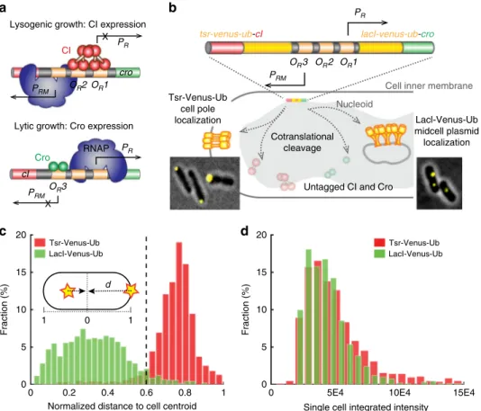
composed of two mutually repressive TFs, CI and Cro (Fig.
1
a);
the expression of CI but not Cro confers lysogenic growth, and
the expression of Cro but not CI confers lytic growth. The
λ
switch has served as a paradigm for studying gene regulation
and cell fate determination
9–11,20,21, but the real-time switching
kinetics and paths between the two distinct, mutually exclusive
gene expression states have not been elucidated experimentally.
To achieve these goals, we developed a dual single-molecule
gene-expression reporting system to follow the stochastic gene-expression
Lysogenic growth: CI expression
Lytic growth: Cro expression OR2 OR1 PR X cro PRM CI PRM X OR3 PR cI RNAP Cro
c
a
b
lacI-venus-ub-cro PR tsr-venus-ub-cIUntagged CI and Cro PRM
OR2 OR3 OR1
d
Normalized distance to cell centroid
0 0.2 0.4 0.6 0.8 1 Fraction (%) 0 5 10 15 20 Tsr-Venus-Ub LacI-Venus-Ub
Single cell integrated intensity
0 5E4 10E4 15E4
Fraction (%) 0 5 10 15 20 Tsr-Venus-Ub LacI-Venus-Ub 1 1 0 d Nucleoid
Cell inner membrane Tsr-Venus-Ub cell pole localization Lacl-Venus-Ub midcell plasmid localization Cotranslational cleavage
Fig. 1 Validation of the dual single-molecule gene-expression reporting system DuTrAc. a Schematic drawing of the two gene expression states of theλ genetic switch. In lysogenic growth, theλ repressor CI binds to its operator sites OR1 and OR2 to stimulate its expression from promoter PRM, and at the same time shuts down Cro expression from promoter PR. In lytic growth, Cro binds to OR3 to repress CI and turns on its own expression. b Schematic drawing of the DuTrAC system. Theλ switch (colored bar) containing tsr-venus-ub-cI and lacI-venus-ub-cro is integrated into the chromosome at the lac operon locus. The expressed fusion polypeptide chain is cotranslationally cleaved by the constitutively expressed deubiquitinase UBP1 at the last residue of the Ub sequence, separating cell-pole-targeting Tsr-Venus-Ub from CI, and mid/quarter-cell-targeting LacI-Venus-Ub from Cro for single-molecule detection. CI and Cro are thus untagged and can bind respective operators to regulate gene expression. Representative cell images of pole- or midcell-localized Venusfluorescence spots are shown as insets. c Histograms of normalized distance (d) of Venus fluorescence spots to cell centroid (inset) in strain XF002 (red, expressing Tsr-Venus-Ub-CI only, Supplementary Figure1, Table1) and XF003 (green, expressing LacI-Venus-Ub-CI only,
Supplementary Figure1, Table1) in the presence of the deubiquitinase UBP1; 92% of Tsr-Venus-Ub spots (n = 993 spots) localized at d ≥ 0.6 (dashed line); 95% of LacI-Venus-Ub spots (n = 1384 spots) localized at d < 0.6, suggesting that the threshold of d = 0.6 could be used to distinguish the identity of the fused protein.d Histograms of integratedfluorescence level of Tsr-Venus-Ub (red) and LacI-Venus-Ub (green) in individual XF002 and XF003 cells. The distributions and mean levels (4.8 ± 2.9 × 10⁴, n = 840 cells for XF002, and 4.2 ± 2.1 × 10⁴, n = 913 cells for XF003, μ ± s.d.) of Venus fluorescence in the two strains were indistinguishable from each other, indicating that both Tsr-Venus-Ub and LacI-Venus-Ub, despite the different mRNA and protein sequences, reported the expression levels of CI equivalently
dynamics of CI and Cro simultaneously in the same cells
(Fig.
1
b).
In the dual gene-expression reporting system, we fused a
fast-maturing yellow
fluorescent protein variant, Venus
22, to one of
two cellular localization tags, Tsr or LacI, to distinguish the
production of CI and Cro in the same cell. The strategy of using
two different subcellular localizations differs from previous
studies using
fluorescent proteins of different colors
23, and avoids
the major disadvantage of temporal mismatches caused by
different
fluorescent protein maturation rates (e.g., ~1 h for red
fluorescent proteins such as mCherry
24and ~5–10 min for
Venus
22,25–28). Tsr is a membrane protein that localizes rapidly
and specifically to cell poles
29. LacI binds specifically to 256 lacO
sites (lacO
256) incorporated onto a multi-copy,
mid/quarter-cell-localizing RK2 plasmid pZZ6
30. With the ability to localize single
fluorescent protein molecules with 30–40 nm precision in live
Escherichia coli cells
27,31, we could distinguish between individual
Tsr-Venus and LacI-Venus molecules based on their subcellular
positions. Using a control strain XF004 expressing Tsr-Venus and
LacI-mCherry independently (Supplementary Figure
1
,
Supple-mentary Table
1
), we demonstrated that there was indeed
minimal spatial overlap (~2%) between the two localization tags
(Supplementary Figure
2
and Supplementary Movie
1
).
Next, to distinguish the expression of CI and Cro in the same
cell while avoiding possible disruptions of their functions due to
the
fluorescent protein fusion, we generated two translational
fusion genes, tsr-venus-ub-cI and lacI-venus-ub-cro, and used the
CoTrAC strategy (CoTranslational Activation by Cleavage
26,27)
to cleave cotranslationally the Tsr-Venus-Ub or LacI-Venus-Ub
reporter from CI or Cro (Fig.
1
b). This strategy ensures a 1:1 ratio
in real-time between localized Venus reporter molecules and the
fused CI or Cro molecules. Using two control strains expressing
only Tsr-Venus-Ub-CI (XF002) or LacI-Venus-Ub-CI (XF003),
we verified that the cellular localization and fluorescence intensity
of cell-pole- and midcell-targeted Venus spots faithfully reported
both the identity and expression level of CI (Fig.
1
c, d,
Supplementary Figure
1
, Supplementary Movies
2
and
3
). We
named this new, dual gene-expression reporting system DuTrAC
(Dual coTranslational Activation by Cleavage).
λ switch exhibits two new CI and Cro expression populations.
To investigate the regulatory dynamics of CI and Cro in the
λ
switch using DuTrAC, we constructed strain XF204. We fused
tsr-venus-ub to a temperature-sensitive CI mutant (cI857
32), and
lacI-venus-ub to cro, replacing the native cI and cro genes in the
genetic switch, similar to what was previously described
(Sup-plementary Figure
1
, Supplementary Table
1
)
27. We then
inte-grated this circuit from O
Lto the end of the cro gene into the
chromosome of E. coli MG1655 strain at the lac operon locus
(Fig.
1
b). Hence, Tsr-Venus-Ub reports the expression of CI857,
and LacI-Venus-Ub reports the expression of Cro. We used the
temperature-sensitive mutant CI857 (A66T
32) in place of
wild-type (WT) CI
WTfor the convenience of using temperature to
tune the fraction of active CI. CI857 has normal DNA-binding
affinity and transcription regulation activity at the permissive
temperature of 30 °C
33. At higher temperatures, an increasing
fraction of CI857 becomes inactivated due to misfolding and
subsequent degradation
33, and therefore temperature can be used
as a convenient
“control knob” to change the fraction of active,
WT CI molecules
8. We verified that the fusion of DuTrAC
reporters to CI and Cro did not change the switching behavior of
the genetic switch (Supplementary Figure
3
A). Furthermore, to
examine the switching behavior across different protein
expres-sion levels, we generated two additional strains XF214 and XF224
(Supplementary Figure
1
, Supplementary Table
1
), in which the
expression level of LacI-Venus-Ub-Cro was reduced in the order
of [Cro]
XF224< [Cro]
XF214< [Cro]
XF204. Using Western blotting,
we confirmed that the expression levels and switching behaviors
of these strains were as expected (Supplementary Figure
3
B). Note
that in the following experiments, for simplicity, we referred
CI857 as CI.
To investigate the switching behavior of the modified λ switch,
we
first quantified the expression levels of CI and Cro in
individual cells of strain XF204, XF214, or XF224 maintained
constantly at different temperatures for >20 generations (Fig.
2
,
Supplementary Figures
4
,
5
and Supplementary Table
2
). We
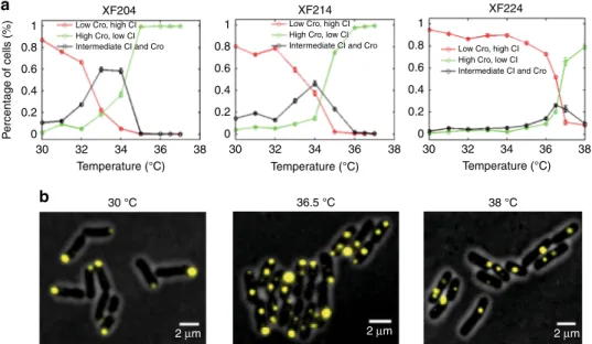
found that consistent with the typical bistable behavior, at a low
temperature (30 °C), cells had few Cro but predominately CI
molecules (hereafter termed [L, H] for low-Cro and high-CI level,
a
b
30 °C 36.5 °C 38 °C P ercentage of cells (%) 1 XF204 XF214 XF224Low Cro, high CI High Cro, low CI Intermediate CI and Cro
Low Cro, high CI High Cro, low CI
Intermediate CI and Cro Low Cro, high CI High Cro, low CI Intermediate CI and Cro 0.8 0.6 0.4 0.2 0 1 0.8 0.6 0.4 0.2 0 1 0.8 0.6 0.4 0.2 0 30 Temperature (°C) Temperature (°C) 32 34 36 38 30 32 34 36 38 Temperature (°C) 32 34 36 38 30 2 µm 2 µm 2 µm
Fig. 2 Expression levels of CI and Cro in strains XF204, XF214, and XF224 at different temperatures showed more than two expected cell populations. a Percentages of cells having CI only (red, copy number ratio r = CI/(CI + Cro) ≥ 0.8), Cro only (green, r ≤ 0.2), or both CI and Cro (black, 0.2 < r < 0.8) in strains XF204, XF214, and XF224 at different temperatures.b Representativefluorescent images of XF224 cells showing CI expression (yellow pole-localized Tsr-Venus-Ub spots) and Cro expression (yellow quarter/midcell-pole-localized LacI-Venus-Ub spots) at low, intermediate, and high temperatures overlaid with phase-contrast cell images (gray)
Fig.
2
a, red curves); at a high temperature (37 °C) the switch was
flipped and cells predominately existed in high-Cro, low-CI level
([H, L], Fig.
2
a, green curves). Interestingly, between the two
extreme temperatures, we observed that a large population of cells
had both CI and Cro [H, H] in the same cells at intermediate
levels (Fig.
2
a, black curves, Supplementary Figure
5
,
Supple-mentary Table
2
). Reduced Cro levels in strains XF214 and XF224
did not abolish the presence of [H, H] cells, but shifted the
temperature at which the percentage of this population of cells
was the highest from 33 °C in XF204 to 34 °C in XF214 and to
36.5 °C in XF224 (Fig.
2
, Supplementary Figure
5
, Supplementary
Table
2
). A fourth population of cells having little CI or Cro ([L,
L]) also existed at these temperatures. A few representative
images of the four cell populations of strain XF224 at low,
intermediate, and high temperatures are shown in Fig.
2
b.
Previous studies probing CI and Cro expression levels
indepen-dently did not observe the presence of the two new populations of
cells
9,34.
Observing the switching of CI and Cro production. The
observation of cells having both CI and Cro indicated that cells
could switch between CI- and Cro-expressing states within each
other’s degradation time scales, and/or there existed a new
expression state in which CI and Cro were expressed
con-currently. The snapshot nature of the above measurement could
not distinguish these possibilities. In addition, the snapshot
measurement of CI was complicated by the fact that at high
temperatures an increasing population of CI857 becomes
inac-tive; hence, the actual steady-state level of active CI (molecules/
cell) is only proportional to the measured level of Tsr-Venus-Ub.
These problems could be circumvented by following protein
production in real time; the number of newly produced protein
molecules per unit time directly reflects promoter activity during
that time without the convolution of any downstream processes.
Therefore, we grew XF224 cells in a precision
temperature-control chamber (T
= 36.5 ± 0.1 °C over the length of the
experiment of ~7 h, Supplementary Figure
6
) on a microscope
stage, and counted the number of newly produced CI and Cro
molecules in individual cells every 5 min for multiple generations
(Supplementary Figure
7
, Supplementary Movies
4
,
5
, mean cell
cycle time
τ = 71 ± 22 min, μ ± s.d., n = 457 cell cycles). We
photobleached Venus molecules after each detection, so that new
fluorescent molecules detected after 5 min of the dark interval
were newly produced during the 5 min
26,27. We chose strain
XF224 because it had the lowest Cro steady-state levels compared
to XF204 and XF214 (Supplementary Figures
3
,
5
), facilitating the
accurate identification and counting of single Venus molecules in
small E. coli cells (Supplementary Figure
8
). We choose to
con-duct the real-time experiment at 36.5 °C because the steady-state
experiment showed that at this temperature XF224 has the largest
population of cells expressing both CI and Cro.
In Fig.
3
, we show four representative time traces of different
XF224 cell lineages. For each colony, we only picked randomly
one cell lineage for analysis in order to avoid double-counting
data. We observed stochastic, anti-correlated production of CI
and Cro (Fig.
3
a–d, Supplementary Figure
9
). Intriguingly, in
many time traces, we observed that there were periods of time in
which neither CI or Cro was produced, or both were produced.
The presence of the four production populations was evident
when we plotted the two-dimensional (2D) histogram of the
number of CI and Cro molecules produced in each 5-min
imaging interval for all time traces (Fig.
3
e). In addition to the
two expected populations of high-CI ([L, H]), and high-Cro ([H,
L]) production states, there were two additional populations. One
resided at [0, 0] where no CI or Cro was produced, and another
centered at ~4 molecules for both CI and Cro, similar to the [L, L]
and [H, H] populations we observed in steady-state
measure-ments (Fig.
2
). The one-dimensional (1D) histograms of CI and
Cro alone showed two-state distributions (Fig.
3
e).
We verified that the presence of the four populations was not
caused by the independent production of CI and Cro from two
copies of the
λ switch due to chromosome replication, because the
four populations existed similarly in young cells where the
chromosomal copy was one before replication (cell age
⩽0.4, less
than 40% of the cell cycle time, Supplementary Figure
10
A).
Furthermore, single-molecule
fluorescence in situ hybridization
(smFISH, Supplementary Table
3
) showed co-existence of cI and
cro mRNA molecules in a significant population of cells (16.3 ±
0.7%, cI and cro mRNAs at 0.9 ± 0.03 and 0.6 ± 0.02 molecules per
cell,
μ ± s.e., n = 2627 cells), irrespective of cell ages
(Supplemen-tary Figure
10
B and Supplementary Table
4
). This result
suggested that cI and cro mRNAs were produced within each
other’s short lifetime window (~1.5 min
35) (Supplementary
Note
4
). Co-existence of cI and cro mRNAs has also been
previously observed in cells growing under a different growth
condition
35. Finally, we verified that the stochastic maturation
process of the Venus
fluorophore only affected the spread, but not
the presence, of each population in the 2D histogram
(Supple-mentary Note
5
, Supplementary Figure
11
). Taken together, these
results suggested that in each 5-min time window, a cell could
produce none, only one or the other, or both proteins.
Quantifying potential landscape and switching kinetics. Using
the experimentally measured 2D distributions of CI and Cro
production levels, we generated the corresponding potential
landscape by calculating the negative logarithm of probabilities
(Fig.
3
f). There were clearly four basins, approximately around at
[0, 0], [4, 0], [0, 4], and [4,4] for produced [Cro, CI] protein
molecule numbers per 5 min (Fig.
3
f). Interestingly, there was one
central peak separating the four basins such that the barrier
height between two opposite basins [L, L] and [H, H], or [H, L]
and [L, H], was higher than that between two adjacent basins [L,
L] and [L, H], or [L, L] and [H, L] (Fig.
3
f). This type of landscape
has not been previously observed for such a genetic circuitry, and
suggested specific switching paths between the basins. For
example, to switch from [H, L] to [L, H], the path going through
the [L, L] or [H, H] basins would have higher probability than the
path of switching directly between the two.
To quantitatively identify possible production states of CI and
Cro corresponding to the observed basins in the potential
landscape, and obtain the associated transition rates between
these states, we used a modified Hidden Markov Model (HMM)
(Supplementary Note
1
), which is commonly used in temporal
pattern recognition
36,37. We found that a four-state HMM ([L, L],
[L, H], [H, L], and [H, H]) matched the observed 2D histogram of
CI and Cro production the best (Supplementary Figure
12
,
Supplementary Note
2
). The mean production levels of Cro and
CI of each state and the corresponding dwell times were
summarized in Table
1
and Supplementary Figure
13
.
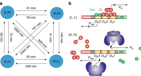
Impor-tantly, HMM allowed us to identify state-switching events in
individual time traces (Fig.
3
a–d, middle panels with colored
bars) and hence the transition time constants (Supplementary
Note
3
, Fig.
4
a, Supplementary Table
5
)
17,38. Similar results were
observed using truncated time traces of only young cells
(Supplementary Figure
14
, Supplementary Tables
5
and
6
).
Because the dynamics of a system is fully determined and
described by its speed and the underlying kinetic processes (or
paths), the transition time constants obtained here can be used to
identify the most likely transition paths and the associated rates of
switching between states, which has not been achieved before. For
example, to switch from the [L, H] state to the [H, L] state, the
most likely path is to go through the [H, H] state instead of
directly switching. We can also determine the time it takes for
switching by the times a cell spent on the two paths (from [L, H]
to [H, H] and from [H, H] to [H, L]). This gives us an insight into
possible mechanisms underlying the kinetic processes in terms of
the speed and the most likely paths, suggesting an unexpected
kinetic route through [H, H] beyond direct switching between CI
and Cro.
Discussion
Theoretical studies have shown that without changing the wiring
configuration of a bistable switch, multistability can arise from
Table 1 Mean production levels of CI and Cro and the corresponding dwell time of each state identi
fied by HMM in time-lapse
experiments
State [Cro, CI] Croa(molecules) CIb(molecules) n (frames) Dwell timec(min) nd(occurrence)
[L, L] 0.0 ± 0.01 0.0 ± 0.0 139 17 ± 2 41
[H, L] 5.2 ± 0.10 0.0 ± 0.0 1173 36 ± 3 162
[L, H] 0.0 ± 0.0 4.7 ± 0.13 1451 27 ± 1 269
[H, H] 4.5 ± 0.04 4.7 ± 0.05 3069 47 ± 3 396
a, b, cValues were expressed as mean ± standard error dNumber of occurrences of each state in all time traces
CI 0 10 20 0 50 100 150 200 250 300 350 Cro 0 5 10 15 CI 0 10 20 0 50 100 150 200 250 300 350 Cro 0 5 10 15 CI 0 10 20 0 50 100 150 200 250 300 350 Cro 0 5 10 15 CI 0 10 20 0 50 100 150 200 250 300 350 Cro 0 5 10 15
Time (min) Time (min)
a
b
c
d
Cro 0 5 10 CI 0 5 10 0 0.01 0.02 0.03 0.04 0 0.2 0 0.2e
) P( nl -[H, H] [L, H] [L, L] [H, L]f
8 9 8.5 8 7.5 6.5 5.5 4.5 3.5 4 5 6 7 7 6 5 4 3 12 10 8 6 CI 4 Cro 2 2 4 6 8 10 12 0 0Fig. 3 Real-time production time traces of CI and Cro in strain XF224 showed four distinct populations at 36.5 °C. a–d Time traces of newly produced CI (top) and Cro (bottom) molecule numbers in the same cells in four representative XF224 cell lineages. The corresponding state-switching time trace identified by HMM is shown in the middle panel of each lineage, with blue corresponding to state [H, L] for high Cro and low CI production, green to [L, H], purple for [H, H] and red for [L, L]. The dashed vertical lines indicate cell division.e 2D histogram of produced CI and Cro protein molecules in individual cells measured at each 5-min frame in time-lapse experiments (n = 6453 frames from 94 time traces). Corresponding 1D histograms of CI and Cro are shown on the right and top of the 2D histograms, respectively. Colors and scale bars indicate fractions of cells.f The potential landscape was calculated using the experimentally measured 2D histogram of CI and Cro expression numbers in every 5-min frame and interpolated
weakened regulatory interactions, which impose fewer constraints
on possible TF binding configurations
13–16. In eukaryotic cells,
epigenetic phenomena such as histone modification and DNA
methylation could reduce the binding rates of TFs to their
tar-geting DNA sites, leading to longer time scale of gene regulation.
In bacterial cells, low TF expression levels
39and high levels of
non-specific binding
40can effectively slow down the binding of
TFs to specific target sites, leading to weakened regulation. Such a
regime in gene regulation is termed non-adiabatic, in contrast to
the classic adiabatic regime, in which protein binding and
unbinding are fast compared to the protein’s production and
degradation time scales with rapid equilibration in a well-mixed
environment
14–16,41. Previous studies under different
experi-mental conditions in which CI was expressed at higher levels than
ours have elegantly demonstrated the adiabatic regime of the
λ
switch
8,42,43.
In strain XF224, both CI and Cro were expressed at much
lower levels than in the WT strain XF204 (Supplementary
Table
2
, Supplementary Figures
5
and
15
). Under this condition,
slow association/dissociation times (in the range of a few minutes
to hours
44,45) and significant levels of non-specific binding for
both CI and Cro (>70%,
40) were previously demonstrated. Our
results showed that the [L, L] state persisted for ~20 min
(Fig.
3
a–d), which is longer than the reactions times typically
associated with relevant biochemical events such as transcription
initiation, mRNA degradation, and TF binding
46. The [L, L]
production state could emerge and remain relatively long-lived
when a combination of CI and Cro occupies all the operator sites,
shutting down the production of CI and Cro simultaneously
(Fig.
4
b).
When either CI or Cro dissociates, and the rebinding is slow,
RNA polymerase (RNAP) can bind to the exposed P
Ror P
RMpromoters, resulting in the [H, L] or [L, H] production states.
RNAP exists at a much higher level (~3000 molecules per cell
under a similar growth condition
47) compared to CI and Cro, and
hence its binding rate would be significantly faster. When both CI
and Cro dissociate from all three operators, which would occur at
a much lower probability than only one of them dissociating,
RNAP can initiate transcription from either one of the two
promoters, resulting in the [H, H] production state (Fig.
4
b).
Consistent with this possibility, we previously measured the basal
expression level of P
RMpromoter in the absence of CI and Cro to
be similar to the CI production level in the [H, H] state
27. In
addition, in vitro studies have demonstrated that both P
Rand
P
RMpromoters on the same
λ switch can be occupied by two
RNAP molecules simultaneously in the absence of CI and
Cro
48,49.
One important aspect of our time-lapse experiments, in
con-trast to an earlier experiment with a similar genetic network
8, is
that we measured the production, but not cellular concentrations,
of CI and Cro. Protein production rates directly measure
pro-moter activities, which reflect propro-moter configurations with
respect to TF and RNAP binding. In the adiabatic regime, protein
concentration changes (and hence changes in protein binding
rates) are instantaneously reflected in protein production rates; in
the non-adiabatic regime, however, promoter activity can lag
protein concentration changes. One concentration state can
correspond to multiple production states, and hence multiple cell
fate potentials. The well-known hysteresis effect of bistable
switches
8is likely a result of the non-adiabatic cellular
environ-ment in which protein binding/unbinding is slow—cells starting
in one state have a tendency to stay in that state before switching
to the other states even when the concentration of a critical
protein has already changed.
Previous studies showed that different wiring conditions of
bistable switches could give rise to a maximum of three states in
the adiabatic regime
17–19. Here we showed that, in the
non-adiabatic regime, four protein production states can emerge from
bistable switches without changing wiring configurations, with
consequences in establishing new cell fates
13–16,50. A living cell, in
which a non-equilibrium state is the norm, could potentially
utilize non-adiabaticity to encode more than two cell fates with
limited circuitry, allowing a high degree of adaptation and
dif-ferentiation. However, from the opposing point of view, this
increased diversity of states in the non-adiabatic regime places
more limits on genetic circuitry that will produce robust binary
switching; hence, avoiding the non-adiabatic regime will be key to
engineer robust, binary genetic switches.
Methods
Bacterial strains and plasmids construction. Strains XF103 and ZH051 were generated using the parental strain BW2511351andλ RED recombination52. (H,H)
41 min 78 min
25 min 249 min
63 min 75 min 41 min 140 min 1660 min 828 min 167 min 2354 min (L,L) (H,L) (L,H) OR2 OR1 PR cro PRM RNAP OR2 OR1 PR X cro PRM CI Cro OR3 X OR3 [L, L] [H, H]
a
b
Fig. 4 Transition time constants (a) and possible underlying molecular events (b) for the four stable production states observed in theλ switch. a Transition time constants were identified using a modified HMM analysis. Longer transition times indicate that direct switching between diagonal states is much less likely than that between side states.b The [L, L] states could arise due to the co-occupancy of the three operators by a combination of Cro and CI. The [H, H] state could arise from the slow association of Cro or CI to the operators, allowing RNAP to bind freely on PRor PRMto initiation transcription. Note that only these two simple possibilities were depicted here but other promoter configurations could also potentially lead to the [L, L] and [H, H] production states
Briefly, plasmid pXF103 carrying the lacI-venus-ub-cI fusion gene was constructed by replacing the tsr-venus sequence of theλ switch in plasmid pZH05127with the lacI-venus sequence amplified from plasmid pVS143 using primers P1 and P2. The full-lengthλ switch sequence (from OLto the end of cro) containing lacI-venus-ub-cI on pXF103, or tsr-Venus-ub-lacI-venus-ub-cI on pZH051, was then PCR amplified (primer pair P3:P4) together with the drug resistance gene kanRon the plasmid and replaced the lacI gene on chromosome usingλ RED recombination. Subsequently, the kanR gene was removed by transforming the FLP recombinase expressing plasmid pCP20 into the host strain to generate thefinal XF103 or ZH051 strain.
Strains XF204, XF016, XF206, XF214, XF224, XF225, and XF226 were generated using the parental strain E. coli K12 MG1655 (Yale University E. coli Genetic Stock Center) and the landing pad approach, which is specific for the chromosomal integration of large genetic constructs53. First, the ind1 and sam7 mutations in the cI857 sequence on plasmid pZH016 were corrected using primers P5 and P6, and the resulting tsr-venus-ub-cI857 was used as a template for subsequent strain constructions. To generate the two-reporterλ switch (tsr-venus-ub-cI in place of cI and lacI-venus-ub-cro in place of cro), lacI-venus-ub was amplified from plasmid pXF103 using primer pair P7:P8 and ligated into pZH016 in front of cro to generate two-reporterλ switch pXF104. The landing pad vector pTKIP (containing LP1 and LP2, gift from Dr. Thomas E. Kuhlman) was opened to add NheI and SalI restriction sites at two ends using inverse PCR (primer pair P9: P10). Plasmid pXF104 was digested with NheI and SalI to release the two-reporter λ switch DNA fragment and ligated into similarly digested pTKIP inverse PCR product to obtain plasmid pXF204. Plasmid pXF204 was then served as a template to generate pXF214 (primer pair P11:P12, ATG start codon of lacI changed to GTG) and pXF224 (primer pair P13:P14, PRpromoter−32 A to G) using site-directed mutagenesis.
To eliminate thefluorescence of Venus, two glycine residues in the tri-peptide of Venus chromophore were mutated to Alanin54using site-directed mutagenesis (primer pair P15:P16) to obtain pXF106 from pZH016. Plasmid pXF106 was digested by BspEI and SalI to release the tsr-venus*(G65A and G67A)-ub-cI857 fragment and ligated with similarly digested vector from pXF204 to obtain pXF206. Plasmid pXF106 was digested by BspEI and SalI to release the tsr-venus*(G65A and G67A)-ub-cI857 fragment and ligated with similarly digested pXF224 to obtain pXF226, which contain the tsr-venus*-ub-cI and the mutated PRpromoter in front of lacI-venus-ub-cI.
To generate pXF016, which was the landing pads version of pZH016, pZH016 was digested with NheI and SalI to release theλ switch DNA fragment containing tsr-venus-ub-cI857 and cro. The fragment was then ligated into NheI- and SalI-digested pXF214 to obtain pXF016. pXF225 was generated from pXF016 using site-directed mutagenesis (primer pair P13:P14) to mutate PRpromoter (−32 A to G).
To prepare the parental strain MG1655 for landing pad integration, the fragment of LP1-tetR-LP2 containing two landing pads LP1, LP2, and a tetracycline resistance gene was amplified from plasmid pTKS/CS (gift from Dr. Thomas E. Kuhlman). The lacI gene on MG1655 chromosome was then replaced with the LP1-tetR-LP2 fragment usingλ RED recombination to obtain strain XF001. To construct strain XF204 using the landing pad approach, plasmids pTKRED (gift from Dr. Thomas E. Kuhlman) and pXF204 were transformed into XF001. Single colonies of XF001(pTKRED/pXF204) transformants were picked and grown in 1 ml LB with 2% arabinose and 4 mM IPTG at 30 °C for 2 h with aeration. Next, 10 μl spectinomycin at a final concentration of 100 μg ml−1was added and the culture was allowed to continue at 30 °C. After 5 h, 1μl kanamycin at a final concentration of 50μg ml−1was added and the culture was kept growing overnight. The next morning, the overnight culture was 1:104diluted using fresh M9 medium, and 50μl of the dilution was plated on LB plate with kanamycin (50μg ml−1) and incubated overnight at 30 °C. Single colonies grown on the kanamycin plate were tested for their failure to grow in tetracycline- or carbenicillin-containing media and subsequently sequenced to obtain strain XF204. Correct colonies were picked into 5 ml LB medium and cultured overnight at 37 °C to eliminate plasmid pTKRED. The other strains (XF016, XF206, XF214, XF224, XF225, and XF226) were constructed following the same procedure using the corresponding helper plasmid (pXF016, pXF206, pXF214, pXF224, pXF225, and pXF226). All the strains were then transformed with the UBP1-expressing plasmid pCG001 (gift from Dr. Roland Baker55) and the lacO256plasmid pZZ6 (gift from Dr. Joe Pogliano30). Note that the 37 °C growth condition to eliminate pTKRED led to a large population of cells expressing high levels of Cro in the presence of the CI857 mutant, especially in the XF204 strain. Therefore, after the elimination of pTKRED, plasmid cells were grown in LB medium at room temperature overnight followed by resteaking on LB plates at 30 °C for another day’s growth. Single colonies that have already switched back to low Cro expression levels (lowerfluorescence level compared to that in high Cro expression levels) were identified using a Halogen lamp equipped with an emissionfilter (ET545/30, Chroma). These colonies were further grown to exponential phase in M9 medium at 30 °C and imaged on the microscope to confirm their expression states. Confirmed cultures were then flash-frozen in liquid nitrogen and stored at−80 °C.
To construct plasmid pXF011 that expressed UBP1 and LacI-mCherry, lacI-mCherry fragment with the pBAD promoter was PCR-amplified from pZH102 using primer pair P19:P20, subsequently restricted with SalI and EagI, and ligated into similarly restricted pCG001 vector to obtain plasmid pXF004. The pBAD promoter in front of lacI-mCherry on pXF004 was then replaced by a constitutive promoter of BBa_J2310356using primer pair P21:P22 to obtain pXF011.
All the strains, plasmids, and primers are listed in Supplementary Table1. Growth conditions. Cells from frozen stocks were streaked on LB plates and incubated at an appropriate temperature overnight. Single colonies were picked the next day and inoculated into M9 medium supplemented with MEM amino acid (Sigma-Aldrich Co. LLC) at appropriate temperature overnight in a precision temperature-controlled shaker (±0.5 °C, MIDSCI IS-300). The next morning, cells were reinnoculated in fresh M9 medium to mid-log phase (OD600≈0.4) before steady-state or time-lapse microscopy experiments. Antibiotics were included in all cultures at concentrations of 50μg ml−1for kanamycin, 25μg ml−1for chlor-amphenicol, and 100μg ml−1for ampicillin when appropriate.
Western blot. Cells were cultured under the same condition as that described for steady-state microscopy measurements. Log phase cells (OD600≈0.4–0.5) were collected and cell numbers counted using a Petroff-Hausser chamber and a plating assay. Cell lysates were prepared by incubating equal number of cells for 10 min at 100 °C followed by 20 min at−75 °C. Protein electrophoresis was carried out using a 4–15% Tris-HCl Precast gradient gel (Bio-Rad) at 100 V for 1.5 h. The gel was then transferred to a PVDF membrane (Bio-Rad) for 2 h at 25 V and 4 °C. Venus bands were detected with 1:2500 mouse antibody to GFP (Clontech, JL-8) and 1:20,000 goat-anti-mouse HRP secondary antibody (BioRad, #170-5047). Immun-StarTM WesternCTMreagents (Bio-Rad) were applied for luminescent visualiza-tion. Images were captured using a Typhoon Scanner (GE Life Sciences). smFISH. CI transcripts were labeled with 30 oligonucleotides (Supplementary Table3) conjugated with TAMRA (Biosearch Technologies). Cro transcripts were labeled with 41 oligonucleotides (Supplementary Table2) conjugated with Quasar 670 (Biosearch Technologies). Because of the short sequence of cro mRNA, only nine of the probes targeted to the cro mRNA sequence and 31 probes targeted to the lacI sequence fused to cro.
Three cultures of XF224 cells were grown using the same procedure and growth medium as that for the time-lapse experiments, but at three different temperatures, 30, 36.5, and 37.5 °C. The 30 °C culture was used as a control to quantify the fluorescence intensity of single cro mRNA molecules, because at 30 °C CI dominated and Cro was transcribed at such a low level that most cro transcripts were single molecules. Similarly, the 37.5 °C culture was used as a control to quantify thefluorescence intensity of single cI mRNA molecules. Cells were fixed at respective growth temperatures and labeled with 1μM CI and 1 μM Cro probes using a protocol as described previously57. Briefly, the cells were fixed for 30 min with 3.7% formaldehyde and were then permeabilized with 70% ethanol for 1 h. Each sample was hybridized overnight in a 40% formamide hybridization solution. Before imaging, the cells were washed 4× with 40% formamide wash solution and then resuspended in 2x SSC for imaging.
Fixed and labeled cells were imaged using simultaneous laser excitation at 561 nm (Coherent, sapphire) and 647 nm (Coherent, obis). Emission was collected using an OptoSplit III with a long-pass (647 nm) beam splitter and emissionfilters ET590/33 and HQ705/55 (Chroma Technology). Each viewfield was imaged at six z planes separated by 200 nm. The projection of the six planes was then used to detectfluorescent spots using custom MATLAB software as previously described30. The total integratedfluorescence intensity of each spot was divided by the mean intensity of corresponding single transcript molecules to obtain the number of transcript molecules in each spot.
Time-lapse imaging. Log phase cell culture (1 ml) was collected and washed twice with fresh M9 medium. Pelleted cells were diluted 1:100 and 0.5μl was spotted onto a gel pad made of 3% low-melting temperature SeaPlaqueTMagarose (Lonze) using M9 medium in a precision temperature-controlled growth chamber (FCS2, Bioptechs). The chamber was locked on an Olympus IX-81 inverted microscope equipped with a 100× oil-immersion objective lens (Olympus Inc., PlanApo 100×NA 1.45). Both the sample chamber and the objective were maintained at 36.5 °C using respective heaters provided by the FCS2 system (Bioptechs). In all time-lapse experiments, a digital thermometer probe was inserted into the sample chamber to record the temperaturefluctuations in real time using a voltage recorder (MadgeTech, VOLT101A-15V). Excitation at 514 nm with an illumina-tion power density of ~1 kW cm−2was provided by an argon ion laser (Coherent Innova I-308). Emission wasfiltered (ET545/30, Chromas Technology Corp), and fluorescent and bright-field images with a time interval of every 5-min were cap-tured by an Ixon EMCCD camera (Andor IXon DU888) using a custom imaging journal in Metamorph (Molecular Devices) as previously described in refs.26,27. Time-lapse image analysis. We used a previously published procedure to seg-ment cells, detectfluorescent spots, and track cell lineages27. We used a custom Matlab code to assign individualfluorescent spots to CI or Cro by measuring the centroid distance of the spot to cell center (estimated as the center of mass of cells in a segmented, binary image) using a threshold of 0.6 (Fig.1c and Supplementary Figure7). For each micro-colony, only one cell lineage time trace with complete cell cycles was used. A total of 96 time traces (6453 frames) were obtained for the XF224 strain.
Steady-state imaging. Log phase cell culture was prepared exactly the same as described in time-lapse imaging except that cells were diluted 1:50 to obtain more cells in each imaging area. All cells were imaged within 1.5 h at room temperature to avoid significant changes of expression levels. For the two-color strain XF004, Venus was excited using an argon ion laser (Coherent Innova I-308) and mCherry was excited by a rhodamine dye laser (Coherent 599) tuned to ~570 nm. Images were split into the yellow and red channels by an Optosplit II adaptor (Andor) using a long-passfilter, and further filtered by ET545/30 and HQ630/60 bandpass filters (Chroma) for the yellow and red channels, respectively.
Steady-state image analysis. To quantify the copy numbers of CI and Cro in microscopy experiments, we measured thefluorescence intensity distribution of single Tsr-Venus-Ub molecules under our imaging condition (Supplementary Figure4) using theλ−strain expressing very low numbers of Tsr-Venus-Ub per cell cycle27. Using a previously described procedure, individual, well-localized Tsr-Venus-Ub or LacI-Tsr-Venus-Ub spots in experimental strains were detected and the correspondingfluorescence intensity was converted to molecule numbers of CI or Cro by dividing by the peak value of the Gaussian distribution of single Venus molecules27. For strain XF224, both Tsr-Venus-Ub and LacI-Venus-Ub molecules were localized to diffraction-limited spots and the procedure described above was carried out. For strains XF204 and XF214, at high temperatures, Cro was expressed at high levels, and hence did not form well-localized spots but nucleoid-covered clouds. Therefore, we measured total integrated cellularfluorescence and sub-tracted thefluorescence of pole-localizing Tsr-Venus-Ub spots. The subtracted fluorescence intensity was then used to calculate the number of expressed Cro copy numbers. The absolute copy numbers of the three strains at different temperatures measured at steady state are plotted in Supplementary Figure5and summarized in Supplementary Table2.
Code availability. The analyses in this study were performed by using custom MATLAB code, which will be made available from the corresponding author upon reasonable request.
Data availability. The authors declare that the data supporting thefindings of this study are available from the corresponding author upon reasonable request.
Received: 23 January 2018 Accepted: 17 April 2018
References
1. Macarthur, B. D., Ma’ayan, A. & Lemischka, I. R. Systems biology of stem cell fate and cellular reprogramming. Nat. Rev. Mol. Cell Biol. 10, 672–681 (2009). 2. Bouldin, C. M. et al. Wnt signaling and tbx16 form a bistable switch to
commit bipotential progenitors to mesoderm. Development 142, 2499–2507 (2015).
3. Schroter, C., Rue, P., Mackenzie, J. P. & Martinez Arias, A. FGF/MAPK signaling sets the switching threshold of a bistable circuit controlling cell fate decisions in embryonic stem cells. Development 142, 4205–4216 (2015). 4. Wang, L. et al. Bistable switches control memory and plasticity in cellular
differentiation. Proc. Natl Acad. Sci. USA 106, 6638–6643 (2009).
5. Jukam, D. et al. Opposite feedbacks in the Hippo pathway for growth control and neural fate. Science 342, 1238016 (2013).
6. Gamba, P., Jonker, M. J. & Hamoen, L. W. A novel feedback loop that controls bimodal expression of genetic competence. PLoS Genet. 11, e1005047 (2015). 7. Ramachandran, G. et al. A complex genetic switch involving overlapping
divergent promoters and DNA looping regulates expression of conjugation genes of a gram-positive plasmid. PLoS Genet. 10, e1004733 (2014). 8. Bednarz, M., Halliday, J. A., Herman, C. & Golding, I. Revisiting bistability in
the lysis/lysogeny circuit of bacteriophage lambda. PLoS ONE 9, e100876 (2014).
9. Schubert, R. A., Dodd, I. B., Egan, J. B. & Shearwin, K. E. Cro’s role in the CI Cro bistable switch is critical for {lambda}‘s transition from lysogeny to lytic development. Genes Dev. 21, 2461–2472 (2007).
10. St-Pierre, F. & Endy, D. Determination of cell fate selection during phage lambda infection. Proc. Natl Acad. Sci. USA 105, 20705–20710 (2008). 11. Zeng, L. et al. Decision making at a subcellular level determines the outcome
of bacteriophage infection. Cell 141, 682–691 (2010).
12. Little, J. W. & Michalowski, C. B. Stability and instability in the lysogenic state of phage lambda. J. Bacteriol. 192, 6064–6076 (2010).
13. Wang, J. Landscape andflux theory of non-equilibrium dynamical systems with application to biology. Adv. Phys. 64, 1–137 (2015).
14. Hornos, J. E. et al. Self-regulating gene: an exact solution. Phys. Rev. E Stat. Nonlin. Soft Matter Phys. 72, 051907 (2005).
15. Schultz, D., Onuchic, J. N. & Wolynes, P. G. Understanding stochastic simulations of the smallest genetic networks. J. Chem. Phys. 126, 245102 (2007).
16. Feng, H., Han, B. & Wang, J. Adiabatic and non-adiabatic non-equilibrium stochastic dynamics of single regulating genes. J. Phys. Chem. B 115, 1254–1261 (2011).
17. Wang, J., Zhang, K., Xu, L. & Wang, E. Quantifying the Waddington landscape and biological paths for development and differentiation. Proc. Natl Acad. Sci. USA 108, 8257–8262 (2011).
18. Ma, R., Wang, J., Hou, Z. & Liu, H. Small-number effects: a third stable state in a genetic bistable toggle switch. Phys. Rev. Lett. 109, 248107 (2012). 19. Huang, S. Hybrid T-helper cells: stabilizing the moderate center in a polarized
system. PLoS Biol. 11, e1001632 (2013).
20. Ptashne, M. A Genetic Switch: Phage Lambda Revisited 3rd edn (Cold Spring Harbor, New York: Cold Spring Harbor Laboratory Press, 2004).
21. Trinh, J. T., Szekely, T., Shao, Q., Balazsi, G. & Zeng, L. Cell fate decisions emerge as phages cooperate or compete inside their host. Nat. Commun. 8, 14341 (2017).
22. Nagai, T. et al. A variant of yellowfluorescent protein with fast and efficient maturation for cell-biological applications. Nat. Biotechnol. 20, 87–90 (2002). 23. Elowitz, M. B., Levine, A. J., Siggia, E. D. & Swain, P. S. Stochastic gene
expression in a single cell. Science 297, 1183–1186 (2002).
24. Merzlyak, E. M. et al. Bright monomeric redfluorescent protein with an extendedfluorescence lifetime. Nat. Methods 4, 555–557 (2007).
25. Yu, J., Xiao, J., Ren, X., Lao, K. & Xie, X. S. Probing gene expression in live cells, one protein molecule at a time. Science 311, 1600–1603 (2006). 26. Hensel, Z., Fang, X. & Xiao, J. Single-molecule imaging of gene regulation
in vivo using cotranslational activation by Cleavage (CoTrAC). J. Vis. Exp. e50042 (2013).
27. Hensel, Z. et al. Stochastic expression dynamics of a transcription factor revealed by single-molecule noise analysis. Nat. Struct. Mol. Biol. 19, 797–802 (2012).
28. Balleza, E., Kim, J. M. & Cluzel, P. Systematic characterization of maturation time offluorescent proteins in living cells. Nat. Methods 15, 47–51 (2017). 29. Ping, L., Weiner, B. & Kleckner, N. Tsr-GFP accumulates linearly with time at
cell poles, and can be used to differentiate‘old’ versus ‘new’ poles, in Escherichia coli. Mol. Microbiol. 69, 1427–1438 (2008).
30. Pogliano, J., Ho, T. Q., Zhong, Z. & Helinski, D. R. Multicopy plasmids are clustered and localized in Escherichia coli. Proc. Natl Acad. Sci. USA 98, 4486–4491 (2001).
31. Hensel, Z., Weng, X., Lagda, A. C. & Xiao, J. Transcription-factor-mediated DNA looping probed by high-resolution, single-molecule imaging in live E. coli cells. PLoS Biol. 11, e1001591 (2013).
32. Lieb, M. Heat-sensitive lambda repressors retain partial activity during bacteriophage induction. J. Virol. 32, 162–166 (1979).
33. Hecht, M. H., Nelson, H. C. & Sauer, R. T. Mutations in lambda repressor’s amino-terminal domain: implications for protein stability and DNA binding. Proc. Natl Acad. Sci. USA 80, 2676–2680 (1983).
34. Baek, K., Svenningsen, S., Eisen, H., Sneppen, K. & Brown, S. Single-cell analysis of lambda immunity regulation. J. Mol. Biol. 334, 363–372 (2003). 35. Zong, C., So, L. H., Sepulveda, L. A., Skinner, S. O. & Golding, I. Lysogen
stability is determined by the frequency of activity bursts from the fate-determining gene. Mol. Syst. Biol. 6, 440 (2010).
36. Baum, L. E. & Petrie, T. Statistical inference for probabilistic functions offinite state Markov chains. Ann. Math. Stat. 37, 1554–1563 (1966).
37. Bohrer, C. H., Bettridge, K. & Xiao, J. Reduction of confinement error in single-molecule tracking in live bacterial cells using SPICER. Biophys. J. 112, 568–574 (2017).
38. Feng, H., Zhang, K. & Wang, J. Non-equilibrium transition state rate theory. Chem. Sci. 5, 3761–3769 (2014).
39. Taniguchi, Y. et al. Quantifying E. coli proteome and transcriptome with single-molecule sensitivity in single cells. Science 329, 533–538 (2010). 40. Bakk, A. & Metzler, R. Nonspecific binding of the OR repressors CI and Cro
of bacteriophage lambda. J. Theor. Biol. 231, 525–533 (2004).
41. Ackers, G. K., Johnson, A. D. & Shea, M. A. Quantitative model for gene regulation by lambda phage repressor. Proc. Natl Acad. Sci. USA 79, 1129–1133 (1982).
42. Arkin, A. P. & Youvan, D. C. An algorithm for protein engineering: simulations of recursive ensemble mutagenesis. Proc. Natl Acad. Sci. USA 89, 7811–7815 (1992).
43. Sepulveda, L. A., Xu, H., Zhang, J., Wang, M. & Golding, I. Measurement of gene regulation in individual cells reveals rapid switching between promoter states. Science 351, 1218–1222 (2016).
44. Kim, J. G., Takeda, Y., Matthews, B. W. & Anderson, W. F. Kinetic studies on Cro repressor-operator DNA interaction. J. Mol. Biol. 196, 149–158 (1987). 45. Johnson, A. D., Pabo, C. O. & Sauer, R. T. Bacteriophage lambda repressor
and cro protein: interactions with operator DNA. Methods Enzymol. 65, 839–856 (1980).
46. Jones, D. L. et al. Kinetics of dCas9 target search in Escherichia coli. Science 357, 1420–1424 (2017).
47. Stracy, M. et al. Live-cell superresolution microscopy reveals the organization of RNA polymerase in the bacterial nucleoid. Proc. Natl Acad. Sci. USA 112, E4390–4399 (2015).
48. Woody, S. T., Fong, R. S. & Gussin, G. N. A cryptic promoter in the O(R) region of bacteriophage lambda. J. Bacteriol. 175, 5648–5654 (1993). 49. Owens, E. M. & Gussin, G. N. Differential binding of RNA polymerase to the
pRM and pR promoters of bacteriophage lambda. Gene 23, 157–166 (1983). 50. Feng, H. & Wang, J. Landscape and global stability of non-adiabatic and
adiabatic oscillations in a gene network. Biophys. J. 102, 1001 (2012). 51. Baba, T. et al. Construction of Escherichia coli K-12 in-frame, single-gene
knockout mutants: the Keio collection. Mol. Syst. Biol. 2, 2006.0008 (2006). 52. Datsenko, K. A. & Wanner, B. L. One-step inactivation of chromosomal genes
in Escherichia coli K-12 using PCR products. Proc. Natl Acad. Sci. USA 97, 6640–6645 (2000).
53. Kuhlman, T. E. & Cox, E. C. Site-specific chromosomal integration of large synthetic constructs. Nucleic Acids Res. 38, e92 (2010).
54. Rekas, A., Alattia, J. R., Nagai, T., Miyawaki, A. & Ikura, M. Crystal structure of venus, a yellowfluorescent protein with improved maturation and reduced environmental sensitivity. J. Biol. Chem. 277, 50573–50578 (2002). 55. Tobias, J. W. & Varshavsky, A. Cloning and functional analysis of the
ubiquitin-specific protease gene UBP1 of Saccharomyces cerevisiae. J. Biol. Chem. 266, 12021–12028 (1991).
56. Anderson, J. Part:BBa_J23103. Registry of Standard Biological Parts http:// parts.igem.org/Part:BBa_J23103(2006).
57. Moreland, R. B., Langevin, G. L., Singer, R. H., Garcea, R. L. & Hereford, L. M. Amino acid sequences that determine the nuclear localization of yeast histone 2B. Mol. Cell Biol. 7, 4048–4057 (1987).
Acknowledgements
We thank Dr. Thomas E. Kuhlman for the gifts of pTKRED, pTKIP, pTKS/CS; Dr. Roland Baker for the gift of pCG001; Dr. Joe Pogliano for the gift of pZZ6. This work was supported by NSF CAREER Award (0746796), March of Dimes Research grant (1-FY2011), NSF EAGER MCB1019000, NSF PHYS 76066, NSFC 91430217, and LISBOA-01-0145-FEDER-007660.
Author contributions
X.F., J.W. and J.X. designed the projects. X.F. engineered the strains, performed immunoblotting experiments, acquired and analyzed thefluorescence images. Q.L., Z.H. and J.X. developed the image data processing method. Q.L., W.H. and J.W. developed the analytical method. C.B. performed smFISH experiment. Q.L., J.W. X.F. and J.X. analyzed the data. X.F., J.X., and J.W. wrote the manuscript.
Additional information
Supplementary Informationaccompanies this paper at https://doi.org/10.1038/s41467-018-05071-1.
Competing interests:The authors declare no competing interests.
Reprints and permissioninformation is available online athttp://npg.nature.com/ reprintsandpermissions/
Publisher's note:Springer Nature remains neutral with regard to jurisdictional claims in published maps and institutional affiliations.
Open Access This article is licensed under a Creative Commons Attribution 4.0 International License, which permits use, sharing, adaptation, distribution and reproduction in any medium or format, as long as you give appropriate credit to the original author(s) and the source, provide a link to the Creative Commons license, and indicate if changes were made. The images or other third party material in this article are included in the article’s Creative Commons license, unless indicated otherwise in a credit line to the material. If material is not included in the article’s Creative Commons license and your intended use is not permitted by statutory regulation or exceeds the permitted use, you will need to obtain permission directly from the copyright holder. To view a copy of this license, visithttp://creativecommons.org/ licenses/by/4.0/.


