PROPOLIS: A NATURAL PRODUCT AS AN ALTERNATIVE FOR DISINFECTION OF EMBRYONATED EGGS FOR INCUBATION
C.O. Vilela1, G.D. Vargas1, G. Fischer1, S. Ladeira2, R.O. de Faria2, C.F. Nunes1, M. de Lima1, S.O. Hübner1, P. Luz1, L.G. Osório2, M.A. Anciuti3
1Universidade Federal de Pelotas, Laboratório de Virologia e Imunologia, CP 354, CEP 96010-900, Pelotas,
RS, Brasil. E-mail: [email protected]
ABSTRACT
During the cooling process of embryonated eggs, there is a natural air flux from the surface
to the inner part of the eggs, carrying contaminants such as bacteria and fungi through the shell’s pores, infecting embryos and resulting in the inability to hatch or poor chick quality. Formaldehyde, a
toxic product, is still the most used disinfectant for embryonated eggs in the aviculture industry. In order to evaluate the antimicrobial activity of the green propolis ethanolic extract as an alternative
to formaldehyde, 140 hatching eggs from laying hens were collected and submitted to disinfection
with five different treatments: T1 - without disinfection; T2 - formaldehyde fumigated eggs; T3, T4 and T5 disinfection by immersion in propolis solution in the concentrations of 2,400 µg, 240 µg and 24 µg, respectively. The contamination levels by total mesophiles and fungi of the egg shells
(Aspergillus sp. and other moulds) after disinfection with propolis were lower than when compared
to the control without disinfection. In comparison with formaldehyde, the 240 µg and 24 µg propolis concentrations did not differ regarding antibacterial activity, but for antifungal activity the 2,400 µg and 240 µg concentrations were more efficient. The 2,400 µg and 240 µg propolis treatments
presented a hatching rate of 94.1%, compared to only 84.6% for the formaldehyde treatment. The
green propolis ethanolic extract presented antibacterial and antifungal activities in embryonated
eggs showing that it can be a new natural disinfectant product substituting formaldehyde. KEY WORDS: Propolis, embryonated eggs, formaldehyde, disinfectants.
RESUMO
PRÓPOLIS: UM PRODUTO NATURAL COMO ALTERNATIVA PARA DESINFECÇÃO DE OVOS EMBRIONADOS PARA INCUBAÇÃO. Durante o processo de resfriamento dos ovos
embrionados, há um fluxo natural de ar da superfície para o interior dos ovos carreando contami -nantes como bactérias e fungos, por meio dos poros da casca, infectando o embrião e resultando
na inabilidade para eclodir e pintinhos de má qualidade. O formaldeído que é um produto tóxico ainda é o desinfetante mais utilizado para a desinfecção de ovos embrionados pela indústria avícola. Para avaliar a atividade antimicrobiana do extrato etanólico da própolis verde, como alternativa ao formaldeído, foram coletados 140 ovos de ninhos de matrizes de frango de corte submetidos à desinfecção com cinco tratamentos: T1 - sem desinfecção; T2 - ovos fumigados com formaldeído; T3, T4 e T5 desinfetados por imersão com solução de própolis nas concentrações de 2.400 µg, 240 µg e 24 µg, respectivamente. Os níveis de contaminação da casca dos ovos por mesófilos totais
e fungos (Aspergillus e outros bolores), após a desinfecção com própolis, foram menores quando
comparados ao controle. Na comparação ao tratamento com formaldeído as concentrações de própolis com 240 µg e 24 µg não diferiram para atividade antibacteriana, já para atividade anti
-fúngica, 2,4 mg e 240 µg foram superiores. Com relação à eclodibilidade dos ovos, após 21 dias de incubação, os tratamentos de própolis (2,4 mg e 240 µg) apresentaram as maiores taxas com 94,11% superando o tratamento com formaldeído. Portanto, o extrato etanólico de própolis verde
apresenta atividade antibacteriana e antifúngica em ovos embrionados podendo ser um novo
produto natural desinfetante em substituição ao formaldeído.
PALAVRAS-CHAVE: Própolis, ovos embrionados, formaldeído, desinfetantes.
2Universidade Federal de Pelotas, Faculdade de Veterinária, Laboratório de Bacteriologia e Micologia, Pelotas, RS, Brasil.
INTRODUCTION
The ideal environment for the embryo development is the same needed for microorganism multiplication. Therefore, contaminated eggs will disseminate microorganisms in incubators and hatchers reducing hatchability and producing low quality chicks (Bramwell, 2000). The practices for keeping the
eggs sanitary quality require frequent collection and mainly adequate cleaning and disinfection. During the process of eggs cooling, there is a natural air
flux from the surface to inside the eggs which carry
contaminants such as bacteria and fungi through shell’s pores, infecting the embryo and resulting in the inability to hatch, poor quality chicks or sick birds during growing stage (Scott; Swetnam, 1993; cony et al., 2008). Therefore, the eggs should go as quick
as possible through disinfection after being laid, by adequate methods and compounds (SeSti, 2005).
Formaldehyde fumigation method is the disinfectant most frequently used by the poultry industry.
Formalin (formaldehyde 40%) is mixed with potassium permanganate, an oxidant agent to generate a gas. The eggs are then exposed to this gas in a closed cabinet
or in an adequate room (magraS, 1996). Although this method is efficient in keeping incubation with
low levels of contamination and with high levels of hatchability, it is important to highlight that formaldehyde
is toxic, not only to birds but also to human beings.
Formaldehyde fumigation in pre-incubation causes reduction in the size and number of cells from the tracheal epithelium of embryos and from chicks (Zulkifli et al., 1999; Hayretda; kolankaya, 2008).
To human beings, formaldehyde is more danger-ous as it is a carcinogen. Nevertheless, it is still used mainly as preservative and disinfectant. The International Agency for Research on Cancer (international..., 2006) classified it as carcinogen due to
its association with nasopharyngeal cancer in humans and nasal cancer in rodents. Increase in mortality by lymphohematopoietic neoplasm, especially myeloid leukemia and brain cancer, has been observed in anatomists, pathologists and workers from the funeral
industry, for being exposed to formaldehyde
(Hauptmann et al., 2009). There are not yet data on
the literature related to the risk of cancer in workers from the poultry industry, even knowing that these
professionals are constantly exposed to formalde
-hyde at levels considered above the allowed level
(Scott; Swetnam, 1993). Thus, there is a need to
search for alternatives to substitute this disinfectant product, especially in aviculture.
Propolis is a resinous substance collected by honey
bees from exudates from shoots and flower buds
of several plants. It has varied color and consistency and it is used by honey bees to repair honeycombs, to close small openings, to embalm dead insects
as well as to protect the hive against invasion of microorganisms (marcucci, 1995). The chemical
composition of propolis depends on the biodiversity of the region visited by the honey bees (park et al., 2002). Hence, the substances present in the propolis
are directly related to the chemical composition of the resin from the origin plant (caBral et al., 2009). Phenolic compounds, among them the flavonoids,
have been considered as one of the main biologically active components from propolis (li et al., 2009),
together with cinnamic acid derivatives and its esters and diterpenes (luStoSa, 2008).
The complex and variable chemical composition
of propolis is responsible for diverse biological properties such as: antiviral (HuleiHel; iSanu, 2002; ScHnitZler et al., 2010; nolkemper et al., 2010),
anti-bacterial (Sforcin et al., 2000; caBral et al., 2009, cardoSo et al., 2009), antifungal (koc et al., 2005, Quintero et al., 2008; cardoSo et al., 2009), immuno
-modulatory (fiScHer et al., 2007), anti-inflammatory
(paulino et al., 2008), anti-parasitical (Salomão et al.,
2011) and antioxidant properties (caBral et al., 2009;
gregoriS; Stevanato, 2010).
Evaluations of the efficiency of disinfectant
substances that use natural products are scarce in the literature, especially evaluations for usage in eggs that are incubated. With the aim of evaluating the antimicrobial activity of green propolis, the main
objective of this work was to test the use of propolis
as disinfectant for embryonated eggs, as substitution for formaldehyde.
MATERIAL AND METHODS
Green propolis ethanolic extract
A green propolis ethanol extract at 24% produced
by Apis Nativa Produtos Naturais Ltda (PRODA-PYS) and stored at 4º C was used. The propolis was collected in São Paulo state, Brazil.
Eggs disinfection
To evaluate green propolis ethanolic extract
antimicrobial activity, 140 eggs collected from 68 weeks old laying hens of 051-Embrapa lineage, from
Conjunto Agrotécnico “Visconde da Graça” (CAVG),
were used. The eggs were selected and the ones not
fit for incubation (dirty, cracked, faulty eggshell and
too small or too big) were discarded. The eggs were
divided into five treatments with 28 eggs each: T1 – eggs not submitted to any disinfection process, T2 –
eggs fumigated with formaldehyde 91%, T3, T4 and T5 eggs submitted to disinfection using a propolis
minutes (mauldin, 2002). From the total of 140 eggs,
40 eggs (eight eggs from each group) were selected for microbiological analysis at the bacteriology laboratory of the Faculdade de Veterinária of Uni-versidade Federal de Pelotas (UFPel) and the others
were incubated for 21 days in the CAVG incubator.
On day seven eggs were candled and after hatching, the rate (%) of initial embryonic mortality, fertility and hatchability were determined.
Microbiological evaluation
Microbiological evaluation for determining the contamination level and the eggshell disinfection
efficiency was based on the counting of total mesophiles,
according to the methodology proposed by Silva et al. (1997). Initially, material from the eggshells was
collected with a sterile swab previously damped in 1 mL of sterile saline solution. The swab was placed in a falcon tube with a sterile saline solution and homogenized for 30 seconds. Decimal dilution of the samples in saline solution was performed and aliquots of 0.1 mL from the different dilutions were plated on Standard Count Agar. The samples from eggs, which underwent disinfection process were diluted 10-1, 10-2 and 10-3, whereas the egg samples
which were not disinfected were diluted 10-3, 10-4 and
10-5. Finally, the plates were incubated at 37 ºC for
48 h when the reading of bacterial colony forming unit (CFU/mL) was performed.
In order to evaluate antifungal activity, plating as previously described was also performed, however, the dilution used was 10-1 and the culture medium was Agar Sabouraud Dextrose. The plates were incubated at 25º C for five days to allow growth of
filamentous fungi, where CFU counting for Aspergillus
sp., other moulds and total moulds were carried out. The values of CFU counting were converted into log 10 scale. The variance analysis was performed
by General Linear Models - SAS 8.0 (2001) looking for statistically significant differences between the
treatments. The variables that presented statistically
significant differences to the F test were submitted
to Tukey test (P < 0.05) with the aim of identifying differences between the mean of each treatment.
RESULTS
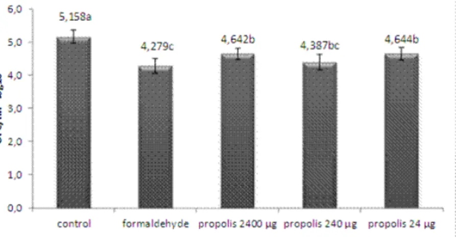
Levels of contamination of the eggshells by
total mesophiles expressed in log10 of CFU/mL
after disinfection can be observed in Figure 1. The control treatment differed from the others (P < 0.0001) showing a higher contamination level. However,
the propolis treatment in the 240 µg concentration
did not differ statistically from the formaldehyde treatment (P > 0.05).
In the evaluation of fungi contamination, Figure
2 shows the contamination by Aspergillus sp., where
there was no significant difference between the
treatments tested (P > 0.05). Yet, propolis treatments allowed a lower contamination than the control
treatment, and the 2,400 µg and 240 µg propolis
concentrations afforded a smaller number of colonies than the formaldehyde treatment. In Figure 3 it can be observed the contamination by other fungi, like
moulds, in which the treatments with 2,400 µg and 240 µg of propolis concentrations did not differ
statistically from the formaldehyde treatment (P > 0.05). On the other hand, these treatments did not differ statistically from the control group. Finally, Fig-ure 4 shows the total fungi contamination (Aspergillus
sp. and other moulds). None of the treatments differed statistically from the control group (P > 0.05).
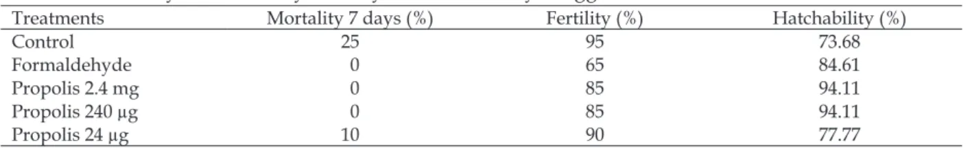
Regarding the embryo diagnosis (Table 1), the control treatment presented higher mortality rate
in the first seven days of incubation. The treatments with 2400 µg and 240 µg of propolis concentrations
and the formaldehyde treatment did not present mortality in this initial period of incubation. Regarding
the eggs hatchability after 21 days of incubation, the propolis treatments (2,400 µg and 240 µg) presented
the highest rates (94.1%), even higher than the eggs treated with formaldehyde. The lowest rate was
observed in the control treatment (77%).
DISCUSSION
Microbiological analyses are common prac-tices in industrial incubators aiming at detecting total mesophiles and fungi like Aspergillus sp. and other moulds. Considering the fact that the control group did not receive any kind of disinfection, it
was expected that this group would present higher
microbiological contamination, which happened when the total mesophiles prevalence was evalu-ated with higher CFU, differing statistically from all the other treatments (P < 0.0001) (Fig. 1). Similar result was observed by cony et al. (2008) when they
evaluated pulverization and immersion techniques with different disinfectants on embryonic eggs. The three treatments with propolis presented a lower contamination when compared to control group (P <
0.0001), and the treatment with 240 µg did not differ
statistically from the treatment with formaldehyde (P > 0.05). This decrease in the level of contamination of eggs makes evident the propolis antibacterial activity. According to Bankova et al. (1999) and marcucci et al. (2001), the propolis antibacterial activity is higher
against Gram positive bacteria, probably due to
flavonoids, acids and aromatic esters present in the
elucidated. Other authors have already reported on the propolis antimicrobial activity on a large variety of bacteria, especially on Gram positive (kujumgiev et al., 1999; miorin et al., 2003; uZel et al., 2005).
Recently, cardoSo et al. (2009) found similar results using green propolis ethanolic extract against
Staphylococcus aureus and Staphylococcus intermedius
isolates.
Regarding antifungal activity, (Figs. 2, 3 and
4) treatments with propolis allowed a lower con-tamination of eggs than the control treatment, and
propolis concentrations of 2,400 µg and 240 µg were more efficient than formaldehyde. The propolis
antifungal (fungistatic and fungicide) activity is attributed to the phenolic acids (cinnamic, feluric and
caffeic acids), terpenes and flavonoids like chrysin,
ermanina, galangin, kaempferol, pinobanskina and mainly pinocembrine (SiQueira et al., 2009). These
substances are found in green propolis (marcucci et
al., 2001; cuSHnie; lamB, 2005). It is worth noting that
in a study performed by our group (fiScHer et al., 2007) aiming at characterizing the immunomodulator
effect of the same green propolis ethanolic extract
evaluated in this study regarding its antimicrobial activity, a chromatographic analysis (High Performance
Liquid Chromatography – HPLC) showed high levels
of phenolic compounds and cinnamic acid and its
derived. In this extract, the flavonoids corresponded
to 22.37% of the dried extract (fiScHer et al., 2007).
Through embryo diagnosis it was possible to observe that the eggs that did not go through disinfection
resulted in a 25% embryonic mortality rate in the first seven days of incubation. This rate is considered extremely high, as in this initial period of incubation
it is accepted rates of up to 3% (roSa; Ávila, 2000). Treatments with propolis in concentrations of 2,400 µg and 240 µg and the formaldehyde treatment did
not present mortality in this initial period, indicat-ing that they provided an effective disinfection. When hatchability was evaluated, treatments with
propolis were more efficient than the others, what
makes evident that in these concentrations, propolis besides having no harmful effect can even increase the eggs hatchability.
Fig. 2 – Mean log10 CFU/mL of Aspergillus from embryonated eggs submitted to disinfection. Different letters represent
statistically significant differences (P < 0.05) by Tukey test.
Fig. 1 – Mean log10 CFU/mL of total mesophiles from embryonated eggs submitted to disinfection. The control treatment differed from the others (P < 0.0001) by Tukey test. In the others treatments different letters represent
Fig. 3 – Mean log10 CFU/mL of other mouldsfrom embryonated eggs submitted to disinfection. Different letters
repre-sent statistically significant differences (P < 0.05) by Tukey test.
Fig. 4 – Mean log10 CFU/mL of total mouldsfrom embryonated eggs submitted to disinfection. Different letters represent
statistically significant differences (P < 0.05) by Tukey test.
Table 1 – Embryonic mortality, fertility and hatchability of eggs submitted to disinfection.
Treatments Mortality 7 days (%) Fertility (%) Hatchability (%)
Control 25 95 73.68
Formaldehyde 0 65 84.61
Propolis 2.4 mg 0 85 94.11
Propolis 240 µg 0 85 94.11
Propolis 24 µg 10 90 77.77
CONCLUSIONS
The green propolis ethanolic extract, when used
as disinfectant by immersion of embryonated eggs, presented antibacterial and antifungal effect besides
not being harmful to the embryo development, allowing high hatchability rates.
The green propolis ethanolic extract is an alternative
ACKNOWLEDGEMENTS
We are thankful to Apis Nativa Produtos Nat-urais Ltda. (PRODAPYS) for supplying the Green
propolis ethanolic extract and to Coordenação de Aperfeiçoamento de Pessoal de Nível Superior (CAPES) for financial support.
REFERENCES
BANKOVA, V.; CHRISTOV, R.; POPOV, S.; MARCUCCI, M.C.; TSVETKOVA, I.; KUJUMGIEV, A. Antibacterial
activity of essential oils from Brazilian propolis. Fitoterapia, v.70, p.190-193, 1999.
BRAMWELL, R.K. Importancia de las prácticas de
manejo de lãs casetas de reprodutoras. Indústria Avícola,
v.47, p.8-18, 2000.
CABRAL, I.S.R.; OLDONI, T.L.C.; PRADO, A.; BEZERRA, R.M.N.; ALENCAR, S.M.D.; IKEGAKI, M.; ROSALEN, P.L. Composição fenólica, atividade antibacteriana e antioxidante da propolis vermelha
brasileira. Química Nova, v.32, p.1523-1527, 2009.
CARDOSO, R.L.; MABONI, F.; MACHADO, G.; ALVES, S.H.; VARGAS, A.C. 2009. Antimicrobial
activity of propolis extract against Staphylococcus coagulase positive and Malassezia pachydermatis of canine otitis. Veterinary Microbiology, v.142, p.432-434,
2009.
CONY, H.C.; VIEIRA, S.L.; BERRES, J.; GOMES, H.A.; CONEGLIAN, J L.B.; FREITAS, D.M. Técnicas de
pulverização e imersão com distintos desinfetantes sobre ovos incubáveis. Ciência Rural, v.38, p.1407-1412,
2008.
CUSHNIE, T.P.T.; LAMB, A.J. Antimicrobial activity of flavonoids. International Journal of Antimicrobial Agents,
v.26, p.343-356, 2005.
FISCHER G.; CONCEIÇÃO F.R.; LEITE F.P.; DUMMER L.A.; VARGAS G.D.; HÜBNER S. DE O.; DELLAGOSTIN. O.A.; PAULINO N.; PAULINO A.S.; VIDOR T. Immunomodulation produced by a green propolis
extract on humoral and cellular responses of mice
immunized with SuHV-1. Vaccine, v.26, p.1250-1256, 2007.
GREGORIS, E.; STEVANATO, R. Correlations between polyphenolic composition and antioxidant activity of
Venetian propolis. Food and Chemical Toxicology, v.48,
p.76-82, 2010.
HAUPTMANN M.; STEWART, P.; LUBIN, J.; FREE
-MAN, L.; HORNUNG, R.; HERRICK, R.; HOOVER, R.; FRAUMENI, J.; BLAIR, A.; HAYES, R. Mortality From
Lymphohematopoietic Malignancies and Brain Cancer
Among Embalmers Exposed to Formaldehyde. Journal of the National Cancer Institute, v.101, p.1696-1708, 2009.
HAYRETDA, S.; KOLANKAYA, D. Investigation of the
Effects of Pre-Incubation Formaldehyde Fumigation on the Tracheal Epithelium of Chicken Embryos and Chicks Turk. Journal of Veterinary Research and Animal Science, v.32, p.263-267, 2008.
HULEIHEL, M.; ISANU, V. Anti-Herpes Simplex Virus effect of an Aqueous Extract of propolis. The Israel Medical Association Journal, v.4, p.923-927, 2002.
INTERNATIONAL AGENCY FOR RESEARCH ON
CANCER - IARC. Formaldehyde, 2-Butoxyethanol and
Propylene Glycol Mono-t-Butyl Ether. Research lyon,
France: IARC Press, 2006. v.88.
KOC, A. N.; SILICI, S.; AYANGIL, D.; FERAHBAS, A.;
ÇANKAYA, S. Comparison of in vitro activities of
antifungal drugs and ethanolic extract of propolis against
Trichophyton rubrum and T. mentagrophytes by using a microdilution assay. Mycoses v.48, p.205-210, 2005.
KUJUMGIEV, A.; TSVETKOVA, I.; SERKEDJIEVA, Y.; BANKOVA, V.; CHRISTOV, R.; POPOV, S. Antibacte -rial, antifungal and antiviral activity of propolis of dif-ferent geographic origin. Journal of Ethnopharmacology,
v.64, p.235-240, 1999.
LI, F., AWALE, S.; ZHANG, H.; TEZUKA, Y.; ESUMI, H.; KADOTA, S. Chemical Constituents of Propolis from Myanmar and Their Preferential Cytotoxicity
against a Human Pancreatic Cancer Cell Line. Journal of Natural Products, v.72, p.1283-1287, 2009.
LUSTOSA, SARAH R.; GALINDO, A.B.; NUNES, L. C.C.; RANDAU, K. P.; ROLIM NETO, P.J. Propolis: atualizações sobre a química e a farmacologia. Revista Brasileira de Farmacognosia, v.18, p.447-454, 2008.
MAGRAS, I.N. Formaldehyde vapour effects in chicken embryo. Anatomia, histologia, embryologia. Journal of Veterinary Medicine, v.25, p.197-200, 1996.
MARCUCCI, M.C. Propolis: chemical composition, biological properties and therapeutical activity. Apidologie, v.26, p.83-99, 1995.
MARCUCCI, M. C; FERRERES, F.; GARCÍA-VIGUERA, C.; BANKOVA, V.S.; DE CASTRO, S.L.; DANTAS, A.P.; VALENTE, P.H.M.; PAULINO, N. Phenolic compounds
from Brazilian propolis with pharmacological activities. Journal of Ethnopharmacology, v.74, p.105-112, 2001.
MAULDIN, J.M. Maintaining hatching egg quality. In: BELL, D.D.; WEAVER, W.D. (Ed.). Commercial chicken meat and egg production. 5.ed. Norwell: Kluwer
Academic Publishers, 2002. p.705-707.
MIORIN, P.L.; LEVY JUNIOR, N.C.; CUSTODIO, A.R.; BRETZ, W.A.; MARCUCCI, M.C. Antibacterial activity
NOLKEMPER, S.; URGENREICHLING, J.; SENSCH, K.; SCHNITZLER, P. Mechanism of herpes simplex virus type 2 suppression by propolis extracts. Phytomedicine,
v.17, p.132-138, 2010.
PARK, Y.K.; ALENCAR, S.M.; SCAMPARINI, A.R.P.;
AGUIAR, C.L. Propolis produzida no sul do Brasil,
Argentina e Uruguai: Evidências fitoquímicas de sua
origem vegetal. Ciência Rural, v.32, p.997-1003, 2002.
PAULINO , N.; ABREU, S. R. L.; UTO, Y., KOYAMA, D.; NAGASAWA, H.; HORI, H.; DIRSCH, V. M.; VOLLMAR, A. M.; SCREMIN, A.; BRETZ, W. A. Anti-inflammatory effects of a bioavailable compound,
Artepillin C, in Brazilian propolis. European Journal of Pharmacology, v.587, p.296-301, 2008.
QUINTERO, M.; OROZCO, A.; HERNÁNDEZ, F.; GAYOSSO, P.; MARTÍNEZ,R.; ZÁRATE, C.; MIRANDA,L.; CARRILLO, G.; TOVAR, C.; SÁNCHEZ, T. Efecto de extractos de propóleos mexicanos de Apis mellifera sobre el crecimiento in vitro de Candida albicans. Revista Iberoamericana de Micologia, v.25, p.22-26, 2008.
ROSA, P.; ÁVILA, V. Variáveis relacionadas ao rendi -mento da incubação de ovos em matrizes de frangos de
corte. CT/246/Embrapa Suínos e Aves, p.1-3, 2000.
SALOMÃO, K.; SOUZA, E.; PONS,H.; BARBOSA, H.;CASTRO, S. Brazilian Green Propolis: Effects In
Vitro and In Vivo on Trypanosoma cruzi. Evidence-based Complementary and Alternative Medicine, v.2011, p.1-11,
2011.
SAS INSTITUTE. SAS User’s guide: Statisics. Version
8.0 Edition. Cary, NC, 2001.
SCHNITZLER, P.; NEUNER, A.; NOLKEMPER, S.; ZUNDEL, C.; NOWACK. Antiviral activity and mode of action of propolis extracts and selected compounds.
Phytotherapy Research, v.24, p.20-28, 2010.
SCOTT, T. A.; SWETNAM, C. Screening sanitizing
agents and methods of application for hatching eggs. II. Effectiveness against microorganisms on the egg shell. Journal Applied Poultry Research, v.2, p.7-11, 1993.
SESTI, L.A.C. Biosseguridade em granjas de reprodu
-toras In: MACARI, M.; MENDES, A.A. (Ed.). Manejo de matrizes de corte. Santos: Facta, 2005. p.244-317.
SFORCIN, J.M.; FERNANDES JUNIOR, A.; LOPES, C.A.M.; BANKOVA, V.; FUNARI, S.R.C. Seasonal effect
on Brazilian propolis antibacterial activity. Journal of Ethnopharmacology, v.73, p.243-249, 2000.
SILVA, N.; JUNQUEIRA, V.C.A.; SILVEIRA, N.F.A. Ma-nual de métodos de análise microbiológica de alimentos. São
Paulo: Varela, 1997. p.295.
SIQUEIRA, A.B.S.; GOMES, B.S.; CAMBUIM, I.; MAIA, R.; ABREU, S.; SOUZA-MOTTA, C.M.; QUEIROZ, L.A.;
PORTO, A.L.F. Trichophyton species susceptibility to green and red propolis from Brazil. Letters in Applied Microbiology, v.48, p.90-96, 2009.
UZEL, A.; SORKUN, K.; ÖNÇAG, Ö., ÇOGULO, D.; GENÇAY, Ö.; SALIH, B. Chemical compositions and
antimicrobial activities of four different Anatolian propolis samples. Microbiology Research, v.160,
p.189-195, 2005.
ZULKIFLI, I.; FAUZIAH, O.; OMAR, A.R.; SHAIPU
-LLIZAN S.; SITI SELINA, A.H. Respiratory epithelium, production performance and behaviour of
formaldehyde-exposed broiler chicks.Veterinary Research Communications,
v.23, p.91-99, 1999.
Received on 15/3/11