PONTIFÍCIA UNIVERSIDADE CATÓLICA DO RIO GRANDE DO SUL FACULDADE DE ODONTOLOGIA
PROGRAMA DE PÓS-GRADUAÇÃO EM ODONTOLOGIA MESTRADO EM ODONTOLOGIA
ÁREA DE CONCENTRAÇÃO EM ENDODONTIA
MICROSCOPIA ELETRÔNICA DE VARREDURA E ANÁLISE MICROBIOLÓGICA DE CANAIS DE DENTES BOVINOS INFECTADOS PELO
E. FAECALIS E SUBMETIDOS À TERAPIA FOTODINÂMICA.
JANAÍNA GUZZO ZECHIN KUFNER
Dissertação apresentada como parte dos requisitos obrigatórios para obtenção do
título de Mestre em Odontologia, área de concentração em Endodontia.
Linha de pesquisa
Etiopatogênese e Tratamento das Doenças Periodontais e Periapicais
Prof. Dr. José Antonio Poli de Figueiredo Orientador
Dados Internacionais de Catalogação na Publicação (CIP)
K95m Kufner, Janaína Guzzo Zechin
Microscopia eletrônica de varredura e análise microbiológica de canais de dentes bovinos infectados pelo E. faecalis e
submetidos à terapia fotodinâmica / Janaína Guzzo Zechin Kufner. – Porto Alegre, 2011.
45 f.
Diss. (Mestrado) – PUCRS. Faculdade de Odontologia. Programa de Pós-Graduação em Odontologia. Área de
concentração: Endodontia. Linha de Pesquisa: Etiopatogênese e Tratamento das Doenças Periodontais e Periapicais.
Orientador: Prof. Dr. José Antonio Poli de Figueiredo. 1. Odontologia. 2. Endodontia. 3. Tratamento de Canal Radicular. 4. Terapia Fotodinâmica. 5. Microscopia Eletrônica. 6. Bactérias. I. Figueiredo, José Antonio Poli de. II. Título.
CDD 617.634
Aos meus pais, Genir e Nilsa, que fizeram a Odontologia ser uma realidade na minha vida; um agradecimento especial à minha mãe por ter sido uma incansável companheira e amiga em toda a minha trajetória: escola, faculdade, especialização e mestrado.
Ao meu marido, Cleverson Evandro, nenhuma palavra conseguirá transmitir a ideia de quanto a sua presença foi importante nos momentos difíceis e nos momentos felizes; “...você é o amor da minha vida, você é a escada da minha subida...”.
À minha grande amiga e colega, Vanessa Schmitt por ter participado de cada etapa do meu crescimento profissional e principalmente pela seguinte frase: “Tu já pensaste em fazer mestrado?”. Esse título de mestre será um pouco seu também.
Ao meu orientador Prof. Dr. José Antônio Poli de Figueiredo, sua sabedoria e humildade são fontes inspiradoras para minha vida profissional e pessoal; muito obrigada!
À minha amiga e colega, Grasiela, por ter me mostrado que a perseverança, a seriedade e a amizade nos fazem ultrapassar qualquer obstáculo; nunca mais esquecerei esse momento em que convivemos juntas.
À professora Dra. Silvia Dias de Oliveira, cuja participação foi fundamental para a realização dessa pesquisa.
Aos meus colegas do Mestrado e Doutorado em Endodontia, pois a minha vida teve uma separação bem clara, o antes e o depois da nossa convivência; um agradecimento especial ao Carlos Wolle e à Caroline Marca.
À minha colega, Alessandra Trindade por ter iluminado o meu caminho quando eu mais precisei no início do Mestrado.
Aos meus amigos e colegas de profissão, Juliana e Davison, pela paciência e apoio durante esse período.
Aos meus colegas de trabalho do Sindicato dos Trabalhadores Rurais, pelo companheirismo e compreensão durante esses dois anos.
A todos os amigos que compreenderam a minha ausência e me deram apoio incondicional nesses dois anos; um agradecimento especial a Claudia Dal Pozzo e a Marelice Dalla Libera por terem participado efetivamente desse trabalho.
Aos funcionários da Secretaria de Pós-Graduação da Faculdade de Odontologia, pela atenção e carinho em todos os momentos.
Aos técnicos do Centro de Microscopia Eletrônica e Microanálises da PUCRS, Maurício, Eduardo e Miriam, pelo profissionalismo e por terem tornado aquelas horas mais agradáveis.
O objetivo deste estudo foi testar a influência da utilização da fibra óptica e de diferentes tempos de pré-irradiação (PIT) na terapia fotodinâmica com LED, na desinfecção de canais radiculares de dentes bovinos infectados, in vitro, com Enterococcus faecalis. Cento e vinte dentes bovinos foram inoculados com E. faecalis, permanecendo em cultivo por 60 dias. Os dentes foram divididos em seis grupos (n=20): Grupo 1- água destilada; Grupo 2- 1 minuto PIT/sem fibra óptica; Grupo 3- 1 minuto PIT/fibra óptica; Grupo 4- 5 minutos PIT/sem fibra óptica; Grupo 5 - 5 minutos/com fibra óptica; Grupo 6 –hipoclorito de sódio 2%. Foram realizados testes microbiológicos e análise em SEM. A análise microbiológica mostrou que o grupo 6 obteve os melhores resultados, com diferença estatística entre este e os grupos 1, 2, 3, 4 e 5. A análise da microscopia eletrônica de varredura, nos três terços da parede do canal mostrou os melhores resultados para o grupo 6 (com diferença estatisticamente significativa com os grupos 1, 3, 4 e 5) e o grupo 2, entre os grupos da terapia fotodinâmica. Na área de túbulos expostos, no terço apical os grupos com melhores resultados foram os grupos 1 e 3; no terço médio grupos 6 e 2; e no terço cervical grupos 6 e 3. Não foi encontrada diferença na desinfecção dos canais radiculares quando a fibra óptica foi usada na aplicação da terapia fotodinâmica. Esta não pode ser usada de forma isolada na desinfecção dos canais radiculares, o seu valor pode estar na complementação da desinfecção obtida por outros protocolos de limpeza.
ABSTRACT
This study evaluated the use of optic fiber and different pre-irradiation times (PIT) in phototherapy with LED for the root canal disinfection of bovine teeth infected in vitro with Enterococcus faecalis. One hundred twenty bovine teeth were inoculated with E. faecalis and incubated for 60 days. The teeth were divided into six groups (n=20): Group 1 – distilled water; Group 2 – 1 minute PIT and no optic fiber; Group 3 – 1 minute PIT and optic fiber; Group 4 – 5 minute PIT and no optic fiber; Group 5 – 5 minutes PIT and optic fiber; Group 6 – 2% sodium hypochlorite. Specimens were evaluated using microbiological tests and SEM analysis. Microbiological analysis revealed that group 6 had the best results and there were statistically significant differences between this group and groups 1, 2, 3, 4 and 5, there were no statistically significant differences between groups 1, 2, 3, 4 and 5. SEM analysis of canal wall, in the three thirds, revealed group 6 had the best results (there were statistically significant differences between this group and groups 1, 3, 4 and 5) and group 2 had the best results between groups of photodynamic therapy. In exposed tubules, group 1 had the best results in the apical, and group 3 had the best results between groups of photodynamic therapy. Group 6 had the best results in the middle and coronal third, and between groups of photodynamic group 2 had the best results in the middle third and group 3 in the coronal third. No differences in disinfection of root canals were found when optic fiber were used for photodynamic therapy. Photodynamic therapy should not be used alone in the disinfection of root canals, but it may be valuable as a complement for disinfection performed using different cleaning methods.
1 INTRODUÇÃO ... 7
2 ARTIGO ... 10
3 DISCUSSÃO GERAL ... 28
4 REFERÊNCIAS BIBLIOGRÁFICAS ... 31
ANEXOS ... 36
ANEXO A - Ofício de aprovação do projeto de pesquisa no Comitê de Ética para o Uso de Animais. ... 37
ANEXO B - Sequência de diluição ... 38
1 INTRODUÇÃO
Dentre os objetivos do tratamento endodôntico está sanificar e modelar o canal radicular, visando à eliminação dos micro-organismos, baseando-se a sua permanência ou não no sistema de canais radiculares relacionada com o sucesso da terapia endodôntica (1,2).
Miller (3) demonstrou a importância dos microrganismos na etiologia das doenças da polpa e periápice. Já o trabalho clássico de Sundqvist (4) verificou a presença e a importância de micro-organismos anaeróbios nas infecções endodônticas.
A própria anatomia dos canais radiculares, com canais secundários, acessórios e delta apical, não favorece a ação mecânica das limas e a ação química das substâncias auxiliares, podendo permanecer micro-organismos neste sistema e favorecer a recolonização do canal radicular (5,6).
O Enterococcus faecalis, coco Gram-positivo anaeróbio facultativo, possui importante papel no insucesso da terapia endodôntica sendo frequentemente encontrado no sistema de canais radiculares de dentes portadores de lesões periapicais crônicas nos retratamentos endodônticos (4,7,8). A virulência deste tem relação à sua resistência às medicações intracanais mais comumente utilizadas e à habilidade de sobrevivência em canais radiculares sem o suporte de outra bactéria (9,10,11,12).
Segundo alguns estudos, o hipoclorito de sódio é eficaz na eliminação de E. faecalis do interior dos túbulos dentinários (13,14,15). Porém, possui algumas desvantagens como a instabilidade ao armazenamento e inativação por matéria orgânica (16), é extremamente citotóxico quando extravasado no interior dos tecidos perirradiculares (17), diminui a resistência à fratura dos dentes e a resistência de união dos materiais restauradores à dentina (18).
Foi verificado nos estudos de Kairalla (19) e Gutnecht et al. (20) que a redução microbiana pode ultrapassar 99% com a utilização de lasers de alta potência (Er:YAG, Er,Cr:YSGG, Nd:YAG, Ho:YAG e diodo), provocando a morte do micro-organismo por elevação da temperatura, através da desnaturação protéica. Entretanto, a utilização de lasers de alta potência, sem haver controle do fator térmico, pode gerar riscos de injúrias aos tecidos dentais e vizinhos, como carbonização da dentina, promoção de anquilose, derretimento de cemento, reabsorção radicular e necrose perirradicular (19,21).
a luz desencadeando uma cascata de eventos químicos, resultando na produção de espécies reativas de oxigênio, tóxicas às células tumorais, bactérias e fungos (22,23,24,25,26).
O termo LED é um acrônimo para Light Emiting Diode, onde dois diferentes compostos semicondutores emitem luz ao reagir com uma determinada tensão aplicada. Pode-se ter diferentes cores de LED: vermelho, infravermelho e azul; Pode-sendo que cada cor aprePode-senta o seu respectivo comprimento de onda (27).
Os lasers de baixa potência não promovem alterações morfológicas na estrutura dentária e adjacentes (19); bem como o LED promove pequena variação de temperatura (28).
A desinfecção dos canais radiculares através da terapia fotodinâmica tem sido estudada nos últimos anos, trazendo resultados positivos, principalmente sobre o E. faecalis (19,24,25,29).
A PDT tem sido um método efetivo na redução da contaminação intracanal, porém, com variação do protocolo quanto ao uso dos fotossensibilizadores: o azuleno 25%, azul de ortotoluidina 0,0125% e azul de ortotoluidina 0,001%. Já no que se refere à forma de entrega da irradiação intracanal observou-se uma concordância nas pesquisas, sendo o feixe de luz emitido através de fibra óptica (26,30,31).
Contrariando estes resultados, alguns estudos concluíram que a associação entre o fotossensibilizador e o laser teve um efeito bactericida, entretanto, não foi capaz de eliminar totalmente o biofilme dos canais radiculares. Nestes estudos a concordância no protocolo da terapia fotodinâmica ocorreu em relação ao uso do azul de ortotoluidina, havendo a variação quanto ao uso ou não da fibra óptica (29,32).
É necessário um tempo de exposição prévia do tecido alvo ao corante antes da exposição à radiação luminosa (tempo de pré-irradiação) quando se utilizam os corantes azul de metileno e azul de ortotoluidina (23). Alguns autores adotaram 5 minutos de tempo de pré-irradiação em seus trabalhos (19,30,33); já outro, 2 minutos de tempo de pré-pré-irradiação (24).
2 ARTIGO
Comparative analysis of microbial reduction using photodynamic therapy optic fiber in bovine teeth infected in vitro with Enterococcus faecalis.
Comparative analysis of microbial reduction using photodynamic therapy optic fiber in bovine teeth infected in vitro with Enterococcus faecalis.
Janaina Zechin Kufner1, Liviu Steier2, Giampiero Rossi-Fedele1,2, Grasiela Grundling1, Silvia Dias de Oliveira3, Daniela Sá3, Amanda Guwzinski3, Alessandra Cesar Trindade1, José
Antônio Poli de Figueiredo1
1 School of Dentistry, Pontifícia Universidade Católica do Rio Grande do Sul, Porto Alegre, RS, Brazil.
2 Warwick Dentistry, Warwick Medical School, Warwick, United Kingdom.
ABSTRACT
Introduction: This study evaluated the use of optic fiber and different pre-irradiation times (PIT) in phototherapy with LED for the root canal disinfection of bovine teeth infected in vitro with Enterococcus faecalis. Material and Methods One hundred twenty bovine teeth were inoculated with E. faecalis and incubated for 60 days. The teeth were divided into six groups (n=20): Group 1 – distilled water; Group 2 – 1 minute PIT and no optic fiber; Group 3 – 1 minute PIT and optic fiber; Group 4 – 5 minute PIT and no optic fiber; Group 5 – 5 minutes PIT and optic fiber; Group 6 – 2% sodium hypochlorite. Specimens were evaluated using microbiological tests and SEM analysis. Results: Microbiological results revealed that group 6 had the best results and there were statistically significant differences between this group and groups 1, 2, 3, 4 and 5, there were no statistically significant differences between groups 1, 2, 3, 4 and 5. SEM analysis of canal wall, in the three thirds, revealed group 6 had the best results (there were statistically significant differences between this group and groups 1, 3, 4 and 5) and group 2 had the best results between groups of photodynamic therapy. In exposed tubules, group 1 had the best results in the apical third, and group 3 had the best results between groups of photodynamic therapy. Group 6 had the best results in the middle and coronal third, and between groups of photodynamic group 2 had the best results in the middle third and group 3 in the coronal third. Conclusions: No differences in disinfection of root canals were found when optic fiber were used for photodynamic therapy. Photodynamic therapy should not be used alone in the disinfection of root canals, but it may be valuable as a complement for disinfection performed using different cleaning methods.
Key words: biofilm, Enterococcus faecalis, scanning electron microscopy, photodynamic therapy.
INTRODUCTION
Photodynamic therapy (PDT) has been described as an alternative for the disinfection of root canals. It uses low density light (laser or LED) associated with exogenous photosensitizers that absorb light and trigger chemical events that result in the production of oxygen reactive species, which are toxic to tumor cells, bacteria and fungi (8-12).
Low-intensity laser does not promote morphological changes in tooth or adjacent structures (13), and LED produces only a small temperature variation (14).
Disinfection of root canals using PDT has been studied in the last years, and results have been positive, particularly when used against E. faecalis (10, 11, 13, 15).
PDT is effective in reducing intracanal contamination, but when used according to different protocols in which different photosensitizers were applied: 25% azulene, 0.0125% ortho-toluidine blue, and 0.001% ortho-toluidine blue. Also, in these cases, the light beam may be emitted using an optic fiber (12, 16, 17).
Contrary to those results, some studies found that the combined use of photosensitizer and laser had a bactericide effect, but was not capable of completely removing biofilm from root canals. Those studies were similar to those that applied PDT with ortho-toluidine blue as a photosensitizer, but differed in whether they used or not an optic fiber (15, 18).
The target tissues should be previously exposed to dyes before exposure to light radiation, called pre-irradiation time (PIT), and methylene blue and ortho-toluidine blue have been used for this purpose (9). Some authors adopted a 5 minute PIT (13, 16, 19, 20) and others used 2 minute (10, 21) or 10 minute PIT (22).
Several LED or laser units do not have an optic fiber delivery mode. In these cases, and if an optic fiber is proven to affect the results, these units would not have any use in endodontic treatments. However, if the laser beam or LED has enough penetration to reach the photosensitizer even from a distance, the use of optic fibers would be unnecessary, the chances of using PAD would increase, and costs would be reduced. Therefore, it is necessary to know how this treatment would act on consolidated biofilm with microorganisms that are resistant to traditional therapies.
Moreover, there is no consensus in the literature about which protocol to use in adjuvant photodynamic therapy during conventional endodontic treatment. This study compared the effect of the use of an optic fiber and different PIT using red light LED to clean root canals infected with E. faecalis.
This study was approved by the Ethics Committee for the Use of Animals (CEUA) of Pontifícia Universidade Católica do Rio Grande do Sul (PUCRS) under number 10/00163 (Annex A).
Tooth collection and preparation
One hundred and twenty bovine incisors were obtained from animals slaughtered for commercial purposes. Teeth were removed from the mandibles immediately after slaughtering and were stored in 1% sodium hypochlorite (ASPER, Indústria Química Ltda, São Caetano do Sul, Brazil) for no longer than 48 h. Dental crowns and 1 mm of the apical region were sectioned so that all roots measured 15 mm long. To remove the pulp and have a uniform canal diameter, each root was prepared using a 21-mm # 60 instrument (DentsplyMaillefer, Ballaigues, Switzerland) and irrigation with 2% sodium hypochlorite (2% Virex Plus, Johnson Diversey Brasil Ltda, São Paulo, Brazil). After that, the roots were kept immersed in 17% EDTA (Farmashop, Porto Alegre, Brazil) for 5 minutes under agitation to remove the smear layer.
The 126 teeth were fixed in a polypropylene microtube (Genuine Axygen Quality, CA) with cyanoacrylate (Super Bonder, São Paulo, Brazil) to keep it vertical with the coronal side up. Each sample was randomly placed in 6 autoclave polypropylene boxes (Heathrow, Vernon Hills, IL). After that, the orifice to each root was sealed with autoclave tape (3M do Brasil, Sumaré, Brazil), and a hole was made in the microtube cap to make it possible to change BHI (Brain Heart Infusion - Difco Laboratories, Detroit, USA) broth during incubation. After mounting, the set formed by the box and tubes with specimens were autoclaved (Kavo, Joinville, Brazil) at 121o C for 15 minutes.
Sterilization control
the sterilization of the material. The specimen used for sterilization control was not reused, and there was a total of 20 teeth per box.
Culture and preparation of inoculation material
The E. faecalis (ATCC 29212) was obtained and cultured, in BHI broth for 18 to 24 hours at 37O C in an incubator, in the Immunology and Microbiology Laboratory of the School of Biosciences, PUCRS.
The number of colony forming units (CFU/ml) in the inoculation suspension was determined by counting them in blood agar dishes. For that, the E. faecalis suspension was serially diluted up to 10-8 in 0.85% saline solution, and 0.1 ml of the 10-6, 10-7 and 10-8 concentration suspensions were seeded in duplicate on blood agar using a sterilized Drigalsky rod. The plates were incubated at 37o C for 24 hours, after which CFU/ml were counted in the plates that had 15 to 150 colonies. Bacterial density ranged from 4.0 x 10⁸ to 7.2 x 10⁸.
The 120 samples were inoculated with 100 µl of E. faecalis suspension injected into the root canal using a 1 mL insulin syringe (BD, Curitiba, Brazil). Sterile BHI broth was then added to the plastic tube so that was completely filled with culture medium. E. faecalis was culture was incubated for 60 days for the formation of biofilm, and one third of the BHI broth volume was replaced every 48 hours. All teeth were handled under aseptic conditions under a laminar flow hood. Once a week, an aliquot of BHI of the teeth was Gram stained, cultured in blood agar, and submitted to catalase and esculin tests to confirm the exclusive presence of E. faecalis.
Group classification
Teeth were loosened from the tubes and mounted in a utility wax base (Wilson, Polidental, Cotia, Brazil) to avoid overflowing of the irrigating solution and photosensitizers through the apical foramen.
Working length was established at 14 mm, and the groups were divided as described below:
at 30 second intervals. Results of this group were used as bacteria counts before treatment of root canals of the other groups.
Group 2 (n=20): canals were irrigated using a 5 mL disposable plastic syringe with 2 mL of distilled water that was later aspirated also using a 5 mL disposable syringe. The canals were then filled up with 0.001% ortho-toluidine blue (tolonium chloride) viscous photosensitizer using a 5 mL disposable syringe. After 1 minute, tolonium chloride was stirred with a 21-mm-long #50 file at 14 mm without touching the walls of the root canal. The LED beam was activated for 120 seconds using a PAD Plus unit (Denfotex Light Systems Ltd, Inverkeithing, United Kingdom) and 630-nm red light; the optic fiber was not coupled to the LED tip, and the pointer was placed at the canal entrance.
Group 3 (n=20): The procedure was the same as for group 2, but an optic fiber was coupled to the LED tip. The stationary technique was used: the irradiation is distributed along the three thirds of the root canals using preprogrammed pullbacks at three time points, also at a total of 120 seconds.
Group 4 (n=20): the procedure was similar to that followed in group 2, but PIT was 5 minutes and the ortho-toluidine blue photosensitizer was not stirred.
Group 5 (n=20): The procedure was again the same as in group 2, but PIT time was 5 minutes, the photosensitizer was not stirred and the optic fiber was coupled to the LED tip; the stationary technique was the same as in group 3.
After irradiation, the canals in each group were irrigated again with 2 mL distilled water using a 5 mL disposable plastic syringe and aspirated also with a 5 mL disposable plastic syringe.
Group 6 (n=20): irrigation with 2% sodium hypochlorite that filled up the root canals and was kept inside the canal for one minute; all the solution was then aspirated using a 5 mL disposable plastic syringe (BD, Curitiba, Brazil). Each sample received four irrigations at 30 second intervals.
Microbiological analysis
the canal and placed into a tube with 450 µl of sterile 0.85% saline solution. The material was homogenized and diluted to 10-3. Aliquots of 100 µl saline solution and diluted concentrations were seeded in duplicate in blood agar using a Drigalsky rod and incubated for 18 to 24 hours at 37o C. After incubation, the number of CFU was counted in the plates that had 15 to 150 colonies.
Preparation for SEM
The other 10 teeth from each group were immersed in a fixing solution (2.5% glutaraldehyde) immediately after treatment for 7 days for later analysis under scanning electron microscopy (SEM) in the Electronic Microscopy and Microanalysis Center of PUCRS.
Roots were rinsed three times for 30 minutes each time in a 0.2 M phosphate buffer and distilled water at a 1:1 ratio. After that, they were dehydrated by immersion in 30, 50, 70, 90 and 100% acetone. Longitudinal buccolingual grooves were produced on the free surfaces of the roots using a diamond bur (Dhpro, Rhadartrade, Paranaguá, Brazil); care was taken not to penetrate the interior of the root canal. Root fracture was completed using a #50 spatula (SS White, Rio de Janeiro, Brazil) to obtain two halves that were mounted in stubs with the root canal end up. After that, the samples were spurted-coated with gold for electron conduction.
A scanning electron microscope (Phillips XL-30, Eidhoven, Holland) was used for evaluations of roots according to thirds (coronal, middle and apical) at 500 X to 20000X magnification. First, areas of greater concentration of biofilm were selected at a lower magnification; recordings were then made at 5000 X magnification. Backscattering (BSE) was used for image capture.
One single observer blinded to the experimental groups classified images according to the presence of bacteria and using the criteria of position ranks.
Using PowerPoint (Microsoft, Washington, USA), each image was placed in one slide; the less contaminated tooth was classified as position 1, and the most contaminated, as position 60. This classification was used for each third (coronal, middle and apical) and according to the position in the image (canals wall or area of exposed tubules). Therefore, for each third and each site in the image, the mean position of the group was calculated.
a 90% statistical power and level of statistical significance of 5%, a number of 10 specimens was calculated for each group.
Data on levels of contamination measured with electronic microscopy were ranked within thirds. One-way ANOVA was applied to ranks (23) with a robust significance, followed by the Tukey post hoc test to detect differences.
In the microbiology evaluation, all data were log transformed. One-way ANOVA was followed by the Tukey post hoc test.
The level of significance was set to α=0.05. Data were analyzed using the SPSS 17.0.
RESULTS
Results are shown in Figure 1, which summarizes findings of microbiological analysis and SEM; Table 1 shows only SEM findings.
Microbiological analysis
Group 1 had a mean count of 5.47 log CFU/mL; Group 2: 5.51 log CFU/mL; Group 3: 5.33 log CFU/mL; Group 4: 5.80 log CFU/mL; Group 5: 5.41 log CFU/mL and Group 6: 1.07 log CFU/mL.
Group 6 had the best results and there were statistically significant differences between this group and groups 1, 2, 3, 4 and 5 (p = 0,000). There were no statistically significant differences between groups 1, 2, 3, 4 and 5.
SEM analysis
Table 1 shows the results according to the position ranks.
In the exposed tubules, group 1 had the best results in the apical third, there were statistically significant differences between this group and groups 2, 4 and 5 (p<0,001). In this third, group 3 had the best results between groups of photodynamic therapy but there were no statistically significant differences with other groups.
In the exposed tubules, group 6 had the best results in the middle and coronal third. There were statistically significant differences between group 6 and groups 3, 4 and 5 in the middle third (p<0,001), and groups 1, 3, 4 and 5 in the coronal third (p=0,011). Between groups of photodynamic group 2 had the best results in the middle third and group 3 in the coronal third. There were statistically significant differences between group 2 and groups 4 and 5 in the middle third (p<0,001), and between group 3 and groups 1, 4 and 5 in the coronal third (p=0,011).
Figure 1 - Graph a: microbiological tests: Co (group 1) = control with distilled water; FT1Wo (group 2) =
treatments applied to root canals of bovine incisors.
Variable Group 1 Group 2 Group 3 Group 4 Group 5 Group 6 P
Canal Wall n=10 n=10 n=10 n=10 n=10 n=10
Apical third 31(14.3) b.c 23.8(10)a.b 30.2(17.2)b.c * 41.6(10.2)c 44.2(16.4)c 9.2(7.6)a < 0.001
Middle third
29.8(15.3)b.c 22.4(11.2)a.b 33.8(13.7)b.c 38.7(12.9)c.d 50(9.1)d 8.3(8)a < 0.001
Coronal third
38.7(12.5)cd** 15.5(9.8)a.b 28(17.2)b.c 46(7.9)d 42.8(12.9)c.d 11.9(7.7)a < 0.001
Exposed tubules
n=10 n=10 n=10 n=10 n=10 n=10
Apical third 10.5(5.2)a*** 32.8(15.3)bc* 22.5(8.8)a.b.c* 37.5(8.3)c 37(16.5)c 17.7(15.9)a.b < 0.001
Middle third
28.7(20.2)a.b 29(9.6)a.b* 32.6(19)bc 37.6(8.9)cd 41.9(16.2)d 10.1(5.4)a < 0.001
Coronal third
33(20.6)cd** 27.9(12.8)ab 24.3(18.9)bc 40.8(11.2)d 36.4(13.5)cd 15.2(13.2)a 0.011
Data are presented as mean ranks (standard deviation). P = robust significance (Brown-Forsythe) obtained using one-way ANOVA of ranks. Different letter indicate significant differences, in the same line, according to Tukey post hoc test. [*]: sample size reduced to n=9. [**]:sample size reduced to n=8. [***]: sample size reduced to n=6.
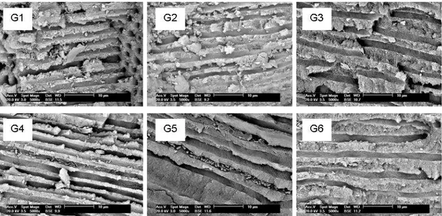
Figure 3- SEM photomicrographs (5000x – canal wall – middle third) of groups under study – G1: group 1; G2: group 2; G3: group 3; G4: group 4; G5: group 5; G6: group 6.
DISCUSSION
Photodynamic therapy has gradually expanded from Medicine to Dentistry, and one of its uses is to be an alternative to cleaning and microbial reduction within root canals. In this study, photodynamic therapy was applied to a biofilm of E. faecalis grown in roots of extracted bovine teeth.
Bovine teeth were chosen because it has already been demonstrated that there are no differences in physical properties between bovine and human dentin in permanent teeth (24). Moreover, it was possible to obtain samples of similar age and dentin properties, and it was possible to distribute teeth of a single animal into several experimental groups, which reduced the number of variables in clinical trials.
Biofilm found in vivo in teeth with apical periodontitis is mature, strongly adhered to the substrate and penetrating into dentinal tubules, which make it more resistant to cleaning and shaping (25). To obtain consistent and organized biofilm samples, E. faecalis was incubated for 60 days. A previous study determined that there is marked invasion of E. faecalis into dentinal tubules at 56 days (26).
The sodium hypochlorite concentration chosen was 2% because that is what is used in the School of Dentistry of PUCRS for the clinical treatment of endodontic infection.
eliminating bacteria and fungi (28). Methylene blue and ortho-toluidine blue were tested to find out which one would have a greater bactericide action against different types of bacteria, both Gram positive and Gram negative. Both were efficient when red-emitting laser was used, but ortho-toluidine blue had the best bactericide effect (29). Therefore, ortho-toluidine blue (tolonium chloride) was the photosensitizer of choice for this study.
The amount and distribution of bacteria on the surface of biofilm may be compared between treatments when the samples are analyzed using SEM (25,30,31,32). For that purpose, SEM was used in our study. A large number of SEM analyses were made to ensure that the areas under study were representative of biofilm distribution on the whole surface of the root canal (25). However, SEM cannot demonstrate bacteria viability. To address this problem, bacterial counts were performed before and after the treatment for each group.
Microbiologic and SEM analyses used different specimens to avoid changes in the results; otherwise, the amount and quality of bacterial biofilm might have been affected by the collection of saline solution from the root canal, which might compromise SEM.
Microbiological results revealed that there were no differences when different photosensitizer PITs were used: 1 minute with photosensitizer stirring, as recommended by the PAD Plus manufacturer, and 5 minutes according to literature (13, 16, 19). When compared the six groups together, the group 6 (sodium hypochlorite 2%) had the best results. No differences were found in canal disinfection when the optic fiber was coupled to the LET tip. The stationary technique was chosen because the comparison of three different techniques, the stationary and the helicoidal techniques and application without optic fiber, did not reveal any differences between them (10).
SEM results revealed superiority of group 6 and group 2. Between groups of photodynamic therapy group 2 had the best results, except in the apical e coronal thirds of exposed tubules that group 3 had this results.
More important than our results, which are contributions to the establishment of a photodynamic therapy protocol, this study confirmed the effectiveness of a protocol of contamination, culture and material collection for later bacterial count.In this study, samples were extracted bovine teeth, but this protocol can also be used with extracted human teeth (33).
photodynamic therapy. However when different photosensitizer PIT were tested, there were statistically significant differences between groups of 1 minute PIT and groups of 5 minutes PIT.
A bactericide effect was found when the photodynamic therapy was associated with cleaning and shaping of root canals using sodium hypochlorite as an irrigating solution (34,35), together with inactivation of endodontic pathogens without affecting host cell viability (36). PLGA nanoparticles with photoactive drugs may enhance its antimicrobial use (37) Therefore, our findings confirm that photodynamic therapy alone does not affect a well-structured biofilm, such as the one produced by E. faecalis. Photodynamic therapy is valuable, however, as a complementary disinfection method for other cleaning protocols.
Conclusions: No differences in disinfection of root canals were found when optic fiber were used for photodynamic therapy. It should not be used alone in the disinfection of root canals, but it may be a valuable complementary method for disinfection performed using different cleaning protocols.
REFERENCES
1. Sundqvist G, Figdor D, Persson S, Sjögren U. Microbiologic analysis of teeth with endodontic treatment and the outcome of conservative retreatment. Oral Surg Oral Med Oral Pathol Oral Radiol Endod. 1998;85:86-93.
2. Molander A, Reit C, Dahlén G, Kvist T. Microbiological Status of root-filled teeth with apical periodontis. Int End J. 1998;31(1):1-7.
3. Love RM. Enterococcus faecalis – a mechanism for its role in endodontic failure. Int Endod J. 2001;34(5):399-405.
Endod J. 1992;25(2):97-106.
6. Byström A, Sundqvist G. The antibacterial action of sodium hypochlorite and EDTA in 60 cases of endodontic therapy. Int Endod J. 1985;18(1):35-40.
7. Peters LB, Wesselink PR, Moorer WR. The fate and the role of bacteria left in root dentinal tubules. Int Endod J. 1995;28(2):95-9.
8. Konopka KE, Goslinski T. Photodynamic Therapy in Dentistry. J Dent Res. 2007;86(8):694-707.
9. Wainwright M. Photodynamic antimicrobial chemotherapy (PACT). J Antimicrob Chemother. 1998;42(1):13-28.
10. Cavalheiro FM. Assessment of bacterial reduction in contaminated root canals comparing three irradiation techniques with low power laser associated with a photosensitizer, an in vitro study [Dissertation]. São Paulo: Dental School of USP; 2007.
11. Williams JA, Pearson GJ, Colles MJ. Antibacterial action of photoactived disinfection {PAD} used on endodontic bacteria in planktonic suspension and in artificial and human root canals. J Dent 2006;34(6):363-71.
12. Silva Garcez A, Núñez SC, Lage-Marques JL, Jorge AO, Ribeiro MS. Efficiency of NaOCl and laser-assisted photosensitization on reduction of Enterococcus faecalis in vitro. Oral Surg Oral Med Oral Pathol Oral Radiol Endod. 2006;102(4):e337-41.
13. Kairalla EC. Study of the intracanal microbial reduction using a low-power laser combined with a photosensitizer and a high-power laser. [Dissertation]. São Paulo: Dental School of USP;2006.
14. Mollica FB, Rocha DM, Travassos AC, Valera MC, Araújo MAM. Temperature variation in pulp chamber during dental bleaching in presence or absence of light activation. J of Dental Sci. 2010; 25(4):382-385.
16. Fonseca MB, Júnior PO, Pallota RC, Filho HF, Denardin OV, Rapoport A, et al. Photodynamic Therapy for Root Canals Infected with Enterococcus faecalis. Photomed Laser Surg. 2008;26(3):209-13.
17. Schlafer S, Vaeth M, Horsted-Bindslev P, Frandsen EVG. Endodontic photoactivated disinfection using a conventional light source: an in vitro and ex vivo study. Oral Surg Oral Med Oral Pathol Oral Radiol Endod. 2010;109(4):634-641.
18. Meire MA, Prijck KD, Coenye T, Nelis HJ, Moor RJGD. Effectiveness of different laser systems to kill Enterococcus faecalis in aqueous suspension and in an infected tooth model. Int Endod Journal. 2009;42:351-359.
19. Garcez Segundo, AS. Low-intensity laser coupled with photosensitizer to reduce bacteria in root canals compared to chemical control. [Dissertation]. São Paulo: Dental School of USP;2002.
20. Soukos NS, Chen PSY, Morris JT, Ruggiero K, Abernethy AD, Som S, Fontana CR, Doukas AG, Stashenko PP. Photodynamic Therapy for Endodontic Disinfection. J Endod. 2006;32:979-984.
21. Souza LC, Brito PRR, Oliveira JCM, Alves FRF, Moreira EJL, Sampaio-Filho HR, Rôças IN, Siqueira Júnior, JF. Phodynamic Therapy with Two Differents Photosensitizers as a Supplement to Instrumentation/Irrigation Procedures in Promoting Intracanal Reduction of Enterococcus faecalis. J Endod. 2010;36:292-296.
22. Fimple JL, Fontana CR, Foschi F, Ruggiero K, Song X, Pagonis TC, Tanner ACR, Kent R, Doukas AG, Stashenko PP, Soukos NS. Photodynamic Treatment of Endodontic Polymicrobial Infection In Vitro. J Endod. 2008;34:728-734.
23. Montgomery, D. C. Design and Analysis of Experiments. New York: Jonh & Wiley Sons, 1976. p. 118.
24. Schilke R, Bauss O, Lisson JA, Schuckar M, Geurtsen W. Bovine dentin as a substitute for human dentin in shear bond strength measurements. American Journal of Dentistry. 1999;12(2):92-96.
by Enterococcus Faecalis. Int Endod J. 2008;41: 873-882.
27. Walsh, LJ. The current status of low level laser therapy in dentistry. Part 2. Hard tissue applications. Australian Dental Journal 1997;42(5):302-6.
28. Gonçalves, L. Effect of the photosensitizers used during the intracanal laser irradiation. [Dissertation]. São Paulo: Dental School of USP;2005.
29. Usacheva MN, Teichert MC, Biel MA. Comparison of the methylene blue and toluidine blue photobactericidal efficacy against gram-positive and gram-negative microorganisms. Lasers Med Surg 2001; 29:165–73.
30. Estrela C, Sydney GB, Figueiredo JAP, Estrela CRA. A Model System to Study Antimicrobial Strategies on Endodontic Biofilms. J Applied Oral Sci. 2009;17:87-91.
31. Ozdemir HO, Buzoglu HD, Calt S, Stabholz A, Steinberg, D. Effect of Ethylenediaminetetraacetic Acid and Sodium Hypochlorite Irrigation on Enterococcus faecalis Biofilm Colonization in Young and Old Human Root Canal Dentin: In vitro Study. J Endod. 2010;36(5):842-46.
32. Liu H, Wei X, Ling J, Wang W, Huang X. Biofilm Formation Capability of Enterococcus faecalis Cells in Starvation Phase and Its Susceptibility to Sodium Hypochlorite. J Endod. 2010;36(4):630-35.
33. Gründling GL. Effect of Ultrasonics on Enterococcus faecalis Biofilm in a Bovine Tooth Model. [MSc Dissertation]. Porto Alegre: Dental School of PUCRS;2011.
34. Souza MA. In vitro effect of photodynamic therapy and sodium hypochlorite on root canal system infected with Enterococcus faecalis. [MSc Dissertation]. Porto Alegre: Dental School of PUCRS;2011.
35. Ng R, Singh F, Papamanou DA, Song X, Patel C, Holewa C, Patel N, Klepac-Ceraj V, Fontana CR, Kent R, Pagonis TC, Stashenko PP, Soukos NS. Endodontic Photodynamic Therapy Ex Vivo. J Endod. 2011;37:217-222.
A terapia fotodinâmica difundiu-se, gradualmente, da Medicina para a Odontologia, sendo que uma das suas atribuições é ser alternativa na limpeza e redução microbiana dos canais radiculares. Uma vez que a complexa anatomia do sistema de canais radiculares não colabora para a total eliminação de microrganismos desse sistema, a terapia fotodinâmica pode alcançar a profundidade dos túbulos dentinários promovendo desinfecção.
Neste estudo, a terapia fotodinâmica foi aplicada em um biofilme de E. faecalis cultivado em raízes de dentes bovinos extraídos; optou-se por dentes bovinos uma vez que já foi demonstrado não haver diferenças de propriedades físicas entre a dentina bovina e a dentina humana de dentes permanentes (34). Além disso, tornou-se viável a obtenção de amostras similares em idade e propriedade de dentina, sendo possível distribuir elementos dentários de um mesmo animal para vários grupos experimentais, reduzindo as variáveis existentes em pesquisas clínicas.
Os biofilmes encontrados em dentes in vivo com periodontite apical são maduros, com forte adesão do substrato e com penetração nos túbulos dentinários, tornando-se, assim, mais resistentes ao preparo químico-mecânico (35). Com o objetivo de se obter um biofilme consistente e organizado, o tempo de cultivo do E. faecalis foi de 60 dias, uma vez que evidenciou-se em estudo prévio a forte invasão de E. faecalis nos túbulos dentinários em um período de 56 dias (36).
Quanto à escolha de concentração para o hipoclorito de sódio, optou-se por 2% tratando-se do protocolo utilizado na Faculdade de Odontologia da Pontifícia Universidade Católica do Rio Grande do Sul para o tratamento clínico de infecções endodônticas.
O protocolo de irrigação dos grupos 1 e 6 foi adotado baseado no tempo médio que a solução irrigadora permanece no canal radicular durante um tratamento endodôntico convencional; já o protocolo de irrigação com água destilada, prévia à terapia fotodinâmica, nos grupos 2, 3, 4 e 5 foi estabelecido segundo às recomendações do fabricante do PAD Plus.
demonstraram que ambos foram eficientes usando laser de emissão vermelha, porém, o azul de ortotoluidina apresentou melhor efeito bactericida que o azul de metileno (39). Por esta razão, o azul de ortotoluidina (cloreto de tolônio) foi o fotossensibilizador de escolha para esta pesquisa.
A quantidade e a distribuição das bactérias na superfície do biofilme podem ser comparadas entre os tratamentos quando se analisa as amostras através da MEV (38, 40, 41, 42); para este fim, a SEM foi um recurso empregado na nossa pesquisa. Realizou-se um número elevado de observações no microscópio eletrônico de varredura para garantir que as áreas observadas fossem representativas da distribuição do biofilme em toda a superfície do canal radicular (38). Entretanto, através da MEV não é possível demonstrar a viabilidade das bactérias. Para que não permanecesse esta lacuna, realizou-se a contagem de unidades formadoras de colônia (CFU/ml) prévia e posterior ao tratamento proposto em cada grupo.
As análises, microbiológica e MEV, foram realizadas em amostras diferentes para que não houvesse alteração dos resultados, uma vez que no momento da coleta da solução salina do canal radicular poderia haver modificação da quantidade e da qualidade do biofilme bacteriano, comprometendo a análise na MEV.
Os resultados microbiológicos demonstraram não ter havido diferença quando se altera o tempo de pré-irradiação do fotossensibilizador: 1 minuto com agitação do fotossensibilizador, indicação do fabricante do PAD Plus, e 5 minutos conforme a literatura (19, 30, 33). Da mesma forma, não foi encontrada diferença de desinfecção dos canais quando usada a fibra óptica na ponta da caneta do LED. A técnica estacionária foi a escolha do estudo pois comparando três diferentes técnicas: técnica estacionária, técnica helicoidal e sem fibra óptica, não foi encontrada diferença entre elas (24). Quando foram comparados os seis grupos entre si, o grupo 6 (hipoclorito de sódio 2%) apresentou os melhores resultados.
Os resultados da análise da microscopia eletrônica de varredura demostraram uma superioridade do hipoclorito de sódio 2% (grupo 6) e da terapia fotodinâmica com 1 minuto de TPI e sem fibra óptica (grupo 2) na desinfecção dos canais radiculares . Quando foram comparados os grupos em que foi aplicada a terapia fotodinâmica, o grupo 2 foi superior aos demais, com exceção dos terços apical e coronal dos túbulos expostos em que o grupo 3 (terapia fotodinâmica com 1 minuto de TPI e com fibra óptica) obteve os melhores resultados.
pode ser aplicado em dentes humanos extraídos (43)
Na análise microbiológica e na MEV, foi possível demonstrar que a terapia fotodinâmica não apresenta um efeito de desinfecção dos canais radiculares quando aplicada isoladamente em canais contaminados com E. faecalis. Não foi encontrada diferença significativa quando houve o uso da fibra óptica na aplicação da terapia fotodinâmica, porém, quanto à variação do tempo de pré-irradiação do fotossensibilizador foi encontrada diferença significativa entre os grupos em que o TPI foi de 1 minuto e os grupos em que o TPI foi de 5 minutos. Este último achado pode estar também relacionado com o fato de que nos grupos em que o TPI foi de 1 minuto o fotossensibilizador foi agitado por esse período.
Houve o efeito antibacteriano quando a terapia fotodinâmica foi associada com o preparo químico-mecânico dos canais radiculares, utilizando o hipoclorito de sódio 2,5% como solução irrigadora (44). Outra vantagem dessa terapia está no aumento insignificante de temperatura proporcionado pela técnica; além de a energia ser transferida ao fotossensibilizador sem causar injúrias aos tecidos circunvizinhos e sem fragilizar a estrutura dentária.
4 REFERÊNCIAS BIBLIOGRÁFICAS
1. Fabricius L, Dahlén G, Sundqvist G, Happonen RP, Möller AJ. Influence of residual bacteria on periapical tissue healing after chemomechanical treatment and root filling of experimentally infected monkey teeth. Eur J Oral Sci. 2006;114(4):278-85.
2. Sundqvist G, Figdor D, Persson S, Sjögren U. Microbiologic analysis of teeth with endodontic treatment and the outcome of conservative retreatment. Oral Surg Oral Med Oral Pathol Oral Radiol Endod. 1998;85:86-93.
3. Miller W.D. An introduction to the study of the bacterio-pathology of the dental pulp. Dent Cosmos. 1894;36:505-507.
4. Sundqvist G. Bacteriological studies of necrotic dental pulps [thesis]. Umea (Suécia): University of Umea; 1976.
5. Oguntebi BR. Dentine tubule infection and endodontic therapy implications. Int Endod J. 1994;27(4):218-22.
6. Siqueira JF Jr, de Uzeda M. Disinfection by calcium hydroxide paste of dentinal tubules infected with two obligate and one facultative anaerobic bacteria. J Endod. 1996; 22:674-6.
7. Molander A, Reit C, Dahlén G, Kvist T. Microbiological Status of root-filled teeth with apical periodontis. Int End J. 1998;31(1):1-7.
8. Love RM. Enterococcus faecalis – a mechanism for its role in endodontic failure. Int Endod J. 2001;34(5):399-405.
9. Orstavik D, Haapasalo M. Disinfection by endodontic irrigants and dressings of experimentally infected dentinal tubules. Endod. Dent. Traumatol. 1990;6(4):142-9.
10. Chong BS, Pitt Ford TR. The role of intracanal medication in root canal treatment. Int Endod J. 1992;25(2):97-106.
12. Peters LB, Wesselink PR, Moorer WR. The fate and the role of bacteria left in root dentinal tubules. Int Endod J. 1995;28(2):95-9.
13. Siqueira JF, Machado AG, Silveira RM, Lopes HP, Uzeda M. Evaluation of the effectiveness of sodium hypochlorite used with three irrigation methods in the elimination of Enterococcus faecalis from the root canal, in vitro. Int Endod J. 1997;30(4):279-282.
14. Gomes BPFA, Ferraz CCR, Vianna ME, Berber VB, Teixeira FB, Souza-Filho FJ. In vitro antimicrobial activity of several concentrations of sodium hypochlorite and chlorhexidine gluconate in the elimination of Enterococcus faecalis. Int Endod J. 2001;34:424-428.
15. Oliveira DP, Barbizam JVB, Trope M, Teixeira FB. In vitro antibacterial efficacy of endodontic irrigants against Enterococcus faecalis. Oral Surg Oral Med Oral Pathol Oral Radiol Endod. 2007;103(5):702-706.
16. Lopes HP, Siqueira Jr. JF. Endodontia – Biologia e Técnica. 2a ed. Rio de Janeiro: Guanabara; 2004.
17. Chang YC, Huang FM, Tai KW, Chou MY. The effect of sodium hypochlorite and chlorhexidine on cultured human periodontal ligament cells. Oral Surg Oral Med Oral Pathol Oral Radiol Endod. 2001;(92):446–50.
18. Cecchin D, Farina AP, Barbizam JVB, Paranhos MPG, Carlini Júnior B. Effects of endodontic irrigants solutions on the adhesive bond strength to dentin. Braz Dent J. No prelo 2009.
19. Kairalla EC. Estudo da redução microbiana intracanal utilizando laser de baixa potência associado a fotossensibilizador e laser de alta potência. [Dissertação]. São Paulo: Universidade de São Paulo, Faculdade de Odontologia, Instituto de Pesquisas Energéticas e Nucleares;2006.
20. Gutknecht N, Moritz A, Conrads G, Sievert T, Lampert F. Bactericidal effect of the ND: YAG laser in vitro root canals. J Clin Laser Med Surg. 1996;14(2):77-80.
21. Hardee MW, Miserendino LJ, Kos W, Walia H. Evaluation of the antibacterial effects of intracanal Nd:YAG laser irradiation. J Endod. 1994;20(8):377-80.
23. Wainwright M. Photodynamic antimicrobial chemotherapy (PACT). J Antimicrob Chemother. 1998;42(1):13-28.
24. Cavalheiro FM. Avaliação da redução microbiana em condutos radiculares contaminados comparando três técnicas de irradiação com laser de baixa potência associado a fotossensibilizador. [Dissertação]. São Paulo: Universidade de São Paulo, Faculdade de Odontologia, Instituto de Pesquisas Energéticas e Nucleares; 2007.
25. Williams JA, Pearson GJ, Colles MJ. Antibacterial action of photoactived disinfection {PAD} used on endodontic bacteria in planktonic suspension and in artificial and human root canals. J Dent 2006;34(6):363-71.
26. Silva Garcez A, Núñez SC, Lage-Marques JL, Jorge AO, Ribeiro MS. Efficiency of NaOCl and laser-assisted photosensitization on reduction of Enterococcus faecalis in vitro. Oral Surg Oral Med Oral Pathol Oral Radiol Endod. 2006;102(4):e337-41.
27. Bastos JLM. Estudo Comparativo de Sistemas a base de Lasers, LEds e Ultra-som (US) de Baixa Intensidade no Reparo Tecidual de Tendão Calcâneo. [Dissertação]. São Paulo: Universidade de São Paulo, Programa de Pós Graduação Interunidades em Bioengenharia – Escola de Engenharia de São Carlos; 2008.
28. Mollica FB, Rocha DM, Travassos AC, Valera MC, Araújo MAM. Temperature variation in pulp chamber during dental bleaching in presence or absence of light activation. Journal of Dental Science. 2010; 25(4):382-385.
29. Seal GJ, NG YL, Spratt D, Bhatti M, Gulabivala K. An in vitro comparison of the bactericidal efficacy of lethal photosensitization or sodium hyphochlorite irrigation on Streptococcus Intermedius biofilm in root canals. Int Endod J. 2002;35(3):268-74.
30. Fonseca MB, Júnior PO, Pallota RC, Filho HF, Denardin OV, Rapoport A, et al. Photodynamic Therapy for Root Canals Infected with Enterococcus faecalis. Photomed Laser Surg. 2008;26(3):209-13.
31. Schlafer S, Vaeth M, Horsted-Bindslev P, Frandsen EVG. Endodontic photoactivated disinfection using a conventional light source: an in vitro and ex vivo study. Oral Surg Oral Med Oral Pathol Oral Radiol Endod. 2010;109(4):634-641.
33. Garcez Segundo, AS. Laser em baixa intensidade associado a fotossensibilizador para redução bacteriana intracanal comparado ao controle químico. [Dissertação]. São Paulo: Faculade de Odontologia da USP;2002.
34. Schilke R, Bauss O, Lisson JA, Schuckar M, Geurtsen W. Bovine dentin as a substitute for human dentin in shear bond strength measurements. American Journal of Dentistry. 1999;12(2):92-96.
35. Bhuva B, Patel S, Wilson R, Niazi S, Beighton D, Manocci F. The effectiveness of passive ultrasonic irrigation on intraradicular Enterococcus faecalis biofilms in extracted single-rooted human teeth. Int Endod Journal. 2010;43:241-250.
36. Chivatxaranukul P, Dashper SG, Messer HH. Dentinal Tubule invasion and adherence by Enterococcus Faecalis. Int Endod J. 2008;41: 873-882.
37. Walsh, LJ. The current status of low level laser therapy in dentistry. Part 2. Hard tissue applications. Australian Dental Journal 1997;42(5):302-6.
38. Gonçalves, L. Efeito de fotoativadores utilizados na irradiação laser intracanal. [Dissertação]. São Paulo: Faculdade de Odontologia da USP;2005.
39. Usacheva MN, Teichert MC, Biel MA. Comparison of the methylene blue and toluidine blue photobactericidal efficacy against gram-positive and gram-negative microorganisms. Lasers Med Surg 2001; 29:165–73.
40. Estrela C, Sydney GB, Figueiredo JAP, Estrela CRA. A Model System to Study Antimicrobial Strategies on Endodontic Biofilms. J Applied Oral Sci. 2009;17:87-91.
41. Ozdemir HO, Buzoglu HD, Calt S, Stabholz A, Steinberg, D. Effect of Ethylenediaminetetraacetic Acid and Sodium Hypochlorite Irrigation on Enterococcus faecalis Biofilm Colonization in Young and Old Human Root Canal Dentin: In vitro Study. J Endod. 2010;36(5):842-46.
42. Liu H, Wei X, Ling J, Wang W, Huang X. Biofilm Formation Capability of Enterococcus faecalis Cells in Starvation Phase and Its Susceptibility to Sodium Hypochlorite. J Endod. 2010;36(4):630-35.
44. Souza MA, Steier L, Rossi-Fedele G, Oliveira SD, Barth Júnior V, Figueiredo JAP. In vitro effect of photodynamic therapy and sodium hypochlorite on root canal system infected with Enterococcus faecalis. [Dissertation]. Porto Alegre: College of Dentistry of PUCRS;2011.
ANEXO B - Sequência de diluição
ANEXO C - Imagens da microscopia eletrônica de varredura
Imagens do grupo 1, terço médio, parede do canal: a) magnificação de 2000x; b) magnificação de 5000x; c) magnificação de 10000x; d) magnificação de 20000x.
a b
Imagens do grupo 2, terço médio, parede do canal: a) magnificação de 2000x; b) magnificação de 5000x; c) magnificação de 10000x; d) magnificação de 20000x.
a b
Imagens do grupo 4, terço médio, parede do canal: a) magnificação de 2000x; b) magnificação de 5000x; c) magnificação de 10000x; d) magnificação de 20000x.
a b
Imagens do grupo 5, terço médio, parede do canal: a) magnificação de 2000x; b) magnificação de 5000x; c) magnificação de 10000x; d) magnificação de 20000x.
a b
Imagens do grupo 6, terço médio, parede do canal: a) magnificação de 2000x; b) magnificação de 5000x; c) magnificação de 10000x; d) magnificação de 20000x.
a b