Printed version ISSN 0001-3765 / Online version ISSN 1678-2690 http://dx.doi.org/10.1590/0001-3765201620150550
www.scielo.br/aabc
Bioprospecting of lipolytic microorganisms obtained from industrial effluents
Greice H.S. Peil, AneliSe V. KuSS, AndréS F.G. rAVe, JoSé P.V. VillArreAl, YoHAnA M.l. HernAndeS and PAtríciA S. nAScente
Departamento de Microbiologia e Parasitologia, Instituto de Biologia, Universidade Federal de Pelotas, Campus Universitário, s/n, 96010-900 Capão do Leão, RS, Brasil
Manuscript received on September 3, 2015; accepted for publication on December 8, 2015 AbStrAct
The lipases have ability to catalyze diverse reactions and are important in different biotechnological applications. The aim of this work was to isolate and characterize microorganisms that produce lipases,
from different food industry effluents localized in Pelotas, RS/Brazil. Bacteria were identified using Gram stain and biochemical tests (Vitek 2®). Fungi were identified according to macro and micromorphology
characteristics. The extracellular lipase production was evaluated using the Rhodamine B test and the
enzymatic activity by titration. Twenty-one bacteria were isolated and identified as Klebsiella pneumoniae ssp. pneumoniae, Serratia marcescens, Enterobacter aerogenes, Raoultella ornithinolytica and Raoultella planticola. Were characterized isolated filamentous fungi by the following genera: Alternaria sp., Fusarium sp., Geotrichum sp., Gliocladium sp., Mucor sp., Paecilomyces sp. and Trichoderma sp. Extracellular lipase production was observed in 71.43% of the bacteria and 57.14% of the fungi. The bacterium that presented better promising enzymatic activity was E. aerogenes (1.54 U/ml) however between fungi there
was not significant difference between the four isolates. This study indicated that microorganisms lipase producers are present in the industrial effluents, as well as these enzymes have potential of biodegradation
of lipid compounds.
Key words: effluent, enzyme activity, extracellular lipase production, lipase enzyme, lipolytic microorganism.
Correspondence to: Greice Hartwig Schwanke Peil E-mail: [email protected]
interesterification reactions (acidolysis, alcoholysis and transesterification) and aminolysis (Carvalho et al. 2003, Patil et al. 2011). The large catalytic
ability of enzymes allows their use in many
biotechnological and industrial applications, such
as food and dairy products, detergents, textile, cosmetics, pharmaceutical field, papermaking, bio surfactants and biopolymers synthesis, as well as in
wastewater treatment (Sharma et al. 2001, Jaeger introduction
and Eggert 2002, Mendes et al. 2005, Verma et al. 2012).
The microbial lipases are very important due to their potential use in industrial applications (Anbu et al. 2011). The large applicability in this area is due to certain properties such as stability, selectivity, large substrate specificity and because they do not need cofactors (Castro et al. 2004), beyond different catalytic activities, genetic manipulation facility and the faster growth of microorganisms in low cost culture media (Hasan et al. 2006). Many microorganisms are lipase producers as well as the main part of commercial lipases is from microbial origin (Veerabagu et al. 2013). In this context, the lack of knowledge of this microbial diversity in effluents motivates to interest in the microorganisms bioprospecting, because they may show biotechnological application potential in different areas. In the environmental area, they can be promising in technologies used in biological systems for the environment treatment with the purpose of eliminating pollutants (Peixoto et al. 2008).
The oily effluents can cause environmental pollution, if not treated appropriately, and they cause different operational problems in wastewater treatment systems, resulting in inefficient processes of treatment (Leal et al. 2002, Sugimori et al. 2002, De Oliveira 2014). The biological treatment, which aim is the removal of fat, oils and greases has been efficient, in addition to improve the operational conditions (Mendes et al. 2005, Gopinath et al. 2013, Odeyemi et al. 2013).
Considering the existing environmental microbial diversity and the various microorganisms capable of producing lipases enzymes, it is highlighted the importance of these industrial effluents for biodegradation of lipid compounds. Therefore, the aim of this present study was to isolate and identify microorganisms that produce these enzymes in industrial effluents, just like evaluation of production extracellular of the
lipase and capacity of hydrolysis produced by microorganisms.
MAteriAlS And MetHodS
SamPleS originand ProceSSing
Effluents samples were collected in three slaughterhouses and in one dairy products industry, localized in the city of Pelotas, state of Rio Grande do Sul, Brazil. The treatment of these effluents in the dairy products industry is realized through the reactor UASB (Upflow Anaerobic Sludge Blanket) and aerated biological reactor. The effluent treatment in the slaughterhouses (1) and (2) is performed by settling tank, and anaerobic and facultative ponds; in slaughterhouse (3), treatment is performed by settling pond, and by aerobic and facultative ponds. The samples collecting was performed using sterile glass bottles that they were carried to the Environmental Microbiology Laboratory in the Biology Institute at the Federal University of Pelotas (UFPEL).
and fungal colonies, which were then replicated in new dishes in order to obtain purified colonies.
microorganiSm’S identification
The obtained bacteria were characterized using the Gram stain (Silva Filho and Oliveira 2007) and then identified with biochemical tests using automatic system Vitek 2®. The isolated filamentous fungi were visualized as to macromorphology, including texture, topography, color and micromorphology, using lactophenol cotton blue, after incubating them for seven days at 30ºC in microcultive of PDA agar, between slide and coverslip.
teStof rHodamine b
The isolated microorganisms were evaluated according to extracellular lipolytic enzymes production, using the Rhodamine B test (adapted from Lima et al. 2004, Moura et al. 2013). The extracellular lipases production is observed when liberated fatty acids and glycerol form a fluorescent complex with Rhodamine B, visualized under UV light. Microorganisms were inoculated in solid minimal media, grown in 1% of Rhodamine B solution (0.1% m/v), at pH final of 7.0.
The bacteria inoculation was made by streaking, and the dishes were incubated at 30ºC for 48 hours, while the filamentous fungi were seeded in the center of dishes and incubated at 30ºC for 168 hours. After that, dishes were visualized at dark chamber with ultraviolet light (UV) 365 nm, and were considered as positives the samples that showed halos of fluorescent orange colored around the microbial colonies.
microbial liPaSe Hydrolytic activity
The lipase enzyme produced by the isolated microorganisms was evaluated for ability to hydrolyze the substrate (olive oil) by titration of fatty acids liberated from the degradation of triacylglycerol (adapted from Burkert et al. 2004).
In the inoculum preparation, bacteria were enriched in 30 ml of BHI medium and cells were recovered by centrifugation using a refrigerated microcentrifuge (washing: 9000 rpm for 15 min at 4°C). The cells were standardized in sterile saline (0. 85%) with an optic density of 1.0 (108 UFC/ ml), and fungi were adjusted from 0.15 to 0.17 (0.4 x 10⁴ a 5 x 10⁴ UFC/ml) in spectrophotometer (540 nm). After the inoculum preparation, 500 μL of the content were inoculated in 30 ml of liquid minimum medium (added of olive oil) and incubated at 30ºC under stirring at 120 rpm. Bacteria were incubated for 24 hours and the fungi during 72 hours. Enzymatic extract of microorganisms was obtained by centrifugation of 10 ml of the incubated material.
For lipolytic enzymes activity evaluation an emulsion of olive oil with gum arabic at 7% (25% of olive oil and 75% of gum arabic) was used. For each isolate, a mixture having 5 ml of emulsion and 2 ml of phosphate buffer (10 mM – pH 7.0) was prepared. To this mixture was added 1 ml of enzymatic extract of each microorganism, and then this mixture was incubated at 37ºC on a rotary shaker (120 rpm), for 30 minutes.
The hydrolyze reaction was discontinued by addition of 10 ml of mixture of acetone and ethanol (1:1 v/v), and fatty acids titrated with a NaOH 0.05 M solution, using phenolphthalein as indicator. The blanks received enzymatic extract after incubation. Next was added a mixture of acetone:ethanol and finally blanks were titrated with NaOH solution.
The enzymatic activity (U/ml) was calculated according to following equation:
Activity = Va - Vb x M x 1000
t x Vcconcentration of NaOH solution; t corresponds to the reaction time in minutes; and 1000 is constant dilution rate.
Enzymatic activity unit is defined as enzyme quantity that liberates 1 μmol of fatty acid per minute of reaction. Activities are expressed in U/ ml, being 1 U/ml=1 μmoles/ml.min. The samples were evaluated with four repetitions and data was submitted to variance analysis and averages were compared by the Duncan test (P<0.05).
reSultS And diScuSSion
Twenty-one bacteria and seven filamentous fungi were selected and then isolated from effluent samples of five industries in the region of Pelotas, RS, Brazil. The bacteria identified through the automatic system Vitek 2®, belong to five different species. Meanwhile the filamentous fungi were evaluated for macro and micromorphology and characterized in seven genera.
Among the twenty-one bacteria, 13 (61.90%) were identified as the following: Klebsiella pneumoniae ssp. pneumoniae. The other bacteria were characterized as the following: Serratia marcescens (two isolates), Enterobacter aerogenes (two isolates), Raoultella ornithinolytica (two isolates) and Raoultella planticola (two isolates). The identification of isolated bacteria from effluents and the production of extracellular lipases enzymes evaluated by Rhodamine B test are presented in Table I.
The isolation of these different species in industrial effluents indicates that diverse microorganisms with potential of enzyme lipase production can survive in the physical-chemical and environmental conditions of these effluents. Studies have been performed in order to improve the production of this enzyme by microorganisms growth in the most diverse culture media and in different environment conditions, in addition to
the isolation of these microorganisms from diverse places.
Odeyemi et al. (2013) identified 32 lipolytic bacteria, among them Klebsiella and Serratia, isolated from wastewater of a restaurant. According to the authors, the bacteria isolated from effluents enriched with foodborne oil feature a new resource to obtain environmental enzymes, and with possibilities of commercial and environmental applications in domestic environment. Lin et al. (2012) also identified many strains of Klebsiella spp., besides four strains of Enterobacter spp., between others species, from fifty-four lipase producer bacteria. These bacteria were isolated from 198 samples obtained from different places (ground, wastewater and polluted water).
The species R. planticola, isolated from effluent in our study, was also isolated from ground by Sugimori et al. (2013), showing potential lipase production. The bacterium was efficient for treatment of the effluent at the restaurant, consuming 70% of lipids, in 48 hours. Many bacterial species with lipases enzyme production ability were characterized, including the ones that were isolated in our study (Sharma et al. 2001, Tebaldi et al. 2008, Messias et al. 2011, Ivanova et al. 2013).
The isolation of new microorganisms with enzyme potential production is very important because they present a wide applicability and are efficient for bioremediation processes of contaminated places and due to its efficient treatment in effluent with a high content of oils and fats (Gopinath et al. 2013).
tAble i
Origin, identification and extracellular lipase production from bacteria obtained from effluents of dairy products and slaughterhouses industries in Pelotas/rS, brazil.
id origin bacterial species Extracellular Lipase
11 DP Klebsiella pneumoniae ssp. pneumoniae +
13 DP K. pneumoniae ssp. pneumoniae +
14 DP K. pneumoniae ssp. pneumoniae
-15 DP K. pneumoniae ssp. pneumoniae
-16 DP K. pneumoniae ssp. pneumoniae +
17 DP K. pneumoniae ssp. pneumoniae
-18 DP K. pneumoniae ssp. pneumoniae +
19 DP K. pneumoniae ssp. pneumoniae
-20 DP K. pneumoniae ssp. pneumoniae +
25 SH1 K. pneumoniae ssp. pneumoniae
-27 DP K. pneumoniae ssp. pneumoniae
-29 DP K. pneumoniae ssp. pneumoniae +
30 DP K. pneumoniae ssp. pneumoniae +
10 DP Serratia marcescens +
12 DP S. marcescens +
21 DP Enterobacter aerogenes +
23 DP E. aerogenes +
22 DP Raoultella ornithinolytica +
24 SH1 R. ornithinolytica +
26 SH1 Raoultella planticola +
28 DP R. planticola +
DP: dairy products industry, SH: slaughterhouse, +: positive reaction, - : negative reaction.
lipolytic production in an extracellular form is due to facility to obtain enzymes and lower cost for
isolation (Saxena et al. 2003, Martins et al. 2008). Several factors can influence the extracellular microbial production, mainly because of carbon source availability, physical-chemical factors (temperature, pH and oxygen), agitation during growth process of microorganisms in culture medium and incubation time (Colla et al. 2012, Kumar et al. 2012, Veerapagu et al. 2013). Enzymatic production in extracellular way consists in a mechanism of adaptation to extreme conditions
of environment, as well as favoring its trophic niche.
Therefore, the ability of microorganisms of produce extracellular enzymes is of great importance for their survival (Gopinath et al. 2005).
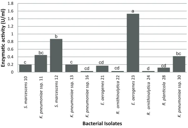
Concerning quantification of enzymatic activity was observed significant variations between bacterial species, from the averages resulting of the titration, analyzed by Duncan test (Figure 1). The
bacteria presented negative results for extracellular lipase production (Rhodamine B test) were not evaluated for enzymatic activity.
E. aerogenes (23) presented the highest efficiency, indicating a higher quantity of free fatty acids (1.54 U/ml) in the medium. However, the bacterial isolates 11, 12 and 30, did not present significant differences between them, as well as the isolates 10, 13, 16, 21, 22 and 28.
The hydrolysis evaluation of olive oil using the enzymatic extract from the isolates 18, 20, 26 and 29 did not present a significant result, and was observed negative values for the lipase activity. This result indicates that the reaction of olive oil hydrolysis did not occur or was not efficient during the incubation time.
There are diverse substances with enzymatic inhibition potential that can associate to the active site or another site of enzyme, resulting in a speed decrease or paralyzing the reaction. Temperature can influence enzymatic reaction speed
because high temperatures can result in enzyme denaturalization (Champe et al. 2006). In this experiment, it is considered that the temperature was not unsatisfactory once other microorganisms presented positive and satisfactory values. Lipases enzymes also can catalyze various reactions, like as the esterification, which is an inverse reaction to hydrolysis. Therefore, during incubation time another reaction could be catalyzed, resulting in reduction of free fatty acids in reaction medium.
Zaki and Saeed (2012) evaluated the enzymatic activity of 12 strains of Serratia marcescens. The lipase originated by the strain that presented best enzymatic activity was assessed for degradation of triacylglycerol in various vegetable fats (soy, coconut, sesame, olive and sunflower). The analysis in spectrophotometer showed an enzymatic activity of 112 U/ml, when olive oil was used as substrate. 0 0.2 0.4 0.6 0.8 1 1.2 1.4 1.6 1.8 S . m a rc e sc e n s 10 K . p n e u m o n ia e ss p . 11 S . m a rc e sc e n s 1 2 K . p n e u m o n ia e ss p . 13 K . p n e u m o n ia e ss p . 16 E . a e ro g e n e s 21 R. o rn it h in o ly ti ca 22 E . a e ro g e n e s 23 R. o rn it h in o ly ti ca 2 4 R. p la n ti co la 28 K . p n e u m o n ia e ss p . 30
E
n
zy
m
a
ti
c
a
c
ti
v
it
y
(
U
/m
l)
Bacterial Isolates
c bc b c cd cd cd a d cd bcFigure 1 - Extracellular lipase enzymatic activity of bacteria obtained from effluents of dairy products and slaughterhouses industries located in Pelotas/RS, Brazil.
However, a higher activity was observed when was used sesame oil (122 U/ml). In this present study, we observed an inferior activity in both S. marcescens strains that can be related to different variables. The enzyme used by Zaki and Saeed was extracted and purified, and the evaluation of enzymatic activity was realized by spectrophotometry. However, in our study was used enzymatic extract produced by S. marcescens and determination of activity was realized by titration of free fatty acids in reaction medium.
Kumari et al. (2009) observed lipolytic activity in Enterobacter aerogenes (27.25 U/ml) in culture medium containing 3% of coconut oil, incubated for 60 hours. The highest enzymatic activity, when compared to results of our study, can be related to type of carbon source, seeing that we used olive oil. The incubation time can also be increase lipase production, which in our study was of 24 hours. In addition, another factor whether intrinsic or environmental, are important in enzymatic production.
According to Immanuel et al. (2008), higher activity of enzymes is related with triglycerides, which are important substrates in lipase enzyme production and may present function of inducing or inhibiting. Studies show that substrates like the olive oil increase the lipase production, presenting inductive function in enzymatic production (Rajendran et al. 2007, Tavares et al. 2011).
Others factors interfere in enzymatic production are medium conditions like temperature, pH and agitation (Willerding et al. 2011, Thakur et al. 2014). In this present study, once the bacteria have been isolated from industrial effluents, we opted to define typical conditions of environmental microorganisms, and so the incubation temperature for isolation and production of enzyme was 30ºC.
The isolate filamentous fungi from effluents were identified as the following genera: Alternaria sp., Fusarium sp., Geotrichum sp., Gliocladium sp., Mucor sp., Paecilomyces sp., and Trichoderma
sp.. Extracellular lipase production was observed in 57.14% of isolated fungi, presenting positive results in the Rhodamine B test the following fungi: Fusarium sp., Geotrichum sp., Gliocladium sp. and Mucor sp. Table II presents extracellular lipolytic production by the filamentous fungi.
The production of lipases enzymes by fungal microorganisms is reported in literature, including the isolated fungi in this study (Gunasekaran and Das 2005, Nagy et al. 2006, Fernandes et al. 2012, Gopinath et al. 2013).
Mukunda et al. (2012) isolated more than 200 fungi from ground and evaluated the ability of production of various hydrolytic enzymes, including lipases. The fungi genera Trichoderma, Fusarium, Pacillomyces, Gliocladium, Alternaria and Mucor did not present extracellular lipase production, seeing that they did not present the transparent halo around the colonies, using Tween as the substrate. In our study, the species Fusarium sp., Gliocladium sp. and Mucor sp. produced extracellular lipase, which is an important factor for further studies and applications in treatment systems.
tAble ii
Lipase enzymatic activity of filamentous fungi obtained from effluents of dairy products and slaughterhouses
industries located in Pelotas/rS, brazil.
origin Fungal Species Extracellular lipase
SH3 Alternaria sp.
-SH2 Fusarium sp. +
SH3 Geotrichum sp. +
DP Gliocladium sp. +
SH3 Mucor sp. +
SH3 Paecilomyces sp.
-SH3 Trichoderma sp.
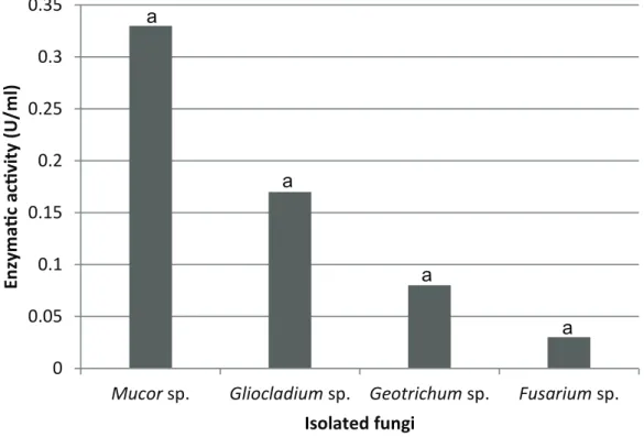
Concerning lipase enzyme activity in the olive oil degradation, the fungi did not present significate differences according to the Duncan test. Figure 2 shows the enzymatic activity of fungi that presented positive values for the Rhodamine B test.
Nwuche and Ogbonna (2011) isolated 12 fungi from the industry effluent of palm oil, including the genera Aspergillus, Penicillium, Trichoderma and Mucor. Using similar methodology to our study, Mucor sp. presented enzymatic activity of 5.72 U/ml, in culture medium containing olive oil. This higher activity can be due to longer incubation time (72 hours) or being in different species, when compared to our study. Through the spectrophotometry, the activity for Mucor griseocyanus lipolytic varied between 0.04 to 0.1 U/ml, using different substrates such as olive oil, coconut oil, sunflower oil, among others (Armas et al. 2008). Another specie, Mucor geophillus,
presented maximum activity of 44.56 U/ml by the titration method, having as substrate the olive oil (Naqvi et al. 2011). Incubation time (72 hours) was the same for both species.
Burkert et al. (2004), with Geotrichum sp., obtained enzymatic activity of 17 U/ml, after 50 hours of incubation, meanwhile Carvalho et al. (2005), observed 12.8 U/ml, with Geotrichum candidum, after 72 hours. Both studies used olive oil as carbon source and quantified the fatty acids by titration. The superior enzymatic activity of the species in these studies may be because the species have isolated in this study are different, as well as the incubation time of our work was only 48 hours.
According to Odeyemi et al. (2013), the biologic oily effluents treatment, had been the most efficient, with degradation of lipids compounds in miscible molecules. According to Gopinath et al. (2013), domestic and industrial residuals may have
0
0.05
0.1
0.15
0.2
0.25
0.3
0.35
Mucor
sp.
Gliocladium
sp.
Geotrichum
sp.
Fusarium
sp.
E
n
zy
m
a
ti
c
ac
ti
v
it
y
(U
/m
l)
Isolated fungi
a
a
a
a
Figure 2 - Lipase enzymatic activity of filamentous fungi obtained from effluents of dairy products and
slaughterhouses industries located in Pelotas/RS, Brazil.
fungi with higher potential degradation of fatty and oils, eliminating wastes and can produce beneficent substances due to its activity.
Metabolism of microorganisms shows the ability of waste elimination and the production of useful substances. Therefore, biodegradation consists in an important process to minimize the environment oil pollution, reducing the environmental impacts (Gopinath et al. 2013). In this way, diverse microorganisms may be efficient in the treatment of contaminated areas through their biotechnological applications.
concluSionS
This study demonstrated that there are microorganisms that produce lipases enzymes in industrial effluents, with ability of extracellular lipase production, a positive factor to obtaining enzymes and their use in future applications. The produced lipases presented biodegradation of lipid compound, being effective in effluents from treatment systems in various sectors.
reSuMo
As lipases têm a capacidade de catalisar diversas reações e são importantes em diferentes aplicações biotecnológicas. Este trabalho teve como objetivo isolar e caracterizar microrganismos produtores de lipases,
provenientes de diferentes efluentes de indústrias
alimentícias da região de Pelotas, RS. As bactérias foram
identificadas quanto à coloração de Gram e a provas
bioquímicas (Vitek 2®). Os fungos foram identificados
quanto às características de macro e micromorfologia.
A produção de lipase extracelular foi avaliada pelo teste rodamina B e a atividade enzimática, por titulação. Foram
isoladas 21 bactérias, identificadas como Klebsiella pneumoniae ssp. pneumoniae, Serratia marcescens, Enterobacter aerogenes, Raoultella ornithinolytica e Raoultella planticola. Os fungos filamentosos isolados foram caracterizados aos gêneros Alternaria sp., Fusarium sp., Geotrichum sp., Gliocladium sp., Mucor sp., Paecilomyces sp. e Trichoderma sp.. A produção de lipase extracelular foi observada em 71,43% das
bactérias e 57,14% dos fungos. A bactéria que apresentou atividade enzimática mais promissora foi a E. aerogenes (1,54 U/ml), entretanto, entre os fungos, não houve
diferença significativa entre os quatro isolados.O estudo revela que assim como microrganismos produtores de
lipases estão presentes em efluentes industriais, estas
enzimas apresentam potencial de biodegradação de compostos lipídicos.
Palavras-chave: efluente, atividade enzimática, produção de lipase extracelular, enzima lipase, microrganismo lipolítico.
reFerenceS
almeida AF, tauk-torniSieloSM and carmona EC.
2013. Acid lipase from Candida viswanathii: Production, biochemical properties, and potential application. Biomed Res Int 2013: 1-10.
anbu P, noH M, kim D, Seo J, Hur B and minKH. 2011. Screening and optimization of extracellular lipases by Acinetobacter species isolated from oil-contaminated soil in South Korea. Afr J Biotechnol 10: 4147-4156.
armaSJC, mendozaJCD and Hernandez JLM. 2008. Mucor griseocyanus lipase production, characterization and study of some catalytic properties of the immobilised enzyme. Food Technol Biotechnol 46: 195-201.
burkert FM, maugeri F and rodrigueS MI. 2004.
Optimization of extracellular lipase production by
Geotrichum sp. using factorial design. Bioresour Technol 91: 77-84.
carvalHoPO, calafatti SA, maraSSiM, da Silva
DM, conteSiniFJ and bizaco R. 2005. Potencial de biocatálise enantiosseletiva de lipases microbianas. Quím Nova 28: 614-621.
carvalHoPO, camPoSPRB, noffSMD, oliveiraJG,
SHimizuMT and da SilvaDM. 2003. Aplicação de
lipases microbianas na obtenção de concentrados de ácidos graxos poli-insaturados. Quím Nova 26: 75-80.
caStroHF, mendeS AA and SantoS JC. 2004. Modificação
de óleos e gorduras por biotransformação. Quím Nova 27: 146-156.
cHamPe PC, HarveyRA and ferrier DR. 2006. Bioquímica
ilustrada. 3ª ed., Porto Alegre: Artmed, 544 p.
cHernicHaro cal. 1997. Princípios do tratamento biológico
de águas residuárias. Vol. 5, Belo Horizonte: DESA/ UFMG, 379 p.
colla LM, reineHr CO and coSta JAV. 2012. Aplicações e produção de lipases microbianas. Revista CIATEC - UPF 4: 1-14.
de oliveira JP, antuneS PWP, Pinotti LM and caSSini
oleosos do saneamento e dos óleos e graxas extraídos visando a conversão em biocombustíveis. Quím Nova 37: 597-602.
fernandeS EG, valério HM, feltrin T and van
der SandST. 2012. Variability in the production of extracellular enzymes by entomopathogenic fungi grown on different substrates. Braz J Microbiol 43: 827-833.
goPinatH SCB, anbu P and Hilda A. 2005. Extracellular
enzymatic activity profiles in fungi isolated from oil-rich environments. Mycoscience 46: 119-126.
goPinatHSCB, anbu P, lakSHmiPriyaT and Hilda
A. 2013. Strategies to characterize fungal lipases for applications in medicine and dairy industry. Bio Med Res Int 2013: 1-10.
gunaSekaran V and daSD. 2005. Lipase fermentation:
progress and prospects. Indian J Biotechnol 4: 437-445. HaSan F, SHaHAA and HameedA. 2006. Industrial
applications of microbial lipases. Enzyme MicrobTechnol 39: 235-251.
immanuel G, eSakkiraJ P, JebadHaS A, iyaPParaJ P and
PalaveSamA. 2008. Investigation of lipase production by
milk isolate Serratia rubidaea. Food Technol Biotechnol 46: 60-65.
ivanovaIA, nikolovaS, yemendzHievH, koniarSka
A and aleXieva Z. 2013. Microflora of Bulgarian oil
contaminated environments. J Cell & Plant Sci 4: 12-17.
Jaeger KE and eggert T. 2002. Lipases for biotechnology.
Curr Opin Biotechnol 13: 390-397.
kumar D, kumar L, nagar S, raina C, ParSHad R and guPta VK. 2012. Screening, isolation and production of lipase/esterase producing Bacillus sp. strain DVL2 and its potential evaluation in esterification and resolution reactions. Arch Appl Sci Res 4: 1763-1770.
kumariA, maHaPatra P and banerJee R. 2009. Statistical
optimization of culture conditions by response surface methodology for synthesis of lipase with Enterobacter aerogenes. Braz Arch Biol Technol 52: 1349-1356. leal MCMR, cammarota MC, freire DMG and
Sant’anna Jr GL. 2002. Hydrolytic enzymes as coadjuvants in the anaerobis treatment of dairy wastewaters. Braz J Chem Eng 19: 175-180.
limaVMG, kriegerN, mitcHellDA, baratti JC, filiPPiS
I and fontana J. 2004. Evaluation of the potential for
use in biocatalysis of a lipase from a wild strain of Bacillus megaterium. J Mol Catal B: Enzyme 31: 53-61.
lin JF, lin Q, Li J, fei ZA, li XR, Xu H, qiao DR and cao Y. 2012. Bacterial diversity of lipase-producing strains in different soils in southwest of China and characteristics of lipase. Afr J Microbiol Res 6: 3797-3806.
martinSVG, kalil SJ and coSta JAV. 2008. Co-produção
de lipase e biossurfactante em estado sólido para utilização em biorremediação de óleos vegetais e hidrocarbonetos. Quím Nova 31: 1942-1947.
mendeS AA, caStroHF, PereiraEB and furigo JR.
A. 2005. Aplicação de lipases no tratamento de águas residuárias com elevados teores de lipídeos. Quím Nova 28: 296-305.
meSSiaSJM, coStaBZ, limaVMG, gieSeEC, dekker
RFHand barboSa AM. 2011. Lipases microbianas: Produção, propriedades e aplicações biotecnológicas. Semina: Tech Ex 32: 213-234.
moura LFWG, oliveira MV, Lô MM, mota JGSM,
magalHãeS EA, lima MCL and magalHãeS FEA.
2013. Bioprospecção de atividade lipolítica de fungos anemófilos isolados do Centro Vocacional Tecnológico (CVT) de Tauá-CE. Rev Bras Prod Agroind 15: 157-165.
mukundaS, onkaraPPaR and kekuda PTR. 2012.
Isolation and screening of industrially important fungi from the soils of western ghats of Agumbe and Koppa, Karnataka, India. Sci Technol Arts Res J 1: 27-32. nagyV, tõke ER, keongLC, SzatzkerG, ibraHim
D, omar IC, SzakácS G and PoPPe L. 2006. Kinetic resolutions with novel, highly enantioselective fungal lipases produced by solid state fermentation. J Mol Catal B: Enzymatic 39: 141-148.
naqvi SH, daHotMU, ali A, kHan MY and rafiq M, 2011. Production and characterization of extracellular lipase secreted by Mucor geophillus. Af J Biotechnol 10: 19598-19606.
nwucHe CO and ogbonnaJC. 2011. Isolation of lipase
producing fungi from palm oil mill effluent (POME) dump sites at Nsukka. Braz Arch Biol Technol 4: 113-116.
odeyemiAT, aderiyeBI and bamideleOS. 2013. Lipolytic
activity of some strains of Klebsiella, Pseudomonas and Staphylococcus spp. from restaurant wastewater and receiving stream. J Microbiol Res 3: 43-52.
Patil KJ, cHoPda MZ and maHaJanRT. 2011. Lipase biodiversity. Indian J Sci Technol 4: 971-982.
PeiXoto RS, roSado AS and taketani RG. 2008.
Bioprospecção da diversidade microbiana cultivável e não cultivável. In: De Melo, Itamar S, De Azevedo and João L. 2ª ed., Microbiologia Ambiental. Jaguariúna: Embrapa Meio Ambiente, p. 83-106.
raJendran A, tHirugnanam M and tHangavelu
V. 2007. Statistical evaluation of medium components by Plackett–Burman experimental design and kinetic modeling of lipase production by Pseudomonas fluorescens. Indian J Biotechnol 6: 469-478.
SaXena RK, SHeoran A, giri B and davidSon WS. 2003.
Purification strategies for microbial lipases. J Microbiol Meth 52: 1-18.
SHarma R, cHiStiYand banerJee UC. 2001. Production,
Silva filHoGNand oliveiraVL. 2007. Microbiologia: manual de aulas práticas. 2ª ed., Florianópolis: UFSC, 157 p.
Sugimori D, nakamura M and miHara Y. 2002.
Microbial degradation by Acinetobacter sp. Strain SOD-1. J Biosc Biotechnol Biochem 66: 1579-1582.
Sugimori D, watanabe M and utSue T. 2013. Isolation
and lipid degradation profile of Raoultella planticola strain 232-2 capable of efficiently catabolizing edible oils under acidic conditions. Appl Microbiol Biotechnol 97: 871-880.
tavareS LLP, naScimento AE, okada K and Silva
CAA. 2011. Seleção de diferentes meios para produção de lipase a partir de Bacillis licheniformis (UCP 1014). Exacta 9: 309-316.
tebaldiVMR, oliveiraTLC, boariCA and Piccoli
RH. 2008. Isolamento de coliformes, estafilococos e enterococos de leite cru provenientes de tanques de refrigeração por expansão comunitários: identificação, ação lipolítica e proteolítica. Ciênc Tecnol Aliment 28: 753-760.
tHakur V, tewari R and SHarma R. 2014. Evaluation of
production parameters for maximum lipase production by
P. stutzeri MTCC 5618 and scale-up in bioreactor. Chin J Biol 2014: 1-14.
veeraPagu M, narayanan AS, Ponmurugan K and
Jeya KR. 2013. Screening selection identification production and optimization of bacterial lipase from oil spilled soil. Asian J Pharm Clin Res 6: 62-67.
verma N, tHakur S and bHattAK. 2012. Microbial lipases: Industrial applications and properties: A review. Int Res J Biol Sci 1: 88-92.
willerding AL, oliveira LA, moreiraFW, germano
MGand cHagaS Jr AF. 2011. Lipase activity among
bacteria isolated from Amazonian soils. Enzyme Res 2011: 1-5.