Sónia da Silva Sousa Machado
Visual Brain Areas in Obsessive
Compulsive Disorder – A Diffusion
Tensor Imaging Approach
Universidade do Minho
Escola de Psicologia
V
isual Brain Areas in Obsessive Compulsive Disorder – A Dif
fusion T
ensor Imaging Approach
Sónia da Silva Sousa Mac
Dissertação de Mestrado
Mestrado Integrado em Psicologia
Área de Especialização em Psicologia Clínica
Trabalho realizado sob a orientação da
Doutora Adriana da Conceição Soares Sampaio
Sónia da Silva Sousa Machado
Visual Brain Areas in Obsessive
Compulsive Disorder – A Diffusion
Tensor Imaging Approach
Universidade do Minho
É AUTORIZADA A REPRODUÇÃO PARCIAL DESTA TESE APENAS PARA EFEITOS DE
INVESTIGAÇÃO, MEDIANTE DECLARAÇÃO ESCRITA DO INTERESSADO, QUE A TAL SE
COMPROMETE;
Acknowledgements
Finalizing this dissertation is the zenith of a long 5-year academic progress. It was an interesting investigation, although challenging, worth it. I feel satisfied professionally and personally for this achievement. Nevertheless, I have to acknowledge some key-persons who had an important contribution in the pursuit of my work.
I would like to acknowledge Prof. Óscar Gonçalves, lab director, for the primary incentive to investigate white matter microstructure in OCD patients with this innovative imaging technique that is Diffusion Tensor Imaging (DTI).
I would like to acknowledge Prof. Adriana Sampaio, my adviser, for the lessons, availability, pressure and elevated degree of exigency throughout my work, plus, for being her self a source of inspiration.
I would like to acknowledge Jorge and Rosana, my lab colleagues, for their always-prompt availability in case help would be needed.
I would like to acknowledge Catarina, my lab colleague and friend, for the friendship, kindness and for being my “prospective memory”.
I would like to acknowledge Cristiana, my sister, for being my time schedule advising about the time I still got to finish my dissertation but always believing I would make it.
I would like to acknowledge Miguel, my boyfriend, for being present hour-to-hour in these last writing days, for the encouragement, opinions, great sense of humor and love.
Finally, I have to acknowledge Fernando Pessoa, Portuguese poet, for the written words, by which I feel inspired every day:
"Para ser grande, sê inteiro: nada TEU exagera ou exclui.
Sê todo em cada coisa. Põe quanto és no mínimo que fazes.
Assim em cada lago a lua toda Brilha, porque alta vive."
Mestrado Integrado em Psicologia da Universidade do Minho Área de Especialização de Psicologia Clínica
Áreas cerebrais visuais na Perturbação Obsessivo-Compulsiva – uma abordagem de Imagiologia por Tensão de Difusão
Sónia Sousa
Orientador: Adriana Sampaio
Resumo
A Perturbação Obsessivo-Compulsiva (POC) é uma condição psiquiátrica pertencente ao espectro das perturbações de ansiedade, sendo caracterizada por obsessões e compulsões. Obsessões são definidas como pensamentos, imagens, ideias ou impulsos recorrentes cujo conteúdo causam um elevado grau de ansiedade no indivíduo, levando-o a executar ações especificas ou rituais mentais de modo a reduzir a ansiedade, i.e. compulsões, (DSM-IV-TR, APA, 2000). A POC é uma psicopatologia bastante debilitante com uma taxa de prevalência de 1 a 2,5% na população adulta, com diversas facetas no que diz respeito às suas características clínicas: comportamentais, emocionais e neurocognitivas. Com especial atenção, défices ao nível do processamento visual e um viés atencional são descritos como uma significante manifestação desta perturbação, desempenhando um papel importante na ineficiente apreensão de estímulos sociais, um importante característica desta perturbação. Em concordância, alterações cerebrais estruturais e funcionais têm sido associadas a este fenótipo. Deste modo, o principal objectivo é analisar a integridade da microestrutura de substância branca em pacientes com POC, através de técnicas de Imagiologia por Tensor de Difusão (do inglês Diffusion Tensor Imaging), usando uma metodologia baseada na segmentação e tractografia, de modo a estabelecer uma relação entre estes padrões de conexão cerebral e a sua sintomatologia clínica. Considerando os défices cognitivos e alterações cerebrais da POC, nomeadamente ao nível das áreas frontais (orbitofrontal e dorsolateral) e occipitais, pretendemos continuar investigações prévias da nossa equipa de investigação e examinar a integridade da substância branca dos feixes da circunvolução frontal inferior (CFI), lobo occipital e área V1, regiões cerebrais relacionadas com o processamento visuo-perceptivo, que hipotetizamos estarem alteradas na população com POC (Gonçalves et al., 2010). Recorrendo a uma inovadora técnica de imagiologia (DTI), foram avaliadas as medidas de integridade dos feixes de substância branca (FA, MD, AD e RD) das áreas da CFI, lobo occipital e V1 devido ao seu papel no processamento visual e perceptivo. Catorze pacientes com POC (idade média,M=32,64±11,41) e dez participantes no grupo controlo (idade média M=30,70±9,73), emparelhados em idade, sexo e lateralidade, participaram neste estudo. Os nosso resultados demonstraram diferenças entre pacientes e controlos em três medidas de DTI nas regiões occipitais e V1. Especificamente, o grupo com POC apresentou menor integridade dos feixes de substância branca nestas áreas cerebrais que estão estritamente relacionadas com o processamento visual. No entanto, não foram encontradas diferenças de grupo no que diz respeito às medidas de integridade da matéria branca no feixe da CFI. Os resultados sugerem que feixes de substância branca envolvidas no processamento precoce de estímulos visuais podem estar associadas às manifestações clínicas da POC, nomeadamente o viés de processamento de estímulos emocionais.
Mestrado Integrado em Psicologia da Universidade do Minho Área de Especialização de Psicologia Clínica
Visual Brain Areas in Obsessive Compulsive Disorder – A Diffusion Tensor Imaging Approach Sónia Sousa
Orientador: Adriana Sampaio
Abstract
Obsessive Compulsive Disorder (OCD) is a psychiatric disorder classified within the spectrum of anxiety disorders, being characterized by obsessions and compulsions. Obsessions are defined as thoughts, images, ideas or recurrent impulses which content causes an elevated degree of anxiety on the individual, impelling him to execute specific actions or mental rituals in order to reduce anxiety, i.e. compulsions, (DSM-IV-TR, APA, 2000). OCD is a very disabling psychopathology with a prevalence rate of 1 to 2,5% in the adult population, with several facets concerning its clinical features: behavioral, emotional and neurocognitive. Of special note, visual processing deficits and an attentional bias has been described as a significant manifestation of this disorder, playing an important role in their inefficient social stimuli aprehension, a important characteristic of this disorder. In agreement, structural and functional brain alterations in the occipital, parietal and other visual areas have been associated with this phenotype. Therefore, the main objective of the present study is to analyze white matter microstructure in OCD patients through the use of Diffusion Tensor Imaging (DTI) techniques, using a segmentation and tractography based approach, in order to establish a relation between connectivity patterns of these visual brain and OCD’ clinical symptomatology. Considering their neurocognitive phenotype and specific brain abnormalities in OCD, namely in frontal (orbitofrontal and dorsolateral) and occipital areas, we now aim to follow our team previous research and investigate the integrity of the Inferior Frontal Gyrus (IFG), occipital lobe and V1 area, as are related with visuo-perceptive processing, which we hypothesize to be abnormal in OCD population (Gonçalves et al., 2010). Using this innovative imaging technique (DTI) we evaluated white matter DTI-derived measures (FA, MD, AD and RD) of the IFG, occipital and V1 areas taking into account their role on visual processing and perception. Fourteen patients with OCD (Mean age=32,64 ± 11,41) and ten comparison controls (Mean age=30,70 ± 9,73), matched on age, sex and handedness, participated in this study. Our results reported group differences in three DTI indexes (MD, AD and RD) in occipital and V1. Specifically, OCD group displayed decreased integrity of these white matter tracts which are strictly related with visual processing. Nevertheless, we did not find group differences regarding the integrity of the IFG white matter tract. Results suggest that brain fiber tracts enrolled in early steges of visual processing may be associated with the clinical features of OCD, specifically the bias in processing for emotional stimuli.
INTRODUCTION ... 1
SECTION I 1. Obsessive Compulsive Disorder – an Overview ... 2
1.1. Obsessive Compulsive Disorder- Prevalence ... 2
1.2. Obsessive Compulsive Disorder Diagnosis ... 3
1.3. Obsessive Compulsive Disorder – Comorbidity ... 5
1.4. Obsessive Compulsive Disorder – Etiology ... 6
1.4.1. Obsessive Compulsive Disorder - Genetics and Neurochemistry ... 7
1.5. Obsessive Compulsive Disorder – Treatment ... 8
SECTION II 2. Obsessive Compulsive Disorder – Different Facets of the Same Disease ... 9
2.1. Obsessive Compulsive Disorder - Emotional and Behavioral Phenotype ... 9
2.2. Obsessive Compulsive Disorder - Neurocognitive Phenotype ... 10
2.3. Obsessive Compulsive Disorder - Neuroanatomical Phenotype ... 11
Objectives ... 13
Hypothesis ... 14
SECTION III 3. Methods and Materials ... 15
3.1. Participants ... 15
3.2. Psychological Assessment ... 15
3.3. MRI Acquisition and Processing ... 16
3.4. Post-Processing and Regions of Interest Definition (ROI) ... 16
3.4.1 Eddy Current and Motion Correction ... 17
3.4.2 Region of Interest extraction (ROI) - Inferior-Frontal Gyrus (IFG), Occipital and V1 17 3.5 Tractography ... 19
SECTION IV
4. Data Analysis ... 20
4.1 Results ... 21
4.1.1 Demographic Data ... 21
4.1.2 ROI’s White matter Microstructure Analysis ... 21
4.1.3. Correlations ... 23
4.1.4 Discussion ... 24
4.1.5 Limitations and future studies ... 26
Table 1 - Comorbidity rates of OCD with other psychiatric disorders ... 6
Table 2 - Demographic Data ... 21
Table 3 - Mean FA, MD, AD and RD Values by ROI ... 23
Table 4 - Pearson correlations between ROI’ measures and Y-BOCS ... 24
Visual Brain Areas in Obsessive Compulsive Disorder – A Diffusion Tensor
Imaging Approach
Introduction
Obsessive Compulsive disorder (OCD) is a very disabling psychopathology with a prevalence rate of 1 to 2,5% in the adult population. Besides behavioral, emotional and neurocognitive deficits, recent investigation has focused in relating this psychological phenotype with underlying structural and functional alterations. Of special note, visual processing deficits and an attentional bias has been described as a significant manifestation of this disorder, playing an important role in their inefficient social stimuli aprehension, an important characteristic of this disorder. In agreement, structural and functional brain alterations of occipital, parietal and other visual areas have been associated with this phenotype. Therefore, the main objective of the present study is to advance in this field of research, by studying specific white matter microstructure in OCD patients through the use of Diffusion Tensor Imaging (DTI) techniques, using a segmentation based approach, in order to establish a relation between these brain connectivity patterns and their clinical symptomatology. This dissertation will be organized in four sections. The first section gives an overview about this psychopathology, its prevalence, diagnosis, comorbidity, possible factors related to the onset of the disorder and available treatments. The second section will inform about distinctive OCD clusters concerning specific aggregation of symptoms, namely, behavioral, emotional, neurocognitive and neuroanatomical phenotypes. On the third section, method and materials will be characterized in detail. Finally, discussion of the results and concluding remarks will be addressed on the fourth section of this dissertation.
SECTION I
1. Obsessive Compulsive Disorder – an Overview
1.1. Obsessive Compulsive Disorder- Prevalence
Obsessive Compulsive Disorder (OCD) is a psychiatric disorder classified within the spectrum of anxiety disorders, being characterized by obsessions and compulsions. According to American Psychiatric Association [APA, (2000)], obsessions are defined as thoughts, images, ideas or recurrent impulses which content causes an elevated degree of anxiety on the individual, impelling him to execute specific actions or mental rituals in order to reduce anxiety, i.e. compulsions. Conceptually, OCD can be defined as an erroneous cue perception, followed by internal signals of danger and anxiety. In a specific situation, the person believes, even though with no practical evidences, that something is wrong and a harmful event will happen, displaying therefore, a specific ritual to avoid the hypothesized negative results (Aouizerate et al., 2004) – the compulsion.
OCD is a very disabling disorder, being the fourth most frequent disorder, after phobias, substance abuse and major depression (Aouizerate et al., 2004), with a cross-cultural lifetime prevalence of around 1 to 2,5% in the adult population (Torres & Lima, 2005). It can be classified according to different dimensions, namely: a) the content of the obsessions, with the most frequent being contamination (50%), pathological doubt (42%), somatic (33%), need for symmetry (32%), aggressive (31%) and sexual (24%) and; b) the type of compulsions as checking (61%), washing (50%), ordering (28%) and 18% hoarding (Torres et al., 2006).
1.2. Obsessive Compulsive Disorder Diagnosis
One of the most widely used manuals to classify mental disorders is the Diagnostical and Statistical Manual of Mental Disorders (DSM) published by the American Psychiatric Association (APA), that provides information about the standard criteria for the classification of the several mental disorders. According to DSM, a mental disorder is an health condition characterized by a dysfunction, clinically significant, in the individual’s cognitions, emotions and behaviors that leads to disturbance in the psychological, biological or development processes underlying mental functioning, reflecting substantial disability, distress and suffering on the individuals, and thus interfering with daily life quality (DSM-IV-TR, APA, 2000). The attribution of a diagnosis has an important role on Clinical Psychology as it should assist clinicians in defining a treatment plan and establish prognosis for the patients. Therefore, in agreement with DSM, to establish an OCD diagnosis, the following criteria have to be fulfilled:
A. Either obsessions or compulsions: Obsessions as defined by (1), (2), (3), and (4):(1) Recurrent and persistent thoughts, impulses, or images that are experienced at some time during the disturbance, as intrusive and inappropriate and causes marked anxiety or distress (2) The thoughts, impulses, or images are not simply excessive worries about real-life problems (3) The person attempts to ignore or suppress such thoughts, impulses, or images, or to neutralize them with some other thought or action (4) The individual recognizes that the obsessive thoughts, impulses, or images are a product of his own mind (not imposed from outside as in thought insertion). Compulsions are defined by (1) and (2): (1) Repetitive behaviors (e.g., hand washing, ordering, checking) or mental acts (e.g., praying, counting, repeating words silently) the person feels driven to perform in response to an obsession, or according to rules that must be applied rigidly; (2) The behaviors or mental acts aim to prevent or reduce distress or prevent some threatening event or situation; however, these behaviors or mental acts are not connected in a realistic way with what they are considered to neutralize or prevent or are clearly excessive; B. At some point during the course of the disorder, the person has recognized that the obsessions or compulsions are excessive or unreasonable. Note: This does not apply to children; C. The obsessions or compulsions causes marked distress, are time consuming (take more than 1 hour a day), or significantly interfere with the person’s normal routine, occupational (or academic) functioning, or usual social activities or relationships; D. If another Axis I disorder is present, the content of the obsessions or compulsions is not restricted to it (e.g., preoccupation with food in the presence of an Eating
Disorder; hair pulling in the presence of Trichotillomania; concern with appearance in the presence of Body Dysmorphic Disorder; preoccupation with drugs in the presence of a Substance Use Disorder; preoccupation with having a serious illness in the presence of Hypochondriasis; preoccupation with sexual urges or fantasies in the presence of a Paraphilia; or guilty ruminations in the presence of Major Depressive Disorder); E. The disturbance is not due to the direct physiological effects of a substance (e.g., a drug of abuse, a medication) or a general medical condition; Specify if: With insufficient criticism (insight): if during the most part of the actual episode, the individual does not recognize that the obsessions and compulsions are excessive or unreasonable (DSM-IV-TR, APA, 2000).
For further information on symptom presentation and severity of OCD, including the evaluation through treatment intervention, several instruments were developed and are used such as diagnostic interviews, clinician-administered inventories and self-report questionnaires. Specifically, the Anxiety Disorders Interview Schedule (ADIS) is a semi-structured interview based on DSM criteria and includes detailed symptom queries for each anxiety disorder. ADIS is used to establish differential diagnosis mainly among anxiety disorders but also between others that frequently co-occur, as mood or substance abuse disorders (Brown, Di Nardo, Lehman & Campbell, 2001). The Structured Clinical Interview for Axis I Disorders (SCID) is also a semi-structured interview containing questions regarding past and present symptoms that allows a DSM diagnosis (First & Gibbon, 2004). These two instruments have diagnostic utility, giving support on the attribution of OCD disorder according DSM classification. The Leyton Obsessional Inventory-Short Form (LOI-SF) is a 30-item self-report questionnaire using a yes/no response format, which assesses the presence or absence of OCD-related symptoms (Mathews et al., 2004). Finally, one of the most known and widely used clinician rated measures is the Yale-Brown Obsessive-Compulsive scale (Y-BOCS) which is used to assess OCD symptom severity plus frequency, providing information on treatment planning and evaluation. Ratings are based on information provided by patients as well as on clinical observations. The Y-BOCS is a clinician-rated 10-item scale, with each item being clinician-rated from 0 (not symptomatic) to 4 (severely symptomatic). It presents separate subtotals for severity of obsessions and compulsions that are not influenced by the existing type of obsessions or compulsions. This evaluation consists of a self-report questionnaire that addresses the amount of time the individual spends on ritualizing (compulsions), the degree of impairment or distress they experience and the resistance and control level they have over these thoughts or obsessions (Goodman et al., 1989).
1.3. Obsessive Compulsive Disorder – Comorbidity
Commonly, OCD is associated with other psychological disorders, more frequently with disorders within the anxiety cluster (see table 1). Depressive episodes are the most commonly associated with OCD (comorbidity rate - 36,8%), followed by generalized anxiety disorder with 31,4%, agoraphobia or panic disorder with 22,1%, social phobia with 17,3% and specific phobia with 15,1% (Torres et al., 2006).
Additionally, according to specific symptomatology of OCD, other psychiatric disorders may co-occur. Conceptually, we can consider two poles within the spectrum of OCD: a) the compulsivity – risk aversion pole, in which individuals present compulsive or repetitive behaviors they cannot control, with an aversion/fear of risky situations, behaving in a way to avoid or suppress disagreeable situations/sensations and; b) the impulsivity – novelty or risk seeking pole for those individuals that display great difficulty in controlling their impulsivity to behave in an unsafe way, searching for different/danger situations that eventually could be pleasant. Therefore, fitting within the compulsive – risk aversion pole, comorbidity with Body Dysmorphic Disorder (BDD), defined as a preoccupation with an imagined defect in appearance (DSM-IV-TR, APA, 2000) is observed within 8% to 12% of the cases (Brawman-Mintzer et al., 1995;Simeon, Hollander, Stein, Cohen, & Aronowitz, 1995); Hypochondriasis, defined as an excessive fear of having a serious disease, based on body signals or symptoms (DSM-IV-TR, APA, 2000) has a comorbidity rate of 13% (Jaisoory, Reddy, & Srinath, 2003); Anorexia, defined as excessive preoccupation with body weight (DSM-IV-TR, APA, 2000) shows a comorbidity rate of 29,5% (Milos, Spindler, Ruggiero, Klaghofer, & Schnyder, 2002). On the other pole, Trichotillomania, described as recurrent pulling of own hair resulting in great hair loss (DSM-IV-TR, APA, 2000), presents a comorbidity rate of 3% (Jaisoory et al., 2003); finally, Tics syndrome characterized by multiple motor tics, which are sudden, involuntary, rapid, repetitive, non-rhythmic, stereotyped motor movements or vocalizations (DSM-IV-TR, APA, 2000) shows a comorbidity rate between 2,5% and 7%.
Table 1 - Comorbidity rates of OCD with other psychiatric disorders
1.4. Obsessive Compulsive Disorder – Etiology
OCD can be described as a psychiatric disorder characterized by a multivariate origin approach and multifaceted clinical expressions. The OCD has been proposed to be a complex disorder in which morphological and functional alterations in specific brain regions, dysfunction of specific neural pathways, biochemistry deregulation, heritability/genetic and environmental factors, amongst others, have been proposed as underlying the cognitive, emotional and behavioral outcome of the disorder.
Disorder Percentage
Depressive Disorder 36,8%
Generalized Anxiety Disorder 31,4%
Agoraphobia or Panic Disorder 22,1%
Social Phobia 17,3% Specific Phobia 15,1% Dysmorphic Disorder 8% - 12% Hypochondriasis 13% Anorexia 29,5% Trichotillomania 3% Tics syndrome 2,5% - 7%
1.4.1. Obsessive Compulsive Disorder - Genetics and Neurochemistry
Familial studies have demonstrated evidence for the role of heritability in OCD disorder, i.e. support for a familial pattern of disorder transmission between first-degree relatives. Jonnal (2000) reported that heritability was estimated to account 33% for obsessiveness and 26% for compulsiveness symptoms in OCD. Furthermore, results from these studies have shown that between 25% (Swedo, Rapoport, Leonard, Lenane & Cheslow, 1989) to 30% (Lenane et al., 1990) of children and adolescents with OCD have a first-degree relative with OCD. Also, OCD rate in patients first-degree relatives is reported between 10,3% to 11,7 % against 2% to 2,7% in the controls relatives (Pauls, Alsobrook, Goodman, Rasmussen, & Leckman, 1995; Nestadt et al., 2000). Finally, twin studies conducted until now, revealed that the concordance rate for OCD is greater in monozygotic twin pairs (i.e. between 67,5% to 87%) than in dizygotic twin pairs, i.e. between 31% to 50%, (Carey & Gottesman, 1981; Billett et al., 1998), indicating thus the potential genetic contribution for OCD etiology (Pato, Schindler & Pato, 2001).
Genetic influences in OCD is thought to have a significant impact in the developing mechanisms of the pathophysiology, namely at the neurochemical level. Indeed, a serotonin (5HT) and dopamine (DA) imbalance in the brain has been proposed to be associated with OCD symptomatology. Therefore, pharmacological treatment in OCD has been targeting this imbalance in neurotransmission. Namely, according to National Institute for Clinical Excellence (NICE) guidelines (NICE, 2005), serotonin reuptake inhibitors (SSRI’s) have been proved to be effective in symptom reduction in patients with OCD because of its anti-obsessional effect, thus, raising the possibility that serotonin (5HT) may play a role in the pathophysiology of OCD. Indeed, when analyzed the metabolites of patients with OCD following a continuous treatment with SSRI’s, it has been shown that serotonin turnover and neuropeptide expression patterns were modified, engaging functional forebrain/midbrain circuits (Baumgarten & Grozdanovic, 1998).
Furthermore, investigation on the basal ganglia structure, which is intimately linked to dopaminergic innervations and a main structure involved in OCD pathophysiology, has shown that insults on these subcortical areas are associated with the emergence of obsessive-compulsive behaviors (Carmin, Wiegartz, Yunus, & Gillock, 2002). In this line, single-photon emission tomography (SPET) studies have corroborated the DA involvement in the OCD. A study carried out by Denys and collaborators (2004) demonstrated that OCD patients had lower dopamine D2 binding ratios in the left caudate nucleus (Denys, van der Wee, Janssen, De Geus, & Westenberg, 2004) with
dopamine transporter (DAT) binding ratios being increased in the basal ganglia (Kim et al., 2003). Further evidence on the importance of the DA function, is the important role of this neurotransmitter on the two cortico-subcortical pathways pointed as presenting disruptions in OCD, i.e., the fronto-striatal loop and the orbitofrontal loop that are strongly supported by dopaminergic innervations (see Alexander & Crutcher, 1990, Gonçalves et al., 2011).
1.5. Obsessive Compulsive Disorder – Treatment
According to the National Institute for Clinical Excellence (NICE, 2005) the type of treatments for OCD shall be adequate to the stage or level of the disorder, being these recommended treatments empirically validated. The model followed by NICE is the stepped-care model which premise is to adapt the treatment to the individual difficulties, being the protocol as follows: 1) for a high level of functioning, the therapy shall be brief, being Cognitive-Behavioral Therapy (CBT), plus Exposure and Response Prevention (ERP) or less than 10 hours of therapy; 2) for moderate functioning, intensive CBT plus ERP is needed, more than 10 hours of therapy or SSRI; 3) in case of very low level of functioning, combined treatment is required, CBT plus ERP and SSRI.
Selective Serotonin Reuptake Inhibitor (SSRI) is a psychopharmacologic drug that can be used to treat OCD, being the main active substance citalopram or fluoxetine. SSRI function is to inhibit serotonin reuptake, since people suffering of OCD disorder have shown a deficit of this neurotransmitter (NICE, 2005). Cognitive-Behavioral Therapy (CBT) is a psychological intervention based on behavioral and cognitive techniques. Specifically, the main ones used to treat OCD are exposure and response prevention (ERP), combined with cognitive techniques like Socratic questioning. The therapy main goal is confronting the patient with the content of his/her obsessions without being able to perform the rituals. Thus, the objective is to compel the individual to face the anxiety elicited by the obsessions while having a Socratic debate. The main goal is demystify the underlying belief supporting the disorder and should be used as a complementary method.
SECTION II
2. Obsessive Compulsive Disorder – Different Facets of the Same Disease
In the next section, different phenotypes displayed by OCD patients accounting to specific symptom presentation will be characterized. OCD symptoms can be classified according different clusters considering the content of obsessions, thus leading to the execution of specific compulsions. Thus, the emotional content of OCD is formed by the obsessive thoughts and the compulsive actions the individual displays in response to these obsessions, constitutes the behavioral content of the disorder. Additionally, and associated with the emotional and behavioral symptoms, specific neurocognitive deficits have been proposed as underlying the broad spectrum of OCD clinical manifestations. In accordance, structural and functional brain alterations in OCD have been described, with fronto-striatal loop and the orbitofrontal loop abnormalities being linked with the emotional, behavioral symptoms and neurocognitive manifestations of this disorder. Next, we will describe in detail the emotional, behavioral, neurocognitive and neuroanatomical/neurofunctional phenotypes of OCD.
2.1. Obsessive Compulsive Disorder - Emotional and Behavioral Phenotype
Obsessive thoughts trigger high levels of anxiety in the individual and are experienced as intrusive and inappropriate, leading to excessive distress levels. Particularly, their content includes: 1) fear of being contaminated by dirt or germs or contaminate others; 2) fear of causing harm to the self or others; 3) thoughts or images with explicit sexual or violent content; 4) excessive focus on religious or moral ideas; 5) fear of losing or throwing away things they might need; 6) excessive focus on exactness, like doing things in a certain order, line up objects or putting papers together following a specific order and 7) excessive attention to particular cues that can signify luck or misfortune (DSM-IV-TR, 2000; Calamari, Wiegartz, & Janeck, 1999). In response to the different clusters of obsessions, i.e. to reduce anxiety, individuals are compelled to display repetitive actions or behaviors such as: 1) excessive washing and cleaning in response to contamination thoughts; 2) excessive checking of locks or loved ones to make sure they are safe, in response to harm or aggressive thoughts; 3) counting and ordering compulsions in
response to exactness obsessions; 4) hoarding behavior in response to fear of throwing away things that might be important; 5) praying excessively or engaging in rituals due to religious ideas (DSM-IV-TR, APA, 2000; Calamari et. al., 1999).
Additionally, other emotional features are observed, namely: 1) pathological doubt about stimuli properties, situation or action (Rasmussen & Eisen, 1989), which can be linked to memory deficits or a lost of confidence in own memory leading to difficulty in tolerating ambiguous or uncertain situations (Foa et al., 2001); 2) intolerance to uncertainty, mainly observed amongst checker’s cluster, being associated with an excessive concern, mainly because patients with OCD attempt to control all of their actions and ambient conditions in order to avoid ambiguity. Hence, both emotional reactions can be interrelated because pathological doubt can be considered as patients' emotional reaction to feelings of uncertainty (Tolin, Abramowitz, Brigidi & Foa, 2003). Responsibility and perfectionism have also been associated with OCD. These patients face an overestimation of their own responsibility through different daily life events, leading to negative thoughts, needed to be neutralized by the rituals (i.e. compulsions). Perfectionism is seen as an attempt to control threat and reduce the occurrence of negative outcomes, searching for exactness and certainty. Therefore, the link between these two features lays in the fact that patients display compulsions to avoid feelings of responsibility for harmful outcomes while attempting to control “risky” situations. Nevertheless, responsibility is considered as a better predictor of OCD symptoms than perfectionism, because perfectionism can be also observed in several other psychopathological disorders (Rhéaume, Freeston, Dugas, Letarte & Ladouceur, 1995; Frost & Steketee, 1997).
Finally, depressive mood has also been related to OCD, although it cannot be considered as a clinical feature of this disorder since reduction of depressive symptoms does not necessarily imply a reduction in obsessive-compulsive behavior, but instead, it can be viewed as a side effect of OCD symptoms, as proposed by others (Foa, Kozak, Steketee & McCarthy, 1992).
2.2. Obsessive Compulsive Disorder - Neurocognitive Phenotype
Cognitive deficits have been widely reported in patients with OCD, thus contributing for the psychological and behavioral dysfunctions of this disorder. Several studies have evidenced impairments in spatial working memory, spatial recognition, motor initiation and motor execution (Purcell, Maruff, Kyrios & Pantelis, 1998) as well as psychomotor speed slowness (Henry, 2006).
Deficits in executive function have also been reported, mainly in set shifting (Henry, 2006; Lawrence et al., 2006), impaired decision-making (Lawrence et al., 2006), inhibitory control (Page et al., 2009), problem solving (Veale, Sahakian, Owen & Marks, 1996), reversal learning (Remijnse et al., 2006), reward expectancy (Figee et al., 2011), cognitive flexibility, and motor inhibition. In accordance, patients with OCD often display a dysexecutive dysfunction leading to severe cognitive and behavioral difficulties, being easily distracted by other competing stimuli, excessive monitoring and checking of the response to ensure that a mistake does not occur, being more rigid at setting aside a main goal (Veale et al., 1996). These dysexecutive symptoms are proposed as cognitive endophenotypes in OCD, mainly because deficits in these dimensions have also been observed in first-degree relatives of OCD patients (Chamberlain et al., 2007).
Furthermore, our research team has suggested that patients with OCD exhibit a deficit in visuo-perceptive processing of social stimuli, mainly with high emotional intensity (Gonçalves, Marques, Lori, Sampaio & Castelo Branco, 2010). Specifically, visuo-perceptive and visuo-processing abnormalities for biological motion have been reported in OCD. Specifically, patients with OCD exhibit an impaired performance on biological motion tasks whereas their performance in global shape perception was comparable to healthy controls. Thus, these results suggest that patients with OCD have a specific deficit in perceiving biological motion signals but the perception for non-bilogical coherent motion and static global shape is preserved. Taking into account that efficient social stimuli aprehension depends on accurate perception of subtle socially relevant cues, deficits in biological motion perception possibly compromise visuo-processing of socio-emotional stimuli (Kim et al., 2008).
2.3. Obsessive Compulsive Disorder - Neuroanatomical Phenotype
Non-evasive imagiology techniques (MRI, fMRI, DTI) have been a key methodology in mapping the cerebral organization and finding the structural and microstructural idiosyncrasies related with different neuropsychiatric disorders, including OCD. According to the literature, two cortico-subcortical pathways are proposed to be affected in OCD- the fronto-striatal loop (dorsolateral-caudate-striatum-thalamus) and the orbitofrontal loop (orbitofrontal-medial prefrontal-cingulate). The role of both pathways in OCD is linked to deficits in two inhibitory processes that are reported in patients with this disorder, i.e., cognitive inhibitory process, underlying obsessive symptoms and behavioral inhibitory process, underlying compulsive symptoms (Chamberlain et al., 2005). Likewise, the neuropsychological deficits observed in OCD are described as depending upon the abnormal activation of these two cortico-subcortical pathways. Moreover, fronto-striatal and orbitofrontal loop are known to be involved in executive
functioning (Bonelli & Cummings, 2007; Narayanan, 2003), which, as acknowledged before, is impaired in OCD population. Finally, deficits in the orbitofrontal loop are associated with behavioral disinhibition, emotional lability, inattention, distractibility and increased motor activity (Bonelli & Cummings, 2007).
In addition to fronto-striatal loop and orbitofrontal loop abnormalities, several studies have been evidencing other structural and functional brain alterations in OCD. Indeed, evidence of volumetric abnormalities in specific brain areas have been reported, although in an inconsistent ways across studies. Volume reduction was identified on both, gray and white matter, with evidence of specific brain alterations according to each symptom subgroup: orders, washers and checkers. Overall, patients with OCD showed significantly decreased gray matter volume in the left orbitofrontal, left inferior frontal, left dorsolateral and occipital cortexes. Therefore, these specific regional gray/white matter volume alterations diverged according to each OCD subgroup. Specifically, symptoms of order and symmetry were negatively correlated with the right motor cortex, left insula and parietal cortex structural volumes; contamination/washing symptoms showed a negative correlation with the bilateral caudate and the right parietal white matter volumes; finally, checking symptoms were negatively correlated with the bilateral temporal lobe white matter volumes (van den Heuvel et al., 2009; Koprivováa et al., 2009). Additionally, other magnetic resonance imaging (MRI) studies evidenced volume decreases on the left anterior cingulate and bilateral orbitofrontal cortex together with an increase in the thalamus volume, bilaterally (Rotge, et al., 2009). Nevertheless, a Voxel Based Morphometry (VBM) study reported no volume differences between specific white matter brain regions in OCD,, despite the evidences of an overall white matter density decrease (Duran, Hoexter, Valente, Miguel & Busatto, 2009). Moreover, white matter abnormalities evidenced by both MRI segmentation and VBM studies were corroborated by Diffusion Tensor Imaging (DTI) research; specifically, a decrease in fractional anisotropy (FA) values in areas involved in OCD pathophysiology as the anterior cingulate cortex, bilateral parietal, occipital, anterior limb of internal capsule (Cannistraro et al., 2007) and rostrum region of corpus callosum (Saito et al., 2008) have been described in OCD. Additionally, a diffusion spectrum imaging (DSI) study revealed lower generalized fractional anisotropy (GFA) values in the anterior thalamic radiation (ATR) and in the left anterior cingulate bundle (ACB), confirming local changes in the microstructural integrity of those tracts in OCD patients. In accordance, the mean GFA of the left ACB was positively correlated with the obsessive scores on the Y-BOCS corroborating neurobiological and neuropsychological models suggesting a deficit of fronto-striatal circuitry in OCD (Chiu et al., 2011). Indeed, in association with the visual abnormalities observed in OCD, bilateral lower FA values were observed in the corpus
callosum (splenium), cingulate bundle, superior longitudinal fasciculus, optic radiation and in the inferior fronto-occipital fasciculus, highlighting white matter abnormalities regarding coherence and fiber direction in the connection
between frontal and occipital areas, as expected (Garibotto et al., 2010).
All together, these structural data (MRI and DTI) are also consistent with functional data proposing an involvement of visual brain regions, orbitofrontal cortex and insula abnormal activations in the pathophysiology of OCD. Indeed, an increase of regional cerebral blood flow (rCBF) in these areas has been observed in OCD patients, while performing a particular emotional task eliciting disgust (Stein, Arya, Pietrini, Rapoport & Swedo, 2006). Also, functional magnetic resonance imaging (fMRI) studies have showed that these patients exhibit a greater activation on the bilateral orbitofrontal regions and right cingulate, while performing a visuo-emotional eliciting task, whereas healthy people displayed a higher activation in the occipital cortices, bilateral visual areas and inferior frontal gyrus (Mataix-Cols et. al, 2004). Similarly, lower activity in the right inferior parietal lobe was observed in OCD while performing an imagery cognitive control task, confirming a deficit in visuospatial processing (Koçak, Özpolat, Atbaßsoglu & Çiçek, 2011).
Objectives
Considering the neurocognitive output, structural and microstructural abnormalities found in specific areas in OCD patients, namely frontal (orbitofrontal and dorsolateral) and occipital areas, we now aim to follow our team previous research and investigate the integrity of the Inferior Frontal Gyrus (IFG), which connect frontal to posterior areas, namely occipital lobe. Occipital and visual areas microstructure is also of our interest as it relates to visuo-perceptual processing, which we hypothesize being impaired in OCD population (Gonçalves et al., 2010). Therefore, we expect to find differences between OCD patients and controls in white matter microstructural measures, highlighting a deficit in visual processing brain related structures, which are mainly observed for socio-emotional stimuli (thus originating inaccurate cues about those stimuli at a higher level of cognitive processing, for instance, in memory encoding and emotional states). Accordingly, using this innovator imaging technique (DTI) we will evaluate white matter DTI-derived measures (FA, MD, AD and RD) on the Inferior Frontal Gyrus (IFG), occipital and V1 areas, taking into account their role on visual processing and perception.
Hypothesis:
Our research team has previously suggested that emotional arousal deficits observed in OCD patients, may be due to a subcortical overactivation and parieto-occipital deactivation. Specifically, this hyperactivation of fronto-subcortical regions was compensated by a deactivation on parieto-occipital regions associated to visuo-perceptive processes of social stimuli, mainly high emotional intensity stimuli, leading to a bias in their processing, (Gonçalves et al., 2010).
Therefore, we now aim to investigate if it can be due to a disruption of the Inferior Frontal Gyrus (IFG), which connect anterior to posterior brain areas (Gonçalves et al., 2010). Besides IFG tract, we will also search for abnormalities in the occipital and V1 microstructure as it relates to visuo-perceptual processing, which we hypothesize being impaired in OCD population. Specifically, we expect to find low FA values in IFG according to other studies (Garibotto et al., 2010). Namely, these authors found bilateral lower FA values in OCD patients, suggesting that white matter abnormalities on coherence and fiber direction were found in the connection between frontal and occipital areas. Nevertheless, this article has particular limitations as it only included an OCD subpopulation (men washers and checkers) plus some method constrictions. Thus, we hypothesize to find decreased Fractional Anisotropy (FA) values plus high Mean Diffusivity (MD), Axial Diffusivity (AD) and Radial Diffusivity) values in IFG, occipital and V1 regions in the OCD group.
SECTION III
3. Methods and Materials
3.1. Participants
Fourteen patients with OCD (4 females plus 10 males) , with ages between 19 and 60 years old ( M= 32,64 ± 11,41) were recruited from Coimbra Healthcare System and Psychological Service of University of Minho. DSM-IV diagnoses were based on SCID-I interviews (First & Gibbon, 2004), Y-BOCS (Goodman et al., 1989) and information derived from patient medical records. Patients were recruited if they fulfilled the criteria for OCD. Additionally, the
Edinburgh Handedness Inventory [EHI, (Oldfield, 1971)] and Beck Depression Inventory [EHI, (Beck, 1988)] were administered. Exclusion criteria were: the presence of comorbidity with other DSM axis II disorders and medical history of other psychotic or neurologic pathologies. Ten comparison control participants (group-matched controls on age, sex and handedness) were recruited through local advertisements (3 females plus 7 males), with ages between 19 and 60 years old (M= 30,70 ± 9,73) and tested with SCID-I interviews, Y-BOC scale, EHI and BDI inventories. These control participants were included in the study if they met the following criteria: no history of neurological illness, no alcohol or drug dependence, right handedness and an ability and desire to cooperate with the procedures confirmed by written informed consent.
3.2. Psychological Assessment
For each participant, the Structured Clinical Interview for Axis I Disorders (First & Gibbon, 2004) was administered in order to assess past and present OCD symptoms, which allow a DSM diagnosis. Y-BOC scale (Goodman et al., 1989) was used to assess OCD symptom severity plus frequency, allowing the rating of obsessions and compulsions independently. Also, Beck Depression Inventory (Beck, 1988) was administered to evaluate depressive symptomatology such as negative attitudes or thoughts, performance impairment, pessimism, irritability, amongst others. Handedness was assessed with the Edinburgh Inventory (Oldfield, 1971), and only participants with a laterality quotient greater that 0.5, i.e. right handed, were included. All tests were administered and scored
according to standard protocols by trained psychologists.
3.3. MRI Acquisition and Processing
MRI images were obtained on a 3-T Siemens Trio. A sagittal T1-weighted 3D MPRAGE sequence (TR=2300ms, TE=2,98ms, slice thickness=1mm, pixel size=1x1mm, flip angle=9º, FOV=256mm) and an axial T2-weighted images (TR=6000ms, TE=93ms, slice thickness=2mm, pixel size=1x1mm, flip angle=120º, FOV=256mm) were acquired prior to Diffusion Tensor Imaging (DTI) acquisition. DTI is based on a mathematical procedure to follow coherently white matter trajectories, reflecting brain functionality and connectivity between the cerebral areas. This recent imaging technique, derived from classical Magnetic Resonance Imaging (MRI), has been used to visualize and measure white matter tracts in the living brain. Through DTI indexes, is possible to measure and summarize the direction of water molecules diffusion along the axon, defined as fiber orientation. The axon provides signal transmission between neurons, therefore, DTI measures inform about the connectivity and integrity of the communication between several brain areas with different functions. Thus, having the information about fiber integrity can lead to an increase in knowledge about deficits in specific tracts or connection between particular areas, which will highlight pathological phenotypes. Clinically, changes in local diffusion measures are often a sign of alterations on functional, clinical or behavioral measures that offer insight about cellular microstructure of the living brain, (Thomason & Thompson, 2011). Diffusion gradients were applied along 36 directions with one b factor = 0 s/mm2 and a maximum b factor of 700 s/mm2 using a single-shot echo planar imaging sequence. The diffusion protocol parameters were TR=8000ms, TE=82ms, axial acquisition matrix of 128x128, flip angle= 90º and voxel size of 2x2x2mm.
3.4. Post-Processing and Regions of Interest Definition (ROI)
Inferior Frontal Gyrus (IFG) segmentation was based on (Kubicki et al., 2011). IFG was segmented in the transformed volumes (wmaparc.mgz e aseg.mgz), using an automated processing method (http://surfer.nmr.mgh.harvard.edu/). The pipeline was the following: 1) Eddy current distortion (FSL eddy correct); 2) DTI – DTI-Recon (http://surfer.nmr.mgh.harvard.edu/); 3). DTI register in T1 (rigid-body transformation), 3)
Spatial normalization DTI and ROI fiber tracking (CC); 4). FA, MD, Radial and Axial Diffusivity were estimated using 3D slicer (www.slicer.org).
Specifically, this procedure will be further clarified in detail:
3.4.1 Eddy Current and Motion Correction
Participants always move their head inside the scanner; therefore it is necessary correct this motion to realign the voxel in order to correspond to a consistent anatomical coordinate. Eddy current procedure was used because of changes in the magnetic fields during acquisition. This procedure was performed using Software DTIPrep (www.slicer.org).
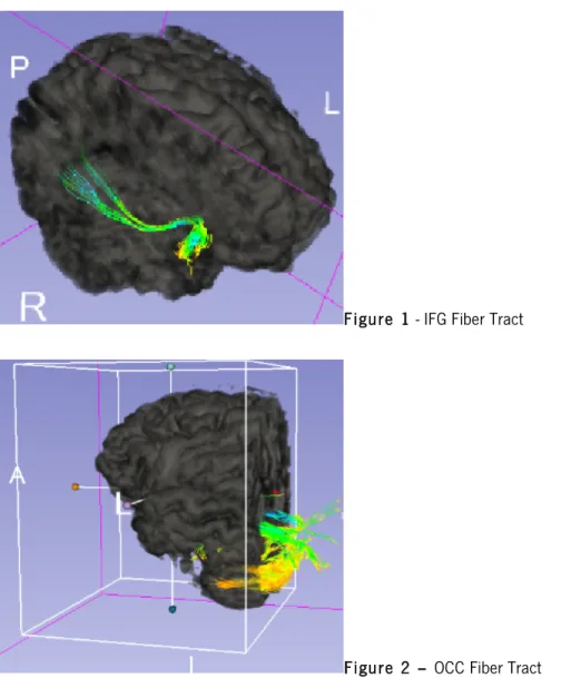
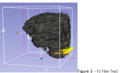
3.4.2 Region of Interest extraction (ROI) - Inferior-Frontal Gyrus (IFG), Occipital and V1 IFG was extracted according to (Kubicki et al., 2011), after ROI extraction using Freesurfer software, anatomical scans, along with corresponding ROIs, were co-registered to DTI space using 3D Slicer. Two additional sets of ROIs were also extracted: Occipital (OCC) lobe and V1 area.
A semiautomatic segmentation of IFG; Occipital and V1 ROI’S was made, using Freesurfer software (http://surfer.nmr.mgh.harvard.edu/) and ROI’s were integrated in Slicer3 (www. Slicer.org). Three anatomical regions derived from Freesurfer parcellation compose IFG tract: pars opercular, pars orbital and pars triangularis. Using FSL (FSL is a comprehensive library of analysis tools for FMRI, MRI and DTI brain imaging data) command line: fslmaths, it is possible to mask the segmentation file in order to create a new file of labels (IFG) based on the three individual ones.
A similar process was made for occipital label that is constituted by four anatomical regions: cuneus, lingual, pericalcarine and lateral occipital, inserted on the cortical parcellation of Freesurfer. These regions were merged and converted in a volume for statistical analysis.
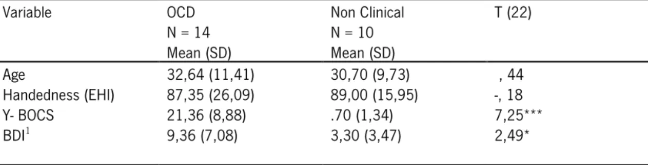
V1 area is a label created automatically by Freesurfer parcellation, which was necessary to convert in a volume using mri.label2vol. command line.
Figure 1 - IFG Fiber Tract
Figure 3 – V1 Fiber Tract
3.5 Tractography - Tractography is a method that uses several algorithms for the construction of fiber tracts with highest diffusion across voxels, i.e. the direction of the eigenvector with the major eigenvalue of the diffusion tensor. Specifically, in our study, was used Streamline Tractography which is based on the assumption that the orientation of fibers is collinear with the direction of the principal eigenvector, (Thomason & Thompson, 2011).
Thus, once Regions of interest (ROIs) were transferred into the DTI space; to generate white matter connections, Streamline Tractography was performed using Slicer 3.6 with the following parameters: a seed step of 2 mm between seed points, and a stopping curvature of 0,7 degrees per mm. A minimum Linear measure for seeding to start was defined with 0,3 and the threshold of FA has stopping criteria is 0.25.
3.6 Diffusion Indexes: Axial, Radial, Fractional and Mean diffusivity- Although the most widely reported measure of DTI is Fractional Anisotropy (FA) value, to better access white matter microstructure and integrity, we are going to use four different DTI indexes, i.e. Fractional anisotropy (FA) and Mean (MD), axial (AD) and radial (RD) diffusivity. All values derived from these three indexes are related to each other because they all derive from the same 3 eigenvalues of the tensor. The principal eigenvalues reflect the places where diffusion values are highest, (Thomason & Thompson, 2011).
FA is a scalar value between 0 and 1 that describes the degree of anisotropy of water diffusion in the brain. Zero represents no preferred direction (isotropic diffusion) and 1 represents unidirectional movement (anisotropic diffusion). Normally high values indicate constrained diffusion in one dominant direction along the fiber tract and are
associated with fiber integrity or connectivity. FA decreases due to a loss of coherence in the preferred direction of movement. Thus, high FA values are associated with healthier white matter pathways, i.e. best density, myelination and coherence in orientation, (Thomason & Thompson, 2011).
MD value is obtained by the mean of 3 eigenvalues, i.e. the mean molecular diffusion (diffusivity). This index reports the presence of barriers to free diffusion in the volume but doesn’t provide information about the direction of movement. In damaged or atrophied white matter, MD values are higher as a result of increased free diffusion, (Thomason & Thompson, 2011).
Axial diffusivity is derived from the largest of the 3 eigenvalues and measures the rate of diffusion in the direction of the fastest diffusion, detecting longitudinal diffusion along the axons. Decreased axial diffusion results from increased barriers to organized diffusion in the primary plane, (Thomason & Thompson, 2011).
Radial diffusivity derives from the average of the second and third eigenvalues, representing the transverse direction of diffusion that is more constrained by cellular structure and myelination. Damage to white matter may result in an increase of radial diffusion through the loss of barriers to perpendicular diffusion, (Thomason & Thompson, 2011).
SECTION IV
4. Data Analysis
Repeated measures ANOVA and independent t-tests were used in order to assess differences between OCD patients and controls in the microstructural white matter measures (FA, MD, AD and RD) in the three ROI under study (IFG, Occipital and V1). Pearson’s correlations were used to analyze the relationship between subjects' ROI microstructural measures (FA, MD, AD and RD) and their clinical profile, quantified by their scores on Y-BOC scale.
4.1 Results
4.1.1 Demographic Data
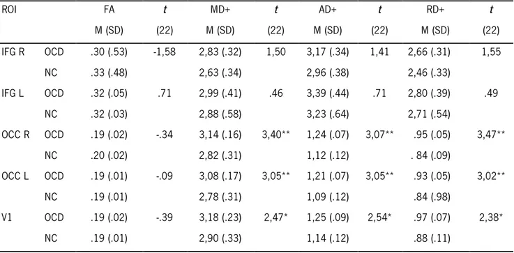
Groups did not differ in age at the time of scan (t (22) =.44, p=n.s.) and handedness (t (22)= -.18, p=n.s). In addition, as expected, scores in Y-BOCS and BDI scores were superior in the clinical group (t (22)= 7,25, p<.001) and (t (22)= 2,49, p<.05), respectively. No association between sex and group was found (χ2 (1) = .006, P= .94) - (see table 2).
Table 2 - Demographic Data
Variable OCD N = 14 Mean (SD) Non Clinical N = 10 Mean (SD) T (22) Age 32,64 (11,41) 30,70 (9,73) , 44 Handedness (EHI) 87,35 (26,09) 89,00 (15,95) -, 18 Y- BOCS 21,36 (8,88) .70 (1,34) 7,25*** BDI1 9,36 (7,08) 3,30 (3,47) 2,49* ***p<.001; *p<.05
4.1.2 ROI’s White Matter Microstructure Analysis
A repeated-measures ANOVA of IFG ROI showed no main effect of Side (Left vs Right) for FA [F (1,22)=.15, p=.71)], but this effect was observed in the other white matter integrity indexes: MD [F (1,22)= 6.80, p=.02)] ; AD [F (1,22)= 8.06, p=.01)]; RD [F (1,22)= 6.28, p=.02)] . No interaction between Side and Diagnosis was found for the 4 assessed indexes: FA [F (1,22)= .60, p=.45)] ; MD [F (1,22)= 1.07, p=.31)] ; AD [F (1,22)= 1.20, p=.29)]; RD [F (1,22)= 1.05, p=.32)]. Finally, no group effect was observed for MD [F (1,22)= .36, p=.55)] ; AD [F (1,22)= .13, p=.72)]; and RD [F (1,22)= .56, p=.46)]. However, a trend was found for a group effect in IFG FA [F (1,22)= 3.86, p=.06)], despite independent t-tests revealed no group differences (see table 3).
1 BDI scores were below cutoff in both groups
A repeated-measures ANOVA of Occipital ROI showed no main effect of Side (Left vs Right) for FA [F (1,22)=.53, p=.47)] and RD [F (1,22)= 2.94, p=.10)] values, but this effect was observed in the other white matter integrity indexes: MD [F (1,22)= 6.06, p=.02)] ; AD [F (1,22)= 10.24, p=.004)]. An interaction between Side and Diagnosis was observed for MD [F (1,22)=10.93, p=.003)] ; AD [F (1,22)=9.76, p=.005)]; RD [F (1,22)=11.15, p=.003)], but no interaction effect was observed for FA [F (1,22)=.07, p=.80)]. No group effect was observed for all white matter indexes: FA [F (1,22)= .11, p=.74)] ; MD [F (1,22)= .30, p=.59)] ; AD [F (1,22)= .13, p=.72)]; RD [F (1,22)= .20, p=.66)]. Independent t-tests revealed significant differences between groups in Occipital tract either in the right or left hemisphere, in 3 of the assessed indexes: rh2 MD [t (22)= 3.40, p=.003)] , lh MD [t (22)= 3.05, p=.006)] ; rh AD [t (22)= 3.07, p=.006)] , lh AD [t (22)= 3.05, p=.006)]; rh RD [t (22)= 3.47, p=.002)], lh RD [t (22)= 3.04, p=.006)], (see table 3).
Independent t-tests analyses of Visual area 1 revealed significant group differences in this tract, in 3 of the assessed indexes: MD [t (22)= 2.47, p=.02)]; AD [t (22)= 2.54, p=.02)]; RD [t (22)= 2.38, p=.03)] - (see table 3).
Table 3 – Mean FA, MD, AD and RD Values by ROI
+ values are represented *1000 for visualization purposes; FA = Fractional Anisotropy ; MD = Mean Diffusivity; AD = Axial Diffusivity; RD = Radial Diffusivity **p<.01; *p<.05
4.1.3. Correlations
Pearson correlations revealed an association between microstructural white matter measures (MD; AD; RD) and psychological measures of Obsessive Compulsive disorder. Higher diffusivity values were associated with higher scores on Y-BOCS scale, mainly in the left occipital lobe in OCD patients - (see table 4).
ROI FA M (SD) t (22) MD+ M (SD) t (22) AD+ M (SD) t (22) RD+ M (SD) t (22) IFG R OCD .30 (.53) -1,58 2,83 (.32) 1,50 3,17 (.34) 1,41 2,66 (.31) 1,55 NC .33 (.48) 2,63 (.34) 2,96 (.38) 2,46 (.33) IFG L OCD .32 (.05) .71 2,99 (.41) .46 3,39 (.44) .71 2,80 (.39) .49 NC .32 (.03) 2,88 (.58) 3,23 (.64) 2,71 (.54) OCC R OCD .19 (.02) -.34 3,14 (.16) 3,40** 1,24 (.07) 3,07** .95 (.05) 3,47** NC .20 (.02) 2,82 (.31) 1,12 (.12) . 84 (.09) OCC L OCD .19 (.01) -.09 3,08 (.17) 3,05** 1,21 (.07) 3,05** .93 (.05) 3,02** NC .19 (.01) 2,78 (.31) 1,09 (.12) .84 (.98) V1 OCD .19 (.02) -.39 3,18 (.23) 2,47* 1,25 (.09) 2,54* .97 (.07) 2,38* NC .19 (.01) 2,90 (.33) 1,14 (.12) .88 (.11)
Table 4 - Pearson correlations between ROI’s measures and Y-BOCS
ROI MD AD RD
r r r
OCC Right OCD .54* .63* .48+
NC -.30 -.27 -.31
OCC Left OCD .75** .76** .72**
NC -.35 -.34 -.35
V1 OCD .45+ .51+ .41
NC -.36 -.38 - .34
*p<.05; **p<.01; +p<.10
4.1.4 Discussion
In this study we analyzed, in OCD and in a healthy control matched group, several white matter microstructural indexes in three regions of interest (IFG, Occipital and V1), brain areas associated with visuo-perceptive processes. Our results showed differences between patients and controls in three different indexes (MD, AD and RD) in occipital and visual ROI’s, which are strictly related with early visual processing. Nevertheless, we did not observed group differences regarding white matter integrity measures in the IFG tract.
These results support our research team hypothesis, namely that a hyperactivation of fronto-subcortical regions is compensated by a deactivation on parieto-occipital regions associated to a disruption on the inferior fronto-occipital tract connecting anterior and posterior areas (Gonçalves et al., 2010). Therefore, we addressed this hypothesis by investigating the integrity of the Inferior Frontal Gyrus (IFG), which connects frontal areas with several brain regions including posterior areas as the occipital lobe and other visual-related areas (e.g. V1). These brain regions are associated with visuo-perceptive processing and are hypothesized to be altered in OCD. Hence, our
OCD and healthy controls in the diffusivity indexes (MD, AD and RD). Specifically, we observed higher diffusivity values (MD, AD and RD) in OCD, suggesting that the clinical group present more damaged or atrophied white matter (Thomason & Thompson, 2011) in the occipital and V1 areas, brain areas that are strictly related with an early stage of visual processing. Moreover, these results are consistent with evidence derived from both neurocognitive and neuroimaging studies.
Neurocognitive studies have shown OCD patients present deficits in visual organization and posterior memory recall, supporting the idea that perception and encoding style also affects memory performance contributing to deficits in OCD population (Rampacher et al., 2010). In line with these findings, Soref, Argov & Meiran, (2008), shown that OCD patients tend to use less parallel processing overall, being less responsive to contexts that encourage shifting to parallel processing, indicating they tend to focus on the target stimuli and are less inclined to process the flankers in parallel with the target, i.e. OCD patients present lesser responsiveness to changing contexts. Also, Kaplan et al. (2006) showed that OCD patients present an inflated Latent inhibition (LI), i.e. LI reflects the ability to ignore irrelevant stimuli, that may reveal a compensatory process in their tendency to be distracted by the irrelevant stimulus by rigidly focusing on the relevant stimulus in the early processing stage. Besides, Olatunji, Ciesielski & Zald (2011), shown that attentional deficits in OCD may be regulated by the emotional content of the distractor thus inhibiting the correct processing of the stimuli. Additionally, a study using a biological motion task corroborated evidence of a visuo-perceptive deficit in OCD, indicating a specific deficit in perceiving biological motion signals, although the perception for non-biological coherent motion and static global shape was intact. The authors suggested that deficits in biological motion perception may compromise visuo-processing of socio-emotional stimuli as efficient social stimuli aprehension depends on accurate perception of subtle socially relevant cues (Kim et al., 2008).
These results are also in line with functional studies (fMRI). Specifically, fMRI studies have shown an abnormal functioning of visual networks in patients with OCD while performing a visuo-emotional eliciting task: patients with OCD exhibited a greater activation of the bilateral orbitofrontal regions and right cingulate whereas healthy people displayed a higher activation in the occipital cortices, bilateral visual areas and inferior frontal gyrus (Mataix-Cols et. al, 2004). These findings may add support to the fact that some OCD symptoms, namely neurocognitive and emotional as emotional arousal, visual memory, attentional and executive can have a bottom-up genesis as it may be due to a deficit/bias in early stage of visual processing. Interestingly, taking into account our correlational analyses, we also found positive correlations between microstructural white matter measures (MD; AD; RD) and psychological measures of Obsessive Compulsive disorder. Specifically, higher diffusivity values were associated with higher scores
on Y-BOC scale, mainly in the left occipital lobe meaning that these regional white matter abnormalities are linked to higher Obsessive Compulsive symptomatology. This is also consistent with Garibotto et al. (2010) data, namely an association between higher Y-BOCS scores and lower FA values in the inferior fronto-occipital fasciculus bilaterally.
Finally, although in our study we have not found differences between OCD and controls in all DTI indexes (FA, MD, AD and RD) of IFG, as reported by others (Garibotto et al., 2010), we suggest that this may be possibly related with: a) different scanner and imaging acquisitions (3-tesla scanner vs 1.5T); b) the fact that diffusion tensor maps were calculated with Slicer and white matter measures were based on segmentation method using Freesurfer software, while Garibotto et al. (2010) used Brainvisa software; c) only man washers and checkers composed the OCD group in Garibotto et al. (2010) study and d) tractography of the overall IFG was performed in our study and not specific fronto-occipital white matter connections.
To sum up, we observed that patients with OCD exhibit white matter abnormalities of the occipital and V1 fiber tracts, which correlates with Y-BOCS scores, corroborating our hypothesis of involvement of these brain regions in visuo-perceptive abnormalities in OCD. These visuo-perceptive deficits may underlie the inaccurate cues OCD patients’ form in their social interactions leading to bias in memory encoding and emotional outputs.
4.1.5 Limitations and future studies
We found out some pertinents limitations that should be taken into account in future investigations. Namely, group size should be increased in order to allow the generalization of these findings. Also, the age range (19-59) of our group of participants is additionally identified as being a critical issue because white matter microstructure is very sensitive to changes throughout developmental stages (Lebel & Beaulieu, 2011), being not evident wether the differences found are exclusively due to the psychopathology or may have a developmental contribution. Although this could be controlled with age in the statistical analysies, this was not possible due to our small group sample. Likewise, we did not control our clinical group concerning the type of obsessive-compulsive cluster (e.g. washers, checkers, orders) underling their symptoms and future studies should increase the sample with different OCD clusters.
Also, future studies may adopt recent tractography metods, e.g. Diffusion Spectrum Imaging (DSI), taking into account evidence showing that tractography based on DSI has the capacity to image crossing fibers in the neural
dissertation. Other DTI analysis method as stochastic tractography should also be explored in future studies. Furthermore, other specific tracts should be analyzed as the IFG connection with occipital lobe and V1 areas that revealed to be altered in our OCD group. Finally, we suggest that future investigations should address an fMRI paradigm using a neurocognitive visual task in order to investigate specific alterations in brain activation patterns of the ROIs under study ( Occipital and V1).
References
1. Alexander, G. E. & Crutcher, M. D. (1990). Functional architecture of basal ganglia circuits: neural substrates of parallel processing. Trends in Neuroscience, 13, 266–271.
2. American Psychiatric Association (2000). Diagnostic and Statistical Manual of Mental Disorders: DSM-IV- TR. 4th ed. Washington, D.C.: American Psychiatric Association.
3. Aouizerate, B., Guehl, D., Cuny, E., Rougier, A., Bioulac, B., Tignol, J., & Burbaud, P. (2004). Pathophysiology of obsessive–compulsive disorder: a necessary link between phenomenology, neuropsychology, imagery and physiology. Progress in Neurobiology, 72, 195–221.
4. Baumgarten, H. G. & Grozdanovic, Z. (1998). Role of serotonin in obsessive-compulsive disorder. British Journal of Psychiatry, 35, 13–20.
5. Beck, A.T. (1988). Psychometric properties of the Beck Depression Inventory: Twenty-five years of evaluation. Clinical Psychology Review, 1, 77–100.
6. Bonelli, R.M. & Cummings, J.L. (2007). Frontal-subcortical circuitry and behavior. Dialogues in Clinical Neuroscience, 9, 141–151.
7. Brawman-Mintzer, O., Lydiard, R.B., Phillips, K.A., Morton, A., Czepowicz, V., Emmanuel, N., Villareal, G., Johnson, M., & Ballenger, J.C. (1995). Body dysmorphic disorder in patients with anxiety disorders and major depression: a comorbidity study. The American Journal of Psychiatry, 152, 1665–1667.
8. Brown, T. A., Di Nardo, P. A., Lehman, C. L., & Campbell, L. A. (2001). Reliability of DSM-IV anxiety and mood disorders: implications for the classification of emotional disorders. Journal of Abnormal Psychology, 111, 49–58.
9. Calamari, J.E., Wiegartz, P.S., & Janeck, A.S. (1999). Obsessive-compulsive disorder subgroups: a symptom-based clustering approach, Behaviour Research and Therapy, 37, 113–125.