From the Departments of Psychiatrics and Neurology, University of São Paulo School of Medicine, São Paulo - Brazil.
REVIEW
RELATIONSHIP BETWEEN
OBSESSIVE-COMPULSIVE DISORDERS AND DISEASES
AFFECTING PRIMARILY THE BASAL GANGLIA
Alex S. S. Freire Maia, Egberto Reis Barbosa, Paulo Rossi Menezes and Eurípedes C. Miguel Filho
RHCFAP/2994
MAIA, ASS et al. - Relationship between obsessive-compulsive disorders and diseases affecting primarily the basal ganglia. Rev. Hosp. Clín. Fac. Med. S. Paulo 54 (6):213-221, 1999.
SUMMARY: Obsessive-compulsive disorder (OCD) has been reported in association with some neurological diseases that affect the basal ganglia such as Tourette’s syndrome, Sydenham’s chorea, Parkinson’s disease, and Huntington’s disease. Furthermore, studies such as neuroimaging, suggest a role of the basal ganglia in the pathophysiology of OCD. The aim of this paper is to describe the association of OCD and several neurologic disorders affecting the basal ganglia, report the existing evidences of the role of the basal ganglia in the pathophysiology of OCD, and analyze the mechanisms probably involved in this pathophysiology.
DESCRIPTORS: Obsessive-compulsive disorder. Neuropsychiatry. Basal ganglia. Parkinson’s disease. Tourette’s syndrome. Sydenham’s chorea. Huntington’s disease.
Although neurology and psychiatry are generally defined as separate medi-cal specialties, disorders affecting the brain are not always restricted in clini-cal expression to only one of these spe-cialties. This is particularly true for dis-orders affecting the basal ganglia, in which the clinical picture is hardly ever limited to symptoms classically de-scribed as neurological or psychiatric. For this reason, a current trend is to describe these disturbances as neurop-sychiatric.
Obsessive-compulsive disorder (OCD) is one example of such a neu-ropsychiatric disorder that has been the object of growing interest in recent years. It affects about 3% of the gen-eral population1 and is the fourth most
frequent psychiatric diagnosis, sur-passed only by phobias, depressions, and drug addiction.1 OCD is
character-ized by obsessions and/or compulsions that are intense enough to interfere in the individual’s normal activities and in his social relations. Obsessions are
characterized by thoughts, ideas, or images (for example repetitive doubts or thoughts about contamination) that invade the individual’s awareness and are distressing and persistent. Although meaningless in themselves, the indi-vidual recognizes the obsessions as the product of his own mind and tries, al-beit unsuccessfully, to extinguish them. Compulsions are intentional repetitive behaviors (for example, hand-washing and rituals like checking doors and windows, and so on) that are often car-ried out in response to an obsession in order to reduce or alleviate unpleasant feelings 2.
OCD has recently been described in association with basal-ganglia-re-lated neurological diseases such as Sydenham’s chorea3,4,5,6, Parkinson’s
disease7, and Huntington’s disease8. In
addition, Tourette’s syndrome also ap-pears to be related to the basal ganglia, and is often found in association with OCD9,10,11.
The aim of this overview is to de-scribe the current evidence of associa-tion of OCD with several neurological diseases primarily affecting the basal ganglia, and to analyze the implica-tions of these structures in the patho-physiology of OCD.
OCD and Tourette’s syndrome
There is much evidence in favor of an association between OCD and TS. Gilles de la Tourette himself reported obsessive-compulsive behaviors in the cases he described in 1885. Several au-thors have described a frequency of obsessive-compulsive behavior in pa-tients with TS greater than that found in the general population9,10,11.
The association between OCD and TS is also borne out in genetic studies. Price et al.13, in a study of 43 pairs of
twins in which one member of each pair presented TS, found obsessive-compulsive symptoms in 83% of a to-tal 86 individuals. These authors also observed a greater frequency of obses-sive-compulsive behavior among monozygotic twins (52%) compared with dizygotic twins (15%).
Pauls & Leckman14 found a greater
frequency of OCD in first-degree rela-tives of TS patients, even in the ab-sence of tics or TS, suggesting that cer-tain forms of OCD could represent a variant of the same genes related to TS. Pauls et al.14, in a segregation analysis
study of 30 families, observed that the spectrum of TS is inherited in accor-dance with a dominant autosomal pat-tern with sex-dependent variable pen-etration.
Studies of patients with primary OCD have also showed a high percent-age of tics, ranging from 7% to 37%
15,16,17,11. Rosário-Campos et al.18
as-sessed 42 patients with OCD of whom 12 met criteria for a diagnosis of tics and/or TS. Interestingly, tics and TS oc-curred most frequently in the sub-group presenting earlier onset of obsessive-compulsive symptoms (before 10 years of age) when compared with later-on-set patients (after 18 years of age). Ad-ditionally, tic-like compulsions were more frequent in the early-onset group than in the late-onset group (even in pa-tients without a history of tics or TS). These results suggest that early-onset OCD is more related to the presence of tics and TS than late-onset OCD.
Miguel et al.11,17 compared the
char-acteristics of the repetitive behavior in patients with OCD +TS in relation to a group of patients with OCD and without tics or TS. It was observed that in the patients with OCD + TS, the re-petitive behaviors were less frequently preceded by cognitions (obsessions) and autonomic anxiety (e.g. dry mouth, tachycardia, sweating, pallor, breath-lessness) than in patients with OCD without TS. Patients with OCD + TS also presented a greater frequency of sensory phenomena (e.g. sensations or feelings of discomfort) preceding their behaviors. Thus, patients with OCD linked to TS also differ from those without tics from a clinical point of view, and it is believed that these vari-ables may be important as predictive factors for treatment. Finally, it should be stressed that McDougle et al.19 have
recently shown that patients with OCD + tics do not respond satisfactorily to classic treatment with selective seroto-nin reuptake inhibitors, and that these patients can benefit from the associa-tion of these antidepressants with neuroleptics. Summing up, there is a fair amount of evidence in the litera-ture suggesting a relationship between OCD and TS. Genetic family studies suggest that some forms of OCD may be the expression of the same genes re-lated to TS. Phenomenological studies show that patients with OCD + TS ex-hibit clinical features different from those patients with OCD without tics or TS. Early-onset OCD patients ap-pear to have several characteristics in common with patients with OCD + TS. Finally, patients with OCD + TS also appear to respond differently to com-mon OCD treatments.
OCD and Sydenham’s chorea
Sydenham’s chorea (SC) is a disor-der affecting the basal ganglia, charac-terized by choreiform movements,
some degree of hypotonia and occa-sionally by emotional instability. SC is considered to be a complication of rheumatic fever, and therefore arthritis and carditis are other commonly asso-ciated manifestations20.
The association between OCD and SC was first established by Chapman3
in a study of 8 children with the dis-ease, 4 of whom presented marked ob-sessive-compulsive symptoms (OCS), including washing rituals.
Grimshaw 4, on the other hand,
comparing the prevalence of neurologi-cal alterations in patients with and without obsessive characteristics, found 6 with a history of SC out of 103 obsessive patients, whereas only 2 of the 105 controls presented positive his-tory for this condition.
In two studies, Swedo et al.5,6
de-scribed interesting links between OCS and SC. In the first5, children and
ado-lescents with previous history of SC presented a greater prevalence of ob-sessive-compulsive symptoms when compared with a control group that had had rheumatic fever (RF) without SC. In the second study 6, 11 children in the
acute phase of SC were assessed in or-der to detect obsessive-compulsive symptomatology. Nine of them pre-sented this type of psychiatric disorder with acute onset; none of them had previous history for OCS. Among these 9 children, 4 met criteria for OCD, 2 presented subclinical OCD, and 3 pre-sented only OCS . The OCS began shortly before the appearance of the movement disorder, peaked during the period of most intense choreiform movements, and generally disappeared before the movements did so.
Since the OCS in these children with SC were identical to those in the children with only OCD, Swedo et al.6
words, in genetically susceptible chil-dren, infection by group A b hemolytic streptococcus might induce the forma-tion of antineuronal antibodies that in certain subcortical structures might lead to dysfunction of the basal gan-glia. This dysfunction, in turn, might manifest itself as SC, OCD, or both.
Mercadante et al.21 recently
con-cluded a study analyzing the preva-lence of psychiatric disorders in 22 pa-tients with RF and SC, 20 with RF but without SC, and 20 controls. A greater (but not statistically significant) fre-quency of OCD was found in this study in the two groups with RF when com-pared with the control group. The fre-quency of OCS in patients with only RF was significantly greater when compared with the control group; dif-ferences between the groups with and without SC with regard to OCD and OCS were not observed. In the same study, an increased frequency of tics and attention deficit disorder with hy-peractivity (ADDH) was found in pa-tients with SC. Based on these results, Mercadante et al.21 suggest a
psycho-pathological vulnerability in patients with RF that manifests itself through a symptomatological continuum includ-ing OCS, ADDH, and movement dis-orders such as tics and chorea.
In conclusion, there is evidence to-day that OCD is not only more fre-quent in patients with SC, but also in patients with RF without SC, suggest-ing that autoimmune mechanisms sec-ondary to streptococcal infection could play an important role in the etiology of certain subtypes of OCD.
OCD and Parkinson’s disease
Parkinson’s disease (PD) is a highly-prevalent neurological disorder. Its major clinical features are akinesia, plastic hypertonia, resting tremor, and postural instability. Autonomic alter-ations and psychiatric disorders such as
dementia and depression may also be present. Prevalence ranges from 150 to 200 cases per 100,000 individuals in the general population, increasing with age so that the prevalence of PD in in-dividuals over 65 is 1% 22.
The main pathophysiologic aspect of PD is a decrease in dopaminergic lev-els owing to degeneration of melanin-containing neurons in the substantia ni-gra (pars compacta), which are respon-sible for production of this neurotrans-mitter. These neurons project into the striatum (caudate and putamen).
In the early twentieth century the presence of OCD in cases of post-en-cephalitic parkinsonism was ob-served23. Lauterbach 24, studying 28
families with family Parkinsonism, found a rate of OCD 5 times greater than that of the normal population.
Tomer et al.7 recently carried out a
systematic study in 30 patients with PD, showing that 17 of them presented a total score for obsessive-compulsive symptoms above that of the controls. An association between motor asym-metry and the presence of obsessive-compulsive symptoms was observed in this study. The intensity of the left-side motor symptoms was associated with cleanliness worries, repetitions, and disturbing thoughts, whereas the sever-ity of right-sided symptoms was sig-nificantly related only to obsessions with order and routine. Despite the fact that some neuroimaging studies have shown differences between the two hemispheres 25,26,27,28, evidence
regard-ing lateralized pathophysiology in OCD is inconclusive as yet.
In a preliminary analysis of a prevalence study for OCS in patients with Parkinson’s disease, Maia et al.29
found OCS in 12 (24%) of 50 patients evaluated. This study also found a pre-ponderance of left-side motor symp-toms among patients with OCS, which supports a hypothesis of a possible lat-eralization in the pathophysiology of OCD 30.
One should remember in analyzing these results that Hardie et al.31 reported
that some patients after treatment with levodopa began to present stereotyped movements, even organized rituals, suggesting that some patients with OCS and Parkinson’s disease may suf-fer their symptoms secondary to the effect of the treatment given.
OCD and Huntington’s Disease
Huntington’s disease (HD) is a dominant autosomal neurodegenerative disease that primarily manifests itself through choreiform movements and progressive dementia. Although there are few reports in the literature of OCD in patients with HD, Cummings & Cunningham 8 reported two cases of
HD with OCD appearing after the on-set of the neurological symptoms of HD. Fear of contamination and wash-ing compulsion were present at first, followed by a compulsion to smoke and drink, without the characteristics of dependency. According to the au-thors, this scarcity of accounts may re-flect either an actual low prevalence of OCS in patients with HD, or a failure to identify its manifestations, or even a tendency for the manifestations to appear late in the course of the disease, when patients are presenting acute cog-nitive disorders, making it difficult to recognize the manifestations8.
OCD in other basal ganglia disorders
Obsessive-compulsive symptoms have also been described in patients with neuroacanthocytosis 32 and
idio-pathic spasmodic torticollis33.
Data from the literature also sug-gest a relation between focal cerebral lesions affecting the basal ganglia and the appearance of obsessive-compul-sive symptoms. Pulst et al.34 have
bilateral caudate lesions after carbon monoxide poisoning. Weilburg et al.35
observed a patient with a history of neonatal hypoxia and atrophy of the left caudate and putamen who devel-oped severe OCD in adolescence. There are also accounts of OCD occur-ring after bilateral caudate infarction
36,37,38 and OCS after globus pallidus
le-sions caused by carbon monoxide poi-soning 39,40 and post-anoxia 39.
The basal ganglia and the pathophy-siology of OCD
The basal ganglia are deep subcor-tical structures represented by the stria-tum (caudate and putamen), globus pallidus, substantia nigra, and the sub-thalamic nucleus of Luys.
The anatomic connections of the basal ganglia provide some evidence of the participation of these structures in non-motor functions. Of particular im-portance are the connections between the basal ganglia and the limbic sys-tem. Many limbic projections into the striatum are already known, mainly de-riving from the hippocampus, cingulate cortex, and amygdala. In addition to the primary motor region, however, other cortical regions – above all the frontal cortex – send projections to the basal ganglia.
In 1983, De Long et al.41 proposed
the existence of two loops involving the basal ganglia. The motor loop, cen-tered in the putamen, received afferent projections from the sensory-motor cortex and sent projections to the pre-motor areas of the frontal cortex via the globus pallidus and thalamus. The complex loop, involving afferent pro-jections from the associative areas of the frontal cortex, projected into the caudate nucleus and striatal–pallidal– thalamic–cortical pathways towards the prefrontal cortex.
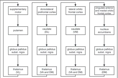
According to studies by Alexander et al.42, it is currently thought that five
parallel circuits exist that are respon-sible for frontal-striatal connections: the motor circuit, the oculomotor cir-cuit, the dorsolateral prefrontal circir-cuit, the lateral orbitofrontal circuit, and the anterior cingulate. Figure 1 shows these circuits, except for the oculomo-tor circuit, which is a variant of the motor circuit.
Analyzing these frontal-striatal cir-cuits, one can observe that cortical ar-eas project with somatotypical organi-zation into the striatum (putamen, cau-date, and nucleus accumbens). These projections are mediated by the exci-tatory neurotransmitter glutamate. The connections between the striatum and the inner globus pallidus–substantia nigra (pars reticulata) complex take two pathways (Figure 2): the direct (in-hibitory) pathway and the indirect pathway, which goes through the exter-nal globus pallidus and the subthalamic nucleus of Luys (excitatory). Efferent GABA-ergic projections leave the glo-bus pallidus–substantia nigra complex for the thalamus. Finally, thalamic nu-clei (lateral ventral, anterior ventral,
and medial dorsal), establish feedback loops by projecting into specific re-gions of the frontal lobe. These thalamo-cortical projections are be-lieved to be excitatory and use aspar-tate or glutamate as the neurotransmit-ter.
The putamen, mainly associated with motor aspects, and the caudate and nucleus accumbens, related to cog-nitive and behavioral aspects, can be distinguished within the striatum. However, given the overlap of differ-ent cortical projections into the stria-tum, this division should be considered a continuous transition without defined limits 43.
Other hypotheses relating to the or-ganization of the striatum exist. Graybiel et al.44,45 propose that the
striatum can be divided into two dis-tinct compartments: the matrix, which is mainly related to sensory-motor pro-cessing, and the striosome, which re-ceives afferent projections from struc-tures associated with the limbic system and associative areas. Both compo-nents of the striatum, putamen and
cau-Figure 1 - Frontal-striatal connections.
date, contain matrix and striosomes. This concept stemmed from studies showing the presence of small acetyl-cholinesterase-poor regions (strio-somes) between acetylcholinesterase-rich areas (matrix). These regions were later found to differ with regard to sev-eral other neurotransmitters45. The
striosome/matrix division is therefore based on neurochemical data, while the individuality of the caudate and puta-men has an anatomic basis.
In addition to anatomic connec-tions, neuroimaging studies have sug-gested the participation of the basal ganglia in the pathophysiology of OCD. Several studies using both cra-nial tomography and magnetic reso-nance imaging have shown the loss of the asymmetry normally observed in caudate nuclei, the left being larger than the right, in patients with OCD or TS 25,26,27,28. On the other hand, other
authors have shown a reduction in size of the lenticular nucleus (globus pallidus and putamen) in magnetic resonance imaging of patients with TS, however without a change in the cau-date volume46,47. The use of functional
neuroimaging enabled Baxter et al.48,49
to identify, through positron emission tomography (PET-scan), an increase in glucose metabolism in the caudate nucleus and the orbitofrontal region of patients with OCD, when compared to controls. Later PET-scan studies 50 and
single-photon emission computed to-mography (SPECT) studies 51 also
showed an increase of metabolic activ-ity in the orbitofrontal region 51, and
Swedo et al.52 observed a correlation
between this increased activity and the intensity of obsessive-compulsive symptoms.
Functional neuroimaging studies of patients with OCD before and after treatment have shown interesting re-sults. Baxter et al.48,53 observed a
rela-tion between decreased metabolic ac-tivity of the caudate nucleus and im-provement of symptoms after treat-ment, and showed later that this de-crease in metabolic activity of the head of the caudate nucleus occurred not only after pharmacologic treatment, but also after behavioral psychothera-peutic treatment. Benkelfat et al.54
found a decrease of metabolism of the
left caudate after treatment with clomi-pramine. Swedo et al.55, also assessing
patients pre- and post-treatment, showed a correlation between an in-crease in activity in the right orbito-frontal region and improvement in symptoms after drug therapy. Based on such data, Baxter et al.53 proposed a
hypothesis that there is an initial alter-ation in the caudate and a later one in the orbitofrontal region.
Interesting PET-scan analyses of cerebral activity have been carried out during provocation of obsessive-com-pulsive symptoms. This provocation was undertaken, for instance, in the case of a patient with obsessions of contamination, by wearing gloves pre-viously contaminated by himself, in comparison with a control state in which the same patient wore sterile gloves. Patterns of cerebral metabolic activity consistent with other patterns already alluded to – hyperactivity of the caudate, thalamus and orbitofrontal cortex – were found in these studies
56,57. Rauch et al.56 have also observed
greater activity of the left anterior cin-gulate cortex.
In addition to neuroimaging data, the classic neuropsychological meth-ods also bear out the hypothesis of the participation of the basal ganglia in the pathophysiology of OCD. One should remember that neuropsychological al-terations can sometimes be detected even in the absence of a detectable structural pathology 58.
Neuropsychological studies with obsessive-compulsive patients have shown similar results to those in pa-tients with Parkinson’s or Huntington’s disease. Studies of OCD have shown impairment of visuospatial ability
59,60,61,62, non-verbal memory 61,63,64, and
executive functions 59,65,66,67,64.
Memory impairment seems to be mainly linked to the implicit mecha-nism of the memory. Storage and re-call of this type of memory is not to-tally dependent on awareness or
cog-Figure 2 - Corticostriate projections.
nitive processes. It consists of a rela-tively slow process that takes place through repetition of tasks, and is char-acterized by a performance improve-ment in the execution of such tasks 68.
Alterations in implicit memory may reflect a dysfunction of the striatum, probably involved with this modality of memory. Rauch et al.58 proposed
that the striatum carries out simpler functions, facilitating good perfor-mance of frontal cortex functions (which are responsible for more com-plex operations), in order to relieve it of some portion of computational work related primarily to processing uncon-scious information. Some authors sug-gest that impairment of executive func-tions alters the planning, organization, and ability to alter “patterns”, leading to visuospatial and memory deficits.58.
On the basis of anatomic knowl-edge and data obtained mainly through neuroimaging studies, Baxter et al.53
have advanced a model that is interest-ing with regard to the participation of the basal ganglia in the pathophysiol-ogy of OCD. According to their model, the cortical-striatal-thalamic-cortical circuits act as filters in order to inhibit irrelevant thoughts.
According to this concept, a dys-function of the caudate nucleus would lead orbital cortex impulses not to be suitably “filtered” (repressed), activat-ing the direct pathway. Thus, the in-hibitory impulses of the caudate over the globus pallidus would increase. This inhibition would result in less in-hibition of the thalamus by the globus pallidus, and consequently, excitatory thalamic impulses to the orbital corti-cal regions would make a reinforce-ment loop. These changes would lead to a failure to inhibit irrelevant worries, which thus receive disproportional
at-tention (obsessions) and trigger repeti-tive, senseless behaviors (compul-sions).
Implications for the treatment of OCD
The knowledge acquired through these studies stimulated proposals for other OCD treatments. In addition to the already existing pharmacologic ap-proaches, such as the use of serotonin reuptake inhibitors (SRIs), there are surgical approaches, and most recently, the emergence of repetitive transcranial magnetic stimulation — rTMS.
With regard to surgical approaches, recent studies suggest that several pro-cedures (e.g. anterior cingulotomy, an-terior capsulotomy, tractotomy of the subcaudate, and limbic leukotomy) may be effective in OCD cases refrac-tory to usual treatment. Baer et al.69,70
have confirmed relative security and effectiveness (around 30% of patients presented marked improvements) in the case of cingulotomy (stereotaxic transection of the tracts from the fron-tal cortex to subcortical sites – caudate) as a treatment for patients with severe OCD that is refractory to other forms of treatment. Capsulotomy (lesion of the internal capsule containing fibers interconnecting the orbitofrontal region and the dorso-medial nucleus of the thalamus and other related areas) has been performed in Europe for three de-cades for refractory anxiety disorders. A promising new technique is the “gamma knife”, which makes a small lesion in the anterior portion of the in-ternal capsule 71. This procedure has
proved effective in a significant per-centage of patients (around 60%), and has advantages including the fact that craniotomy is not necessary and that
the patient does not need a lengthy hos-pital stay after the operation 71.
rTMS is a new non-invasive tech-nique involving direct stimulation of cortical neurons, which can cause al-terations in cerebral activity, and can be used for therapeutic purposes72. It is
believed that specific cerebral circuits, such as those involved in OCD, can be inhibited or stimulated by these tech-niques. Greenberg et al.73 observed that
a single session of rTMS, aimed at the right orbitofrontal cortex, produced a reduction in compulsions for a roughly 8-hour period. The mechanisms by which this technique benefited these patients, however, need deeper study.
In conclusion, therefore, there is evidence in favor of participation of the basal ganglia in the pathophysiology of OCD. These data have diagnostic and therapeutic implications for the clini-cal practice both of neurologists and psychiatrists. In other words, in pa-tients with OCD, the psychiatrist should be aware that the presence of other disorders involving the basal gan-glia, such as Tourette’s syndrome, Sydenham’s chorea, Huntington’s dis-ease, and Parkinson’s disease may be related to this disorder. Similarly, neu-rologists should investigate obsessive-compulsive symptoms when treating patients with these diseases. Finally, it is believed that a greater understanding of the role of the basal ganglia in OCD may be useful in developing new thera-peutic approaches.
ACKNOWLEDGEMENTS: This
RESUMO RHCFAP/2994
MAIA, ASS e col. - Relação entre transtorno obsessivo-compulsivo e doenças neurológicas dos gânglios da base. Rev. Hosp. Clín. Fac. Med.
S. Paulo 54 (6):213-221, 1999.
O transtorno obsessivo-compulsivo (TOC) tem sido reportado em associa-ção com algumas doenças neurológicas que afetam primariamente os gânglios
da base como a síndrome de Tourette , a coréia de Sydenham, a doença de Parkinson e a doença de Huntington. Da mesma forma, estudos de neuro-imagem sugerem a participação dos gânglios da base na fisiopatologia do TOC. O objetivo deste estudo é rever a coexistência de TOC e várias doen-ças que afetam os gânglios da base, as evidências da participação dessas
estru-turas na fisiopatologia do TOC e os mecanismos neurais subjacentes a esse distúrbio psiquiátrico.
DESCRITORES: Transtorno
o b s e s s i v o - c o m p u l s i v o . Neuropsiquiatria. Gânglios da base. Doença de Parkinson. Síndrome de Tourette. Coréia de Sydenham. Doença de Huntington.
REFERENCES
1. KARNO M & GOLDING JM - Obsessive Compulsive Disorder. In: ROBINS LN & REGIER DA eds - Psyschiatric Disorders in America. The Epidemiologic Catchment Area Study. New York, Free Press, 1991.
2. MIGUEL EC - Transtornos do espectro obsessivo-compulsivo: diagnóstico e tratamento. Rio de Janeiro, Guanabara Koogan, 1996. 3. CHAPMAN AH, PILKEY L & GIBBONS MJ - A psychosomatic study of eight children with Sydenham’s chorea. Pediatrics 1958; 21: 582-595.
4. GRINMSHAW L - Obsessional disorder and neurological illness. J Neurol Neurosurg Psychiatry 1964; 27: 229-231.
5. SWEDO SE, RAPOPORT JL, CHESLOW DL et al. - High prevalence of obsessive-compulsive symptoms in patients with Sydenham’s chorea. Am J Psychiatry 1989; 146: 246-249.
6. SWEDO SE, LEONARD HL, SCHAPIRO MB et al. - Sydenham’s chorea: physical and psychological symptoms of St. Vitus dance. Pediatrics 1993; 91: 706-713.
7. TOMER R, LEVIN BE & WEINER WJ - Obsessive-compulsive symptoms and motor asymmetries in Parkinson’s disease. Neuropsych Neuropsychol Behav Neurol 1993; 6:26-30. 8. CUMMINGS JL & CUNNINGHAM K - Obsessive-compulsive disorder
in Huntington disease. Biol Psychiatry 1992; 31: 263-270. 9. CAINE ED, MCBRIDE MC, CHIVERTON P et al. - Tourette’s
syndrome in Monroe County school children. Neurology 1988; 38: 472-475.
10. SINGER HS & ROSENBERG LA - The development of behavioral and emotional problems in Tourette’s syndrome. Pediatr Neurol 1989; 5: 41-44.
11. MIGUEL EC, BAER L, COFFEY, BJ et al. - Phenomenological differences appearing with repetitive behaviours in obsessive-compulsive disorder an Gilles de la Tourette’s syndrome. Br J Psychiatry 1997; 170: 140-145.
12. BRITO GNO - Síndrome de Tourette: Clínica, terapêutica e modelo neurobiológico. In: MIGUEL EC - Transtornos do espectro obsessivo-compulsivo: diagnóstico e tratamento. Rio de Janeiro, Guanabara Koogan, 1996.
13. PRICE RA, KIDD KK, COHEN DJ et al. - A twin study of Tourette syndrome. Arch Gen Psychiatry 1985; 42: 815-820.
14. PAULS DL & LECKMAN JF - The inheritance of Gilles de la Tourette’s Syndrome and associated behaviors. N Engl J Med 1986; 315: 993-997.
15. RASMUNSEN AS & EISEN JL - Epidemiology and clinical features of obsessive-compulsive disorder. In: JENIKE M, BAER L, MINICHELLO W, eds. - Obsessive-compulsive disorder: therapy and management. Chicago, Yr Bk Med Pub, 1990.
16. PITTMAN RK, GREEN RC, JENIKE MA et al. - Clinical comparison of Tourette’s disorder and obsessive-compulsive disorder. Am J Psychiatry 1987; 144: 1166-1171.
17. MIGUEL EC, COFFEY BJ, BAER, L et al.- Phenomenology of intentional repetitive behaviors in obsessive-compulsive disorder and Tourette’s disorder. J Clin Psychiatry 1995; 56: 246-255. 18. ROSÁRIO-CAMPOS MC - Transtorno Obsessivo-Compulsivo de
início precoce e de início tardio: características clínicas, psicopatológicas e de comorbidade. São Paulo, 1998. (Tese -Mestrado, Faculdade de Medicina da Universidade de São Paulo).
19. MCDOUGLE CJ, GOODMAN WK, LECKMAN JF et al. - Haloperidol addition in fluvoxamine-refractory obsessive compulsive disorder. A double-blind, placebo-controlled study in patients with and without tics. Arch Gen Psychiatry 1994; 51: 302-308. 20. ASBAHR FR - Coréia de Sydenham e transtorno
obsessivo-compulsivo. In: MIGUEL, EC - Transtornos do espectro obsessivo-compulsivo: diagnóstico e tratamento. Rio de Janeiro, Guanabara Koogan, l996. p.158-162.
21. MERCADANTE MT et al. - Rheumatic fever and co-morbid psychiatric disorders. Am J. Psychiatry (in print).
22. RAJPUT AH, OFFORD AP, BEAR CM et al. - Epidemiology of Parkinson´s disease: incidence, classification and mortality. Ann Neurol 1984, 16: 78-132.
24. LAUTERBACH EC & DUVOISIN RC - Neuropsychiatric correlates of familial parkinsonism. Neurology 1987; 37 (1).
25. LUXEMBERG J, SWEDO SE, FLAMENT MF et al. - Neuroanatomic abnormalities in obsessive-compulsive disorder detected with quantitative X-ray computed tomography. Am J psychiatry 1988; 145: 1089-1094.
26. ROBINSON D, WU H, MUNNE RA et al. - Reduced caudate nucleus volume in obsessive-compulsive disorder. Arch Gen Psychiatry 1995; 52: 393-398.
27. SCARONE S, COLOMBO C, LIVIAN S et al. - Increased right caudate nucleus size in obsessive-compulsive disorder: detection with magnetic resonance imaging. Psychiatry Res Neuroimaging 1992; 45: 115-121.
28. JENIKE MA, BREITER HC, BAER L et al. - Cerebral structural abnormalities in obsessive-compulsive disorder: a quantitative morphometric magnetic resonance imaging study. Arch Gen Psychiatry 1996; 53:625-32.
29. MAIA AS, PINTO ASS, BARBOSA ER et al. - Obsessive-compulsive symptoms in patients with Parkinson’s disease. In: AMERICAM PSYCHIATRIC ASSOCIATION ANNUAL MEETING, 15th, Toronto, Canadá, 1998. (New Research- Program & Abstracts, NR 137, p.105).
30. PINTO AS, MAIA A, BARBOSA E et al. - Parkinson ‘s disease and obsessive-compulsive symptoms. In: CONGRESSO BRASILEIRO DE NEUROLOGIA, 18th, São Paulo, 1998. Arch Neuro-Psiquiatr, Supl 1(5): 209).
31. HARDIE RJ, LEES AJ & STERN GM - On-off fluctuations in Parkinson’s disease. Brain 1984; 107: 487-506.
32. MIRANDA M, CAMPERO M, TENHAMM, E et al. - Neuro-acantocitosis: comunicacion de 3 casos. Rev Med Chile 1993; 21: 176-179.
33. BIHARI K, HILL JL & MURPHY DL - Obsessive-compulsive characteristics in patients with idiopathic spasmodic torticollis. Psychiatry Res 1992; 42: 267-272.
34. PULST SM, WALSHE TM & ROMERO JA - Carbon monoxide poisoning with features of Gilles de la Tourette’s syndrome. Arch Neurol 1983; 40: 443-444.
35. WEILBURG JB, MESULAM MM, WEINTRAUB S et al. - Focal striatal abnormalities in a patient with obsessive-compulsive disorder. Arch Neurol 1989; 46: 233-235.
36. CROISILE B, TOURNIAIRE D, CONFRAVEUX C et al. - Bilateral damage to the head of the caudate nuclei. Ann Neurol 1989; 25: 313-314.
37. TRILLET M, CROISILE B, TOURNIAIRE D et al. - Perturbations de l’activité motrice volontaire et lésions de noyaux caudés. Rev Neurol 1990; 146: 338-344.
38. WILLIAMS AC, OWEN C & HEATH DA - A compulsive movement disorder with cavitation of caudate nucleus. J Neurol Neurosurg Psychiatry 1988; 51: 447-448.
39. LAPLANE D, LEVASSEUR M, PILLON B et al. - Obsessive-compulsive and other behavioural changes with bilateral basal ganglia lesions. Brain 1989; 112: 699-725.
40. ALI-CHERIF A, ROYERE ML, GOSSET A et al. - Troubles du comportement et de l’activité mentale après intoxication oxycarbonée. Rev Neurol (Paris), 1984; 140: 401-405. 41. DE LONG MR, GEORGOPOULOS AP & CRUTCHER, MD -
Cortico-basal ganglia relations and coding of motor performance. Exp Brain Res 1983; 7: 30-40.
42. ALEXANDER GE, CRUTCHER MD & DE LONG MR - Basal ganglia-thalamocortical circuits: parallel substrates for motor, oculomotor, “prefrontal” and “limbic” functions. Prog Brain Res 1990; 85: 119-146.
43. MELLO LEAM & VILLARES J - Neuroanatomy of the basal ganglia. In: MELLO L et al. - Neuropsychiatry of the basal ganglia. Psychiatric Clinics of North America. Philadelphia, Saunders, 1997. 44. GRAYBIEL AM & RAGSDALE CW - Histochemically distinct compartments in the striatum of human, monkey, and cat demonstrated by acetylcholinesterase staining. Proc Natl Acad Sci SA 1978, 75:5723-5726.
45. GRAYBIEL AM, RAGSDALE CW, YONEOKA ES et al. - An immunohistochemical study of enkephalin and other neuropeptides in the striatum of the cat with evidence that opiate peptides are arranged to form mosaic patterns in register with striosomal compartments visible by acetylcholinesterase staining. Neuroscience 1981; 6:377-397.
46. PETERSON B, RIDDLE MA, COHEN DJ et al. - Reduced basal ganglia volumes in Tourette’s syndrome using three-dimensional reconstruction techniques from magnetic resonance images. Neurology 1993; 43: 941-949.
47. SINGER HS, REISS AL, BROWN JE et al. - Volumetric MRI changes in basal ganglia of children with Tourette’s syndrome. Neurology 1993; 43: 950-956.
48. BAXTER LR, PHELPS ME, MAZZIOTA JC et al. - Local cerebral glucose metabolic rates in obsessive-compulsive disorder. A comparison with rates in unipolar depression and in normal controls. Arch Gen Psychiatry 1987; 44: 211-218.
49. BAXTER L, SCHWARTZ J, MAZZIOTA J et al. - Cerebral glucose metabolic rates in non-depressed patients with obsessive-compulsive disorder. Am J Psychiatry 1988; 145: 1560-1563. 50. NORDHAL TE, BENKELFAT C, SEMPLE W et al. - Cerebral glucose
metabolic rates in obsessive-compulsive disorder. Neuropsychopharmacology 1989; 2: 23-28.
51. RUBIN RT, VILLANUEVA-MEYER J, ANANTH J et al. - Regional xenon 133 cerebral blood flow and cerebral technetium 99m-HMPAO uptake in unmedicated patients with obsessive-compulsive disorder and matched normal control subjects. Determination by high-resolution single photon emission computed tomography. Arch Gen Psychiatry 1992; 49: 695-702.
52. SWEDO SE, SCHAPIRO MB, GADY CL et al. - Cerebral glucose metabolism in childhood-onset obsessive-compulsive disorder. Arch Gen Psychiatry 1989; 46: 518-523.
53. BAXTER LR, SCHWARTZ JM, BERGMAN KS et al. - Caudate glucose metabolic rate changes with both drug and behaviour therapy for obsessive-compulsive disorder. Arch Gen Psychiatry 1992; 49: 681-689.
54. BENKELFAT C, NORDAHL TE, SEMPLE WE et al. - Local cerebral glucose metabolic rates in obsessive-compulsive disorder: patients treated with clomipramine. Arch Gen Psychiatry 1990; 47: 840-848.
55. SWEDO S, PIETRINI P, LEONARD LH et al. - Cerebral glucose metabolism in childhood-onset obsessive-compulsive disorder. Revisualisation during pharmacotherapy. Arch Gen Psychiatry 1992; 49: 690-694.
56. RAUCH SL, JENIKE MA, ALPERT NM et al. - Regional cerebral blood flow measured during symptom provocation in obsessive-compulsive disorder using oxygen 15-labeled carbon dioxide and positron emission tomography. Arch Gen Psychiatry 1994; 51: 62-70. 57. MCGUIRE PK, BENCH CJ, FRITH CD et al. - Functional anatomy
of obsessive-compulsive phenomena. Br J Psychiatry 1994; 164: 459-468.
58. RAUCH SL & SAVAGE CR - Neuroimaging and neuropsychology of the striatum: bridging basic science and clinical practice. Neuropsychiatry of the basal ganglia. Psychiatric clinics of North America. Philadelphia, Saunders, 1997.
59. ARONOWITZ BR, HOLLANDER E, DECARIA C et al. -Neuropsychology of obsessive-compulsive disorder: Preliminary findings. Neuropsych Neuropsychol Behav Neurol 1994; 7:81-86. 60. BEHAR D, RAPOPORT JL, BERG CJ et al. - Computerized tomography and ]neuropsychological test measures in adolescents with obsessive-compulsive disorder. Am J Psychiatry 1984; 141:363-368.
63. CHRISTENSEN KJ, KIM SW, DYSKEN MW et al. - Neuropsycholo-gical performance in obsessive-compulsive disorder. Biol Psychiatry 1992, 31:4-18.
64. ZIELINSKI CM, TAYLOR MA & JUZWIN KR. - Neuropsychological deficits in obsessive-compulsive disorder. Neuropsych Neuropsychol Behav Neurol 1991; 4:110-126.
65. HARVEY NS - Impaired cognitive set-shifting in obsessive-compulsive neurosis. IRCS Med Sci 1986; 14:936-937.
66. HEAD D, BOLTON D, HYMAS N et al. - Deficit in cognitive shifting ability in patients with obsessive-compulsive disorders. Biol Psychiatry 1989; 25:929-937
67. MALLOY P - Frontal lobe dysfunction in obsessive-compulsive disorder. In :PERECMAN E - The Frontal Lobes Revisited. New York, IRBN Press, 1987,
68. KANDEL ER, SCHWARTZ JH & JESSEL TM - Essentials of Neural Science and Behavior. Prent Hall Int 1995.
69. BAER L, RAUCH SL, BALLANTINE HT et al. - Cingulotomy for intractable obsessive-compulsive disorder: prospective long-term follow-up of 18 patients. Arch Gen Psychiatry 1995; 52:384-392. 70. BAER L - Factor analysis of symptom subtypes of obsessive compulsive disorder and their relation to personality and tics. J Clin Psychiatry 1993; 55:[3, suppl] 18-23.
71. MINDUS P & JENIKE MA - Neurosurgical treatment of malignant obsessive-compulsive disorder. Psychiatric Clin North Am 1992; 15(4):921-938.
72. GEORGE MS, WASSERMANN EM & POST RM - Transcranial Magnetic Stimulation: A neuropsychiatric tool for the 21st century. J Neuropsych Clin Neurosci 1996; 8:373-382.
73. GREENBERG BD, MCCANN UD, BENJAMIN J et al. - Repetitive TMS as a probe in anxiety disorders: theoretical considerations and case reports. CNS Spectrums 1997; 2(1): 47-52, 1997.