1. Serviço de Reumatologia e Doenças Ósseas Metabólicas and Rheumatology Research Unit, Hospital de Santa Maria, Centro Hospitalar e Universitário Lisboa Norte, Lisbon Academic Medical Centre, Lisboa, Portugal and Instituto de Medicina Molecular, Faculdade de Medicina, Universidade de Lisboa, Lisboa, Portugal 2. Serviço de Reumatologia, Centro Hospitalar e Universitário de Coimbra, Coimbra, Portugal
3. Serviço de Reumatologia, Unidade Local de Saúde do Alto Minho, Ponte de Lima, Portugal
4. Serviço de Reumatologia, Hospital de Braga, Braga, Portugal 5. Serviço de Reumatologia, Centro Hospitalar S. Francisco, Leiria, Portugal
6. Unidade de Reumatologia e Osteoporose, Hospital de Sant'Ana, SCML, Parede, Portugal
7. Serviço de Reumatologia, Hospital de Egas Moniz, Centro Hospitalar Lisboa Ocidental, Lisboa, Portugal
8. Serviço de Reumatologia, Hospital de Egas Moniz, Centro Hospitalar Lisboa Ocidental, Lisboa, Portugal
9. Serviço de Reumatologia, Cento Hospitalar Médio Tejo, Portugal 10. Serviço de Reumatologia and Faculdade de Ciências da Saúde, Centro Hospitalar e Universitário da Cova da Beira, Covilhã, Portugal and Universidade da Beira Interior, Portugal 11. Unidade de Reumatologia, Hospital dos Lusíadas, Lisboa, Portugal
12. Serviço de Reumatologia and Nova Medical School, Hospital de Egas Moniz, Centro Hospitalar Lisboa Ocidental, Lisboa, Portugal and Faculdade de Ciência Médicas, Universidade Nova de Lisboa, Lisboa, Portugal
13. Serviço de Reumatologia e Doenças Ósseas Metabólicas, Hospital de Santa Maria, Centro Hospitalar e Universitário Lisboa Norte, Lisbon Academic Medical Centre, Lisboa, Portugal 14. Serviço de Reumatologia, Hospital do Divino Espírito Santo, Ponta Delgada, Portugal
Portuguese recommendations for the
use of ultrasound in rheumatology
Polido-Pereira J
1, Serra S
2, Teixeira F
3, Ponte C
1, Cerqueira M
4, Cruz M
5, Araújo F
6, Barros R
1, Costa T
7,
Santos-Faria D
3, Lopes C
8, Madruga-Dias J
9, Oliveira M
10, Teixeira R
1, Vilar A
11, Falcão S
12, Saraiva F
13, Figueiredo G
14group of rheumatologists and rheumatology trainees
with special interest in the subject. The most
impor-tant topics to be addressed were selected and assigned
to subgroups for literature review and draft
recom-mendations. Following an iterative process of
consen-sus, the final recommendations were developed, and
their level of agreement voted anonymously online. A
recommendation was approved when the average le
-vel of agreement was ≥ 7.5 in a 10-point Likert scale.
Results: Fourteen recommendations were produced
regarding nine rheumatology topics: rheumatoid
arthri-tis, spondyloarthriarthri-tis, connective tissue diseases,
polymyalgia rheumatica, vasculitis, crystal-deposition
diseases, soft tissue rheumatism, osteoarthritis and
ul-trasound-guided procedures.
Conclusion: We developed an up-to-date guidance in
the form of recommendations for the use of US in nine
different areas of rheumatology. As US is an important
imaging modality with increasing use in the
rheuma-tology setting, and there are frequent technological
advances in the US machines and probes, in parallel
with continuous associated research, these
recommen-dations should be regularly updated.
Keywords: Ultrasound-guided procedures;
recom-mendations; ultrasound.
IntroductIon
The use of ultrasound (US) for the diagnosis and mana
-gement of rheumatic diseases is relatively recent, when
compared with other areas of medicine, but its use is of
undoubtful usefulness in the diagnosis, disease
activi-ty monitoring, prognosis and treatment of this group of
pathologies. US is a relatively cheap, easily available
and, in many settings, reliable method to improve the
care of rheumatic patients. The use of US in
rheuma-tology clinical practice is very heterogeneous and needs
to be standardized. Recommendations are helpful to
ACTA REUMATOL PORT. 2019;44:7-28AbstrAct
Introduction: Ultrasound (US) is a relatively cheap,
easily available and reliable method to improve the care
of rheumatic patients. However, its use in rheumatolo
-gy practice is very heterogeneous and needs to be
stan-dardized.
Objectives: To develop recommendations for the use
of US in rheumatic diseases endorsed by the Portuguese
Society of Rheumatology.
Methods: A systematic literature review of the availa
-ble recommendations on the use of ultrasound in
rheumatic diseases was performed and presented in a
Portuguese Society of Rheumatology meeting to a
sub-accomplish this goal. This paper aims to develop the
Portuguese recommendations for the use of US by
rheumatologists.
methods
Firstly, the authors reviewed which recommendations
had been already published regarding the use of US in
the setting of rheumatic diseases, particularly focused
on musculoskeletal diseases. SS, FT and JP, with the
help of HD performed a systematic literature review
in PUBMED using the following code
("Muscu-loskeletal Diseases/ultrasonography"[Mesh]) OR
(("Arthritis/ultrasonography"[Mesh]) OR
"Tendinopa-thy/ultrasonography"[Mesh])) Filters: Consensus
De-velopment Conference; Guideline; Practice Guideline;
Systematic Reviews; Meta-Analysis;
Recommenda-tions; Humans; English; Portuguese; Spanish. From
the one hundred and sixty (160) manuscripts
result-ing from this, 147 were excluded after abstract review
and one was excluded after full paper review.
Exclusions were mostly because those papers were not re
-commendations nor guidelines. The resulting 12
manuscripts were then presented in a meeting of the
Portuguese Society of Rheumatology (October 2016)
to a sub-group of rheumatologists and rheumatology
trainees with special interest in US
1-12. It was decided
that the development of recommendations should
fol-low the main areas of rheumatology in which US had
shown greater importance: rheumatoid arthritis (RA),
spondyloarthritis (SpA), connective tissue diseases,
polymyalgia rheumatica, vasculitis, crystal-deposition
diseases, soft tissue rheumatism, osteoarthritis and
ul-trasound-guided (USG) procedures. All these topics
were assigned to different subgroups of
rheumatolo-gists and rheumatology trainees to perform literature
review and draft recommendations.
In a meeting, on May 2017, the published evidence
was presented for each topic to all co-authors for
con-sensus agreement on how the recommendations
should be written. In a final phase, the
recommenda-tions were anonymously voted online to define the
agreement rate among the Portuguese Society of
Rheumatology. For each recommendation voting 0
means total disagreement and 10 total agreement. A
recommendation was approved when the average le
-vel of agreement was ≥ 7.5 in a 0 to 10-point Likert
scale. Due to the broad nature of these
recommenda-tions, the level of evidence was not defined.
results
rheumAtoId ArthrItIs
Recommendation 1 - In rheumatoid arthritis,
ul-trasound is superior to clinical examination in the
detection of joint inflammation and should be used
when there is clinical doubt. Ultrasound may be
used for differential diagnosis between rheumatoid
arthritis and other arthritides.
US provides added value for the detection of synovitis
and can be highly useful in patients with questionable
findings on joint examination or in ca ses requiring a
more accurate assessment of inflammatory activity.
We identified 42 studies comparing US and clinical
examination in the detection of inflammation in va
-rious joints. In general, US detected joint inflammation
more frequently than clinical examination; the mean
detection rate for synovitis at the hand and wrist was
2.18-fold higher for US, regardless of the duration of
RA
1, 13-23.
The presence of synovitis and erosions in US is a
valuable finding for the diagnosis of RA (to differentia
-te from healthy individuals), as is -tenosynovitis,
al-though, in the latter, the number of studies is much
smaller
24,25. On the other hand, the utility of US for the
diagnosis of early undifferentiated arthritis has also
been demonstrated
25. However, the results concerning
the ability to discriminate between RA from other
in-flammatory arthritis are inconsistent
23,26,27.
Neverthe-less, based on clinical experience, the members of the
panel considered that US may be useful in
establish-ing the differential diagnosis with other arthritis.
Recommendation 2 - In rheumatoid arthritis,
ultrasound can detect synovitis even when the di
-sease is in clinical remission. Ultrasound may be
used to assess subclinical inflammation and res
-ponse to treatment.
US can provide added value to physical examination
in patients with RA in remission.
Subclinical synovitis detected in Doppler mode,
even when the disease is in clinical remission, may
pre-dict the development of relapses or new flares over the
short-to-medium term, as well as progression of
struc-tural damage
27-29.
There is a good correlation between different mo
-dels of US evaluation, including comprehensive and
reduced joint counts, in patients with RA in clinical
remission
30.
monitor therapeutic response, regardless of the
firstline therapeutic modalities (synthetic or biological di
sease modifying antirheumatic drugs [DMARD]; di
-sease activity; di-sease duration or the presence of
f actors associated with a good or poor prognosis
22, 31-36.
Recommendation 3 - In rheumatoid arthritis, the
presence of synovitis, tenosynovitis and erosions
detected by ultrasound predicts joint damage and
may be used to assess prognosis.
Baseline synovitis or tenosynovitis detected by US
seems to be predictive of erosive progression at 1 year
(OR 7.18) and 3 years (OR 3.4)
37,38. Baseline erosions
on ultrasound appear to be predictive of further
ero-sions at 6 months
38-41.
Apart from being superior to physical examination
to detect synovitis and tenosynovitis, US is
compable with magnetic resonance imaging (MRI) and
ra-diography to detect erosions and all these findings
pre-dict development and/or progression of structural
damage, which is even more evident when there is
Doppler signal
41,43-45.
spondyloArthrItIs
Recommendation 4 - In spondyloarthritis,
ultrasound may be used for the diagnosis and monito
-ring of arthritis, bursitis, tenosynovitis and
enthe-sitis. There is currently no evidence to recommend
Ultrasound in the assessment of axial disease
in-volvement.
Enthesitis is a major feature of SpA, and US can
im-prove its diagnosis.
Gray scale (GS) findings consist of loss of normal
fibrillar echogenicity of the tendon insertion, with an
increased thickness of the insertion, or intralesional
focal changes of the tendon insertion, such as calcific
deposits, fibrous scars and periosteal changes. These
are often nonspecific and can be found in several cau
-ses of enthesopathy such as mechanic, metabolic and
inflammatory
46-55. Nevertheless, power Doppler (PD)
US
56-62, and its proximity to cortical bone profile
(2mm), are the most discriminative feature
distin-guishing enthesitis of SpA from other inflammatory
and noninflammatory joint diseases, according to
OMERACT consensus
63.
In 9 studies regarding the diagnosis of enthesitis in
SpA, 4 of them in psoriatic arthritis(PsA)/Psoriasis,
sensitivity and specificity ranged from 76% to 98%,
and 48% to 90%, respectively
59,60,62,64-69. The discre
-pancies in methods, the lack of comparison with a gold
standard, such as biopsy, and the lack of evaluation of
a real prognostic value of entheseal lesions detected by
ultrasound, makes it difficult to compare several stu
-dies efficiently. Currently, there is an absence of
con-sensus on the best enthesitis score to use, and whether
different methods should be applied for diagnostic and
monitoring purposes
46,59,65,69-72. However, it is well
known that lower limb enthesis are most commonly
af-fected, and the best diagnostic performance is achieved
by using combined entheseal GS and PD US
modali-ties
73,74.
Regarding the monitoring of disease activity, there
are several literature reports supporting the use of US
in monitoring SpA, namely enthesitis. Many of these
studies showed correlation between GS and PD
find-ings with various aspects used in disease monitoring
such as painful or tender enthesis, Bath Ankilosing
Spondylitis Disease Activity Index (BASDAI), Bath
Ankilosing Spondylitis Functional Index (BASFI),
ery-throcyte sedimentation rate (ESR) and C-reactive
pro-tein (CRP)
61,75-79. Regarding treatment response two
studies showed a significant reduction of PD and GS
enthesis abnormalities (tendon hypoechogenicity
and/or thickening and bursitis) in SpA patients trea ted
with anti-tumor necrosis factor (TNF) drugs. These
studies have the limitation for a relative short time
pe-riod of follow-up (2 and 6 months, respectively)
73,80.
The evidence regarding the assessment of synovitis
is mostly limited to PsA patients
81-83. The SOLAR score,
sonography of large joints in Rheumatology, validated
for rheumatoid arthritis, includes the evaluation of the
shoulder, elbow, hip and knee, can be used for
moni-toring AS and PsA patients with peripheral
involve-ment of medium or large joints
84.
Although there is some scarce evidence on the
po-tential use of US for diagnosing active sacroiliitis,
namely through the use of contrast-enhanced US, the
panel decided that it was not robust enough to re
-commend its use in axial disease
85-88.
Recommendation 5 – Musculoskeletal ultrasound
may be used for the diagnosis and monitoring of
arthritis, bursitis, tenosynovitis or enthesitis in
pa-tients with psoriatic arthritis. It is not
recom-mended to evaluate axial involvement or structural
damage.
Although PsA is a subtype of SpA, the panel found
use-ful to produce a recommendation on PsA, taking its
individual features into account.
demonstrat-ed the usefulness of US in the diagnosis of enthesitis
in PsA patients
63, 67-69.
Regarding arthritis, Milosavljevic J
et al. showed that
US was effective in demonstrating PsA involvement of
the hands and wrists and more sensitive than clinical
examination in detecting pathology
80. Other authors
have shown that US can differentiate RA from PsA in
early arthritis patients, mainly at the
metacarpopha-langeal joint level – PsA patients presented more
evi-dence of extensor peritenon inflammation
82. Lin Z et al.
also showed that US proved valuable in detecting soft
tissue inflammation and enthesitis in the fingers of PsA
patients that were distinctive from RA patients
83.
systemIc lupus erythemAtosus, sjogren’s
syndrome, systemIc sclerosIs And
InflAmmAtory myopAthIes
Recommendation 6 - In systemic lupus
erythe-matosus, Sjögren’s syndrome and systemic
sclero-sis patients, ultrasound may be used to assess
mus-culoskeletal involvement, being more sensitive in
the detection of inflammatory findings than physi
-cal examination.
Regarding systemic lupus erythematosus (SLE), three
systematic reviews, collected evidence on joint and
tendon involvement
89-91. In one review including 610
SLE patients, effusion was identified in 602 (53,5%)
joints, synovial hypertrophy in 150 (13,3%), tenosy
-novitis in 210 (18,7%) and bone erosions in 73 (6,5%)
cases
89. In another review including 459 patients,
mostly asymptomatic, wrist and hands were the most
frequent joints studied, and synovitis and
tenosynovi-tis reported in 25-94% and 28-65%, respectively; PD
in 10-82% and erosions in 2-41% of patients
90. This
evidence suggests a potential role of US in identifying
subclinical disease. Additionally, two studies showed
that US abnormalities depended on the SLE
arthropa-thy subtype (non-deforming, x-ray non-erosive
arthropathy, Jaccoud s arthropathy or Rhupus
syn-drome), with a higher incidence of inflammatory
changes and erosions in the Rhupus sub-group
91. US
has also been used to assess efficacy of therapy in
con-trolling arthritis in patients with SLE under biologic
DMARDs
92,93.
Musculoskeletal involvement in Systemic Sclerosis
(SSc) patients may be underestimated by the
concomitant skin disease, which can make the clinical exa
-mination difficult
94. Three reviews on the use of US in
SSc have shown that: 1) US is superior to conventio
-nal x-ray in identifying digital calcifications and
ero-sions; 2) US is more sensitive in detecting hand and
wrist inflammation than clinical examination; 3)
in-flammatory joint and tendon disease in SSc patients
can be persistent, as showed in a 6-month prospective
study; 4) SSc patients frequently have thicker A1
pul-ley and thicker wrist, knee and ankle retinaculae
thick-ness than healthy subjects
91,95,96. The potential role of
US in the multi-target assessment of SSc, regarding
skin and lung involvement, has been explored recen
-tly
96.
According to a review of five papers, which inclu
-ded 16 to 60 patients with Sjögren’s Syndrome (SjS),
US detected synovitis in 5-76% of patients,
signifi-cantly more prevalent than in healthy controls. The
distribution of joint involvement was similar to RA,
frequently polyarticular and symmetrical, and erosions
were also detected
91,97-101. US can also identify subclini
-cal synovitis in 16% of joints of SjS patients, 2% with
PD
101. Not surprisingly, patients with secondary SjS
with RA are more prone to have synovitis detected by
US than those with primary SjS
100,102. In addition,
pa-tients with SjS and fibromyalgia usually have normal
enthesis and tendons in typical fibromyalgia tendon
tender points
99.
Recommendation 7 - Ultrasound can be used to
as-sess salivary glands’ involvement in Sjögren’s
Syndrome and may be performed to support the dia
-gnosis.
The use of US in the study of salivary glands (SGUS)
has attracted considerable attention given it is an
ac-cessible, safe, noninvasive and reliable technique for
detecting morphological abnormalities in patients with
primary SjS
103-105. SGUS may evaluate parenchyma he
-terogeneity/inhomogeneity, gland size, hypoechogenic
areas, hyperechogenic bands, borders definition, blood
flow changes and the presence of periglandular or
in-traglandular lymph nodes. Of these, inhomogeneity
has the best diagnostic accuracy and was correlated
with disease duration
105-108.
Different SGUS scoring systems, which include one
or more of the US findings described above, have been
developed, but none is validated for use in clinical
practice.
Comparing with other imaging methods, SGUS
showed good correlation to sialography, scintigraphy
and MRI, in terms of diagnostic accuracy
103,109. When
compared to biopsy, US showed lower sensitivity and
similar specificity
107. In a recent meta-analysis,
distinguishing SjS patients from controls was high
(92%), and the pooled sensitivity only moderate
(69%)
110,111. Some studies were also performed in se
-condary SjS, with similar diagnostic sensitivity
108.
Cornec et al. have shown that the addition of a
SGUS score based on glandular echostructure to the
2012 ACR classification criteria notably improved the
diagnostic performance
112,113. There are also some
re-ports on the role of SGUS in prognosis (lymphoma
risk) and response to treatment (rituximab)
114-116.
In conclusion, the SGUS is apparently useful in
de-tecting structural abnormalities of salivary glands in
SjS patients, but we need an international consensual
scoring system to standardize the method; the
intra-and inter-rater reliability must be evaluated in larger
studies; and its role in the follow-up and monitoring
response to therapy is far from established
105,107,111,117,118.
Recommendation 8 - In inflammatory myopathies,
ultrasound may be useful to detect muscle changes
and identify biopsy site, despite the lack of strong
evidence.
Although muscle biopsy is the gold standard to
con-firm the diagnosis of inflammatory myopathies, it can
lead to false-negatives because inflammation may be
spotty
119. US, as other imaging techniques (e.g. MRI),
can detect muscle changes in the acute and chronic
phases of the disease, assess the extension and
severi-ty of muscle damage, and assist in directing the
biop-sy site. MRI is still considered more sensitive than US
in detecting muscle edema and in guiding muscle
biopsy, but it is expensive, less accessible and
con-traindicated in some patients
119-122.
There are few controlled studies reporting the
use-fulness of US in inflammatory myopathies, but some
non-controlled studies have shown that, in the acute
phase, muscles (focally or diffusely) can appear thi
-ckened, and with areas of hypoechogenicity. PD signal
is more common in early disease and correlates with
disease activity. Higher echogenicity and more
pro-nounced atrophy are more common findings in the
chronic stages of myositis
91,123,124.
Contrast-enhanced US allows more accuracy for
muscle perfusion. Two controlled studies showed that
patients with myositis had higher blood velocity, blood
flow and blood volume than healthy controls. The
blood flow was the best measure for diagnosis of
der-matomyositis (DM) and polymyositis (PM), with a
sen-sitivity of 73% and specificity of 91%
119,125.
In 2016, Yoshida et al. determined in 14 patients
with inflammatory myopathies that PD US was useful
for the detection of fasciitis in most of the DM patients
(6/7 patients) and in none of the PM patients. Positive
PD US findings in DM patients were confirmed by
his-tology in all 6 patients and by MRI in 4. In one patient,
PD US was helpful in monitoring response to therapy.
Larger studies are still needed to confirm these findings
and to address whether PD US can replace MRI or
biopsy
126.
polymyAlgIA rheumAtIcA
Recommendation 9 - Ultrasound can be used to
confirm the diagnosis of polymyalgia rheumatica
and to differentiate it from other inflammatory
arthropathies or periarticular diseases.
Three main reviews evaluated the prevalence of US
abnor malities in patients with polymyalgia
rheumati-ca (PMR) and their diagnostic value
127-129. Heterogenei
ty among the included studies was large (numbers va
-ried from 13 to 57 patients) and the most frequent US
findings were subacromial-subdeltoid (SAD) bursitis,
long head of biceps (LHB) tenosynovitis and
gleno-humeral synovitis, in the shoulder, and hip synovitis,
trochanteric bursitis, iliopsoas and ischiogluteal
bur-sitis, in the hip
126-130.
Regarding the shoulder findings, SAD bursitis is the
US abnormality more commonly found, with
preva-lence varying from 65 to 100% and it is considered the
hallmark of PMR, providing the best diagnostic
accu-racy (if bilateral, it is the most specific finding)
130-133].
Lower frequencies found in older studies might be
ex-plained by steroid treatment
134-136. LHB tenosynovitis
and glenohumeral synovitis were less frequent
(60--85% of untreated PMR patients)
137,138.
Regarding hip involvement, US detected hip syno
-vitis in 25-52% PMR patients
130,133,137,138. One study
found trochanteric bursitis in 100% of untreated PMR
patients (90% bilateral), but these results were never
replicated. Iliopsoas bursitis appeared in 30%, and
is-chiogluteal bursitis in 20% of cases
139. Peripheral
arthritis is less often found (18-38%)
130.
Establishing the clinical diagnosis as the
gold-stan-dard, a meta-analysis has shown that SAD bursitis had
80% sensitivity and 68% specificity for the diagnosis
of PMR; the values for bilateral SAD bursitis were 66%
and 89%, for glenohumeral synovitis 62% and 58%,
and for hip synovitis 33% and 78%
129.
US is comparable to MRI in the detection of SAD
bursitis, LHB tenosynovitis, and trochanteric bursitis,
but has lower accuracy for glenohumeral synovitis, hip
synovitis and iliopsoas bursitis
132,133,139.
US also seems to be useful in detecting
inflamma-tory findings in PMR patients with low ESR, and in
de-tecting subclinical findings in patients in clinical
re-mission, therefore it may be superior for monitoring
disease activity when compared with clinical and labo
-ratory markers
132,137,140.
The addiction of US to the PMR classification
crite-ria improves its performance in terms of specificity. US
findings are useful in discriminating PMR patients
from patients with non-RA shoulder conditions, but
less so in discriminating PMR from RA
128,141.
VAsculItIs
Recommendation 10 - In giant cell arteritis a
non--compressible ’halo’ sign is the most important
ul-trasound finding for diagnosis. It is recommended
that patients with suspected giant cell arteritis, or
giant cell arteritis flare, undergo rapid access
ul-trasound of at least the temporal and axillary
ar-teries, performed in a high-quality equipment by
sonographers with expertise in vascular
ultra-sound.
US is a valuable imaging modality for patients with
suspected giant cell arteritis (GCA) or GCA flare
142.
Three meta-analyses have reported a high sensitivity
and specificity for its diagnosis, when compared to
temporal artery biopsy (TAB) or the 1990 ACR
classi-fication criteria
143-145. A recent multicentric study
ana-lyzed 381 patients with newly suspected GCA who
un-derwent both ultrasound of the temporal and axillary
arteries and TAB, within 10 days of starting highdo
-ses of corticosteroids
146. Ultrasound showed superior
sensitivity but lower specificity than TAB for diagno
sing GCA (59% vs. 39% and 81% vs. 100%, respecti
-vely); however, strategies combining clinical
judgement with both tests have shown to be more costeffec
-tive, with higher sensitivity/specificity. Performing
ul-trasound in all cases of suspected GCA, followed by
TAB only in patients with negative ultrasound but
high-risk of having GCA showed a diagnostic
sensi-tivity of 94% and specificity of 77%. Therefore, it is
currently recommended that, in patients with high
clinical suspicion of GCA and positive ultrasound,
there is no need for additional testing to confirm
diagno sis and that, in cases of low clinical probability
and negative ultrasound, alternative diagnoses must
be considered
147.
Ultrasound should be performed in a timely
man-ner and by experienced ultrasonographers
148. A
non--compressible ’halo’ sign, defined as a homogenous,
hypoechoic wall thickening, well delineated towards
the luminal side, visible both in longitudinal and
trans-verse planes, is the most important ultrasound finding
suggestive of GCA
149. The halo sign has been reported
to disappear after a mean of 2-3 weeks following
cor-ticosteroid initiation
150-152and the sensitivity for its
de-tection rapidly decreases under treatment
152. Fast-track
clinics with rapid access to ultrasound are therefore
recommended and have already shown to improve
clinical outcomes, particularly visual loss
153-154.
In around 50% of patients with GCA, ultrasound
assessment has documented large-vessel involvement,
particularly of the axillary arteries, which can occur in
the absence of temporal arteries involvement and
per-sist for a much longer time, therefore increasing the
diagnostic yield for GCA
155-158.
crystAl-relAted ArthrItIdes
Recommendation 11 Ultrasound detects mono
-sodium urate and calcium pyrophosphate
dehy-drate crystals deposition in articular and
periar-ticular structures. It may be used to support the
diagno sis of gout and calcium pyrophosphate
dehydrate crystals deposition disease and for diffe
-rential diagnosis with other arthritides.
Ultrasound is a useful diagnosis method for gout when
the gold standard (demonstration of crystals in syno
-vial fluid) is not available
159-160. The highly sparkling
reflectivity of monosodium urate (MSU) and calcium
pyrophosphate dehydrate (CPPD) crystals can be easi
-ly detected by US, even when on-ly minimal deposits
within cartilage and/or tendon sheets are present
104.
There are both gout non-specific and specific US
findings
161-162. The OMERACT group established
defi-nitions for the specific findings, namely “double
con-tour sign” (DCS), “aggregates” and “tophi”
161-164that
can be found in all gout stages. Several studies and
meta-analysis tested the sensitivity and specificity of
DCS and tophi when compared to direct crystal
ob-servation by synovial fluid analysis. The prevalence of
those US findings ranged from 22-92% for DCS and
from 48 to 80% for tophi presence, depending on the
US technique applied and on the disease stage (more
frequent in longstanding disease)
165. Both DCS and
tophi are highly specific for gout (98-100%)
161, 166,167.
DCS has shown good to excellent intra- and
inter-obser ver agreement and tophi detected by US has
shown good construct validity when compared with
MRI
161,162,167. Tophi and erosions in gout are more easi
-ly identified by US than by radiography
165.
Recently, a collaborative European League Against
Rheumatism (EULAR) and American College of
Rheumatology (ACR) international project developed
new preliminary classification criteria for gout,
in-cluding an imaging domain that improved the
perfor-mance when compared with clinical criteria alone
(sensitivity 92% and specificity 89%, compared with
85% and 78%, respectively)
168.
Regarding gout follow-up, a correlation was found
between uricemia level and US findings through the
vanishing of specific gout signs (mainly tophi and
DCS) after effective urate-lowering therapy
162,165,169.
Considering CPPD disease, the most specific US
findings are: 1) hyperechoic dots or lines within the
medium layer of cartilage (almost pathognomonic of
chondrocalcinosis), rather than on the surface, as seen
in gout; 2) hyperechoic foci (“punctate pattern”) in the
synovial fluid, menisci and triangular fibrocartilage; 3)
linear calcification (often with acoustic shadow) or
ovoid- shaped areas in tendons; and 4) homogeneous
hyperechoic nodular or oval deposits in bursae or
ar-ticular recesses
159,160,170.
In two literature reviews, US sensitivity and
speci-ficity were calculated using the direct observation of
CPP crystals in the synovial fluid as gold standard and
found to be high: 90% and > 95%, respectively
160,170-172.
When compared to conventional radiography, US
showed a good correlation in the detection of
calcifi-cations
159,170.
In conclusion, US in the acute phase of
crystal-re-lated arthritides is useful to identify crystal deposition
in areas of synovitis, tenosynovitis and, and allows
US--guided aspiration of synovial fluid of less accessible
involved structures
159-161. In the intercritical or asym
-ptomatic chronic stages, US can detect specific signs of
gout (DCS, aggregates or tophi) and of CPPD disease
(calcified deposits within cartilage and soft tissues) and
distinguish between them. Moreover, US can help to
differentiate tophi from other subcutaneous nodules
104.
soft tIssue rheumAtIsm
Recommendation 12 - Ultrasound may be used for
the diagnosis and differential diagnosis in patients
with loco-regional symptoms with doubtful clinical
examination. It allows the assessment of periarti
-cular tissues, including muscle, tendon, ligament,
fascia, aponeurosis, retinaculum, bursa, nerves and
subcutaneous tissue.
The use of US for the diagnosis and treatment of
peri-articular disease is broad. Soft tissue rheumatism refers
to non-systemic, focal pathologic syndromes involving
the periarticular tissues, including muscle, tendon, li
-gament, fascia, aponeurosis, retinaculum, bursa, nerve
and subcutaneous tissue
173-175. In this section, we will
review the usefulness of ultrasound in the diagnosis of
soft tissue rheumatism per anatomical area, although,
as agreed by the working group, the recommendation
is broader.
Shoulder: US is mostly used when physical
exami-nation is nonconclusive. It is particularly useful to
diagno se rotator cuff tears, performing better for
full--thickness tears (sensitivity of 95%, and specificity
96%) than for partial-thickness tears (sensitivity of
72%, and specificity 93%). Regarding subacromial
bursitis, sensitivity ranges from 79% to 81%, and
specificity from 94% to 98%. For tendinopathy,
sensi-tivity ranges from 67% to 93%, specificity from 88%
to 100%. Sensitivity for calcifying tendinosis is about
100%, with specificity ranging from 85% to 98%
176.
Evidence is contradictory regarding whether US is
su-perior to MRI for diagnosing partial cuff tears, but
seems inferior to MRI arthrography, using surgery
(open or arthroscopic) as gold standard
177,178. There is
some evidence on the use of US to diagnose
supraspinatus and infraspinatus muscle atrophy, to
evaluate surgical shoulder
179-181and to evaluate
sub-acromial impingement, although the dynamic study is
highly operator dependent
182-184. The experience of the
sonographer seems decisive in the accuracy of the
diagno
sis of rotator cuff tears
185.
Adhesive capsulitis is hardly diagnosed by US, but
coracohumeral ligament thickening is a known mar
-ker of this disease
186.
US can diagnose biceps tendon tenosynovitis and
distinguish inflammatory from noninflammatory
pathologies using PD
187. US can also be used to
diag-nose biceps tendon rupture, dislocation and
tendi-nosis
188,189, and deltoid and pectoralis tears
190.
Although it is usually not used for evaluating
shoulder nerves, US can be useful in the diagnosis of para
-labral cysts compressing the suprascapular nerve and
in detecting teres minor atrophy, frequently related
with axillary nerve entrapment
191,192.
Elbow: There is some evidence of the utility of US
on the diagnosis of several soft tissue rheumatisms,
such as lateral and medial epicondylitis, olecranon
bursitis, triceps tendinosis and enthesopathy
193,194. US
proved useful in identifying the point of maximum
tenderness of the extensor carpi radialis brevis tendon
at the epicondyle insertion
195. For the diagnosis of late
-ral epicondylitis, US is a sensitive (72% to 88%) but
rather nonspecific (36% to 48.5%), inferior to MRI in
an old study
196-198. PD correlates with pain
199.
In a case–control study of medial epicondylitis, US
demonstrated good agreement with physical
exami-nation with 95% sensitivity, 92% specificity, 90%
positi ve predictive value, and 95% negative predictive
value
200.
Although there is evidence that the cross-sectional
area and length of thickening of the ulnar nerve can
correlate with symptoms and electrophysiological
as-pects of ulnar neuropathy
201,202, the role of US for the
diagnosis of this pathology is far from established
203.
The cubital-to-humeral nerve area ratio is a useful
diagnos tic methodology
204. US can demonstrate ulnar
nerve subluxation, a condition predisposing to ulnar
nerve neuropathy
205.
Wrist: Several tendons and tendon sheaths may be
involved in wrist pathology. The most commonly soft
tissue pathology is the De Quervain’s tenosynovitis, for
which US reinforces its diagnosis and eases surgery
planification
206-208. In addition, it is possible to
identi-fy impingement of extensor tendons in screws of
pa-tients with distal radius fracture treated with a volar
plate
209.
There is a widespread use of US for the diagnosis of
carpal tunnel syndrome (CTS)
208-214. The most
fre-quently used US parameter include: increased
medi-an nerve cross section area (CSA), calculation of the
difference between the site of lower CSA (entrapment
area) and greatest nerve swelling or its ratio
215.
Ultra-sound can even be helpful in the diagnosis of CTS in
patients with normal electromyography
216and can also
provide additional diagnostic value in patients with a
bifid median nerve and in rheumatoid arthritis
pa-tients
217-218.
Wrist ganglia can be thoroughly characterized by
US
219.
Hand: Ultrasound can characterize accurately the
flexor and extensor system of the fingers and seems
accurate for specifically diagnosing ganglions and
slightly less for solid lesions such as giant cell tumors
of the tendon sheath
219-222. US also allows the
evalua-tion of the flexor tendon echostructure, being a good
method to characterize trigger fingers
223-227.
Hip: The greater trochanteric pain syndrome is very
frequent, and its etiological diagnosis is sometimes
dif-ficult. Trochanteric bursitis is rare and the role of US
for the diagnosis of gluteal tendinopathy is far from
es-tablished, although it seems the most appropriate
first-line imaging method
228-230. Ultrasound can also be used
to establish adductor tendon disease, tears of the
rec-tus femoris, tendinosis of tensor fascia lata, ischial
bur-sitis and labral lesions
231-237. US is also useful in the
di-agnosis of some extra-articular causes of snapping hip
such as iliotibial band and iliopsoas snapping, which
seem to be the most prevalent cause of this
syn-drome
238-242. Morel-Lavallée lesions appear by US as
hypoechoic or anechoic lesions, compressible, and
lo-cated between the deep fat and overlying fascia
243. US
can also be useful in the diagnosis of hamstring
mus-cles and insertional lesions
244and can be as useful as
MRI in depicting acute hamstring injuries
245.
Knee: US can be useful in the diagnosis of the
Jumper’s knee, namely through the detection of
Doppler signal in the patellar tendon
246and can be
even superior to MRI in diagnosing this pathology
247,
showing high inter-tester reliability
248. It also helps in
the diagnosis of patellar calcifications
249. Quadriceps
and patellar tendon tears can also be easily identified
by US
250-257, as well as enthesitis, although with some
lack of specific etiological findings
258,259. In addition,
meniscal extrusion can also be identified by US,
name-ly in osteoarthritis patients
260. US is also useful in the
diagnosis of medial collateral ligament lesion
261. Ol der
studies show worse diagnostic accuracy in detecting
ligamentous and meniscal knee pathology
262-264. In a
2001 study US demonstrated the presence of Baker’s
cyst with 100% accuracy using MRI as gold
stan-dard
265. There are also some reports on the usage of US
for iliotibial band friction syndrome
266.
Ankle and foot: US could identify tibialis posterior
tenosynovitis with good sensitivity and specificity
when compared with MRI, as well as tendon instabili
-ty
267-269. It has also shown to be useful for diagnosing
instability and anatomical variation of peroneal
ten-dons
270-272. Besides, US seems useful for identifying the
cause of heel pain, particularly Achilles tendinopathy,
according to two case-control studies, and the pre sence
of Doppler findings is useful for diagnosing this
enti-ty
273-277. A meta-analysis proposed that a fascia
plan-taris with a thickness >4 mm is suggestive of
patholo-gy (plantar fasciitis)
278. US also allows the
characteri-zation of ganglia of the ankle and foot
279. US proved
useful in identifying deltoid ligament injuries in
pa-tients with bimalleolar fractures, but mostly to clarify
lateral ligament and syndesmosis lesions
280-288. Accor
-ding to a meta-analysis, for Morton’s neuroma, US
sen-sitivity is equal to MRI
289. In rheumatoid arthritis
pa-tients, US can detect plantar bursitis as a cause of
metatarsalgia
290. In short, for periarticular pathology
of ankle and foot, US represents an accurate, safe and
relatively low-cost technique
291.
osteoArthrItIs
Recommendation 13 - In osteoarthritis, ultrasound
can be used to confirm the diagnosis and
distin-guish it from other arthritides, despite
conven-tional radiography still being the gold standard.
The presence of synovitis or Doppler signal
indi-cates active inflammation. Ultrasound should not
be used as a routine imaging in the follow-up and
prognosis of osteoarthritis.
The diagnosis of osteoarthritis (OA) is clinical and less
prevalent than radiographic OA. The relevance of
ra-diographic asymptomatic OA is unknown. Imaging is
not required to make the diagnosis in patients with
typical presentation of OA nor is a substitute for a
de-tailed clinical history and thorough examination.
Imaging methods should be used when an alternative
diagnosis is considered, or in atypical presentations, to
help confirm the diagnosis and/or make alternative or
additional diagnosis. Nowadays, there is no
sono-graphic definition of osteoarthritis
292-296.
US is useful to analyse inflammatory changes (syno
-vial hypertrophy, fluid, Doppler signal) and structural
changes (osteophytes, cartilage thickness, erosions)
and to differentiate the involvement of articular from
periarticular structures. It can detect more osteophytes
and possibly more erosions than radiography in the
hands but doesn’t visualize subchondral cysts. In the
majority of erosive hand OA inflammation can be
iden-tified. In a swollen knee, the presence of meniscal
extrusion and joint space narrowing can suggest
OA
297-306.
Cartilage quantification by US is objective, reliable
and valid when compared with conventional radio
-graphy, but evidence of its applicability is lacking
301.
In OA there seems to be a weak correlation between
US findings, radiographic grade and symptoms
307.
Re-garding response to treatment, evidence is
contradic-tory. The presence of hip synovitis, and
ultrasound--guided aspiration of Baker’s cyst in patients with knee
OA are predictors of response to local steroids
injec-tion; oppositely there is evidence that the presence of
knee synovitis in knee OA is a negative predictor of
lo-cal steroid injection
308-313. For hand and foot OA,
searching for predictors of response to intraarticular
steroid or hyaluronic acid injection failed
314-316. For
hand OA, inflammatory features do not diminish after
administration of parenteral steroids
317.
As a conclusion, in OA, US seems useful mostly for
differential diagnosis and to identify concomitant soft
tissue rheumatism but less useful to predict treatment
response.
ultrAsound-guIded procedures
Recommendation 14: Ultrasound guidance may
im-prove accuracy of articular and periarticular
injec-tions or aspirainjec-tions, and it is particularly
recom-mended in structures difficult to access.
USG injection of articular or periarticular structures
seems to improve accuracy of the procedure compared
to blinded or landmark guided (LMG) injections. Se
-veral studies compared USG and LMG injections or
aspirations using different accuracy assessments. A
better outcome was found in various meta-analysis,
including studies with shoulder injections (better
re-sults for USG injections of the glenohumeral joint,
acromioclavicular joint and biceps tendon sheath, but
not for the subacromial space), hip joint, knee joint
(injection or arthrocentesis) and elbow joint (in a
sin-gle trial)
318-321.
Pain related to the procedure appears to be smaller
when performing USG procedure, as found in several
studies with knee injection or arthrocentesis and in a
trial with injection of tenosynovitis in different
loca-tions of patients with inflammatory chronic
arthri-tis
320,322.
Regarding efficacy, several meta-analyses showed
greater improvement in pain or function scores with
USG injections of the subacromial-subdeltoid bursa,
biceps tendon sheath, carpal tunnel syndrome, wrist
and plantar fascia
318,323-326. Two trials using injections in
different locations of arthritis or tenosynovitis in
pa-tients with inflammatory rheumatic diseases also
found better results with USG injections
322,327.
However, single studies with injections of the glenohume
-ral joint in patients with adhesive capsulitis, trigger
finger or Morton’s neuroma failed to show advantage
of the USG arm
318,328,329.
Generally, most studies comparing USG with LMG
procedures include a small number of patients, are
methodologically heterogeneous or apply subjective
outcomes. Although in most cases better efficacy is
found in the USG injection arms, this advantage has
not been consistent. Moreover, cost-benefit analyses
have not been performed in most trials. Nevertheless,
studies that evaluated accuracy and applied objective
outcomes, found better results when performing USG
procedures. Therefore, the group recognized that
per-forming procedures guided by US offers some
advan-tages, particularly in injections or aspirations of
struc-tures that are anatomically or technically difficult to
access.
leVel of Agreement
Sixty-six rheumatologists voted anonymously online
and the results are shown in Table I. All but one re
-commendation achieved at least an average of 7.5 of
level of agreement. The recommendation regarding the
use of US to evaluate the muscle in inflammatory myo
-pathies achieved only 6,9 of average level of agreement
and only 48,5% of the voters rated the
recommenda-tion 8 or higher. This may be explained by the fact that
Portuguese rheumatologists, even those performing
US are unfamiliar with the use this technique in this
setting. All other recommendations achieve le vel of
agreement higher or equal to 7.5, however, only one
recommendation had more than 90% responses 8 or
higher, recommendation 14, regarding the use of
ul-trasound guided procedures, which are now widely
used throughout the Portuguese rheumatology
practice. Many recommendations had less than 80% res
-ponses graded 8 or higher (6 out of 14) which may be
related to the fact that, although the recommendations
were produced by US rheumatology experts, the
on-line survey could be responded by any
rheumatolo-gist. This dispersion of responses may be related with
the asymmetrical use of US in rheumatology clinical
practice in Portugal.
conclusIon
The use of US in rheumatology had an enormous
growth in the last decade. It is now part of the optimal
rheumatology care in inflammatory joint diseases, ha
-ving a role in the diagnosis, prognosis and response to
treatment, namely in RA and SpA, but also in other
rheumatic diseases, such as SLE, SjS, SSc and
inflam-matory myopathies. In PMR, US is now included in
the classification criteria. Depending on the clinical
setting, US is determinant for the accurate diagnosis of
loco-regional complains, giving, in most cases, a
pcise anatomical definition of the cause of pain. More
re-cently, this diagnostic method has also shown its
im-portance in crystal-induced arthritides with
distinc-tive, almost pathognomonic, findings that are very
im-portant in the correct differential diagnosis. However,
the role of US in rheumatology now goes beyond the
musculoskeletal system, being increasingly used for
the diagnosis of SjS (characteristic salivary gland fin
-dings) and GCA (typical halo sign in the temporal
and/or axillary arteries). These recommendations tried
to take into account latest literature evidence, but also
the current US practice in the Portuguese rheumato
logy. For this reason, some topics that are in develo
-pment, such as US of the lung and elastography in SSc,
nailfold US in PsA and USG biopsies were not inclu
-ded in this review. The potential deve lopment of these
techniques may determine a revision of the current
recommendations in the future. In addi tion, it is very
important to highlight that US has a very long lear ning
curve; therefore experience in US of the local
rheuma-tologists performing the exam needs to be considered
when applying these recommendations.
correspondence to Joaquim Polido Pereira Rheumatology Research Unit Instituto de Medicina Molecular Av. Prof. Egas Moniz, Lisboa, Portugal E-mail: [email protected] references
1. Colebatch A, Edwards C, Østergaard M, van der Heijde D, Ba-lint PV, D'Agostino MA et al. EULAR recommendations for the use of imaging of the joints in the clinical management of rheumatoid arthritis. Ann Rheum Dis 2013; 72:804–814. 2. Mandl P, Navarro-Compán V, Terslev L, Aegerter P, van der
Heijde D, D'Agostino MA, et al; European League Against Rheumatism (EULAR). EULAR recommendations for the use of imaging in the diagnosis and management of spondy-loarthritis in clinical practice. Ann Rheum Dis. 2015 Jul;74(7):1327-39. doi: 10.1136/annrheumdis-2014-206971. Epub 2015 Apr 2.
3. Brown AK, O'connor PJ, Roberts TE, Wakefield RJ, Karim Z, Emery P. Recommendations for musculoskeletal ultrasono-graphy by rheumatologists: setting global standards for best practice by expert consensus. Arthritis Rheum. 2005 Feb 15;53(1):83-92. Review.
4. Backhaus M, Burmester GR, Gerber T, Grassi W, Machold KP, Swen WA, et al; Working Group for Musculoskeletal Ultra-sound in the EULAR Standing Committee on International Clinical Studies including Therapeutic Trials. Guidelines for musculoskeletal ultrasound in rheumatology. Ann Rheum Dis. 2001 Jul;60(7):641-649
5. Iagnocco A, Porta F, Cuomo G, Delle Sedie A, Filippucci E, Gras-si W, et al; Musculoskeletal Ultrasound Study Group of the Ita-lian Society of Rheumatology. The ItaIta-lian MSUS Study Group recommendations for the format and content of the report and documentation in musculoskeletal ultrasonography in rheu-matology. Rheumatology (Oxford). 2014 Feb;53(2): 367-73.
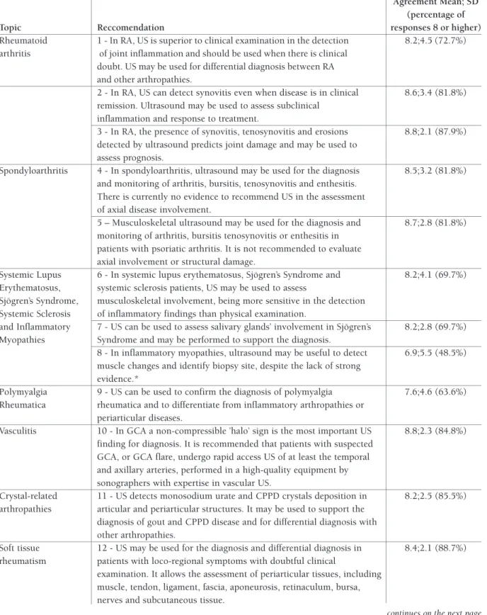
tAble I. portuguese recommendAtIons on the use of ultrAssonogrAphy In rheumAtology
Agreement Mean; SD (percentage of
Topic Reccomendation responses 8 or higher)
Rheumatoid 1 - In RA, US is superior to clinical examination in the detection 8.2;4.5 (72.7%) arthritis of joint inflammation and should be used when there is clinical
doubt. US may be used for differential diagnosis between RA and other arthropathies.
2 - In RA, US can detect synovitis even when disease is in clinical 8.6;3.4 (81.8%) remission. Ultrasound may be used to assess subclinical
inflammation and response to treatment.
3 - In RA, the presence of synovitis, tenosynovitis and erosions 8.8;2.1 (87.9%) detected by ultrasound predicts joint damage and may be used to
assess prognosis.
Spondyloarthritis 4 - In spondyloarthritis, ultrasound may be used for the diagnosis 8.5;3.2 (81.8%) and monitoring of arthritis, bursitis, tenosynovitis and enthesitis.
There is currently no evidence to recommend US in the assessment of axial disease involvement.
5 – Musculoskeletal ultrasound may be used for the diagnosis and 8.7;2.8 (81.8%) monitoring of arthritis, bursitis tenosynovitis or enthesitis in
patients with psoriatic arthritis. It is not recommended to evaluate axial involvement or structural damage.
Systemic Lupus 6 - In systemic lupus erythematosus, Sjögren’s Syndrome and 8.2;4.1 (69.7%) Erythematosus, systemic sclerosis patients, US may be used to assess
Sjögren’s Syndrome, musculoskeletal involvement, being more sensitive in the detection Systemic Sclerosis of inflammatory findings than physical examination.
and Inflammatory 7 - US can be used to assess salivary glands’ involvement in Sjögren’s 8.2;2.8 (69.7%) Myopathies Syndrome and may be performed to support the diagnosis.
8 - In inflammatory myopathies, ultrasound may be useful to detect 6.9;5.5 (48.5%) muscle changes and identify biopsy site, despite the lack of strong
evidence.*
Polymyalgia 9 - US can be used to confirm the diagnosis of polymyalgia 7.6;4.6 (63.6%) Rheumatica rheumatica and to differentiate from inflammatory arthropathies or
periarticular diseases.
Vasculitis 10 - In GCA a non-compressible ’halo’ sign is the most important US 8.8;2.3 (84.8%) finding for diagnosis. It is recommended that patients with suspected
GCA, or GCA flare, undergo rapid access US of at least the temporal and axillary arteries, performed in a high-quality equipment by sonographers with expertise in vascular US.
Crystal-related 11 - US detects monosodium urate and CPPD crystals deposition in 8.2;2.5 (85.5%) arthropathies articular and periarticular structures. It may be used to support the
diagnosis of gout and CPPD disease and for differential diagnosis with other arthropathies.
Soft tissue 12 - US may be used for the diagnosis and differential diagnosis in 8.4;2.1 (88.7%) rheumatism patients with loco-regional symptoms with doubtful clinical
examination. It allows the assessment of periarticular tissues, including muscle, tendon, ligament, fascia, aponeurosis, retinaculum, bursa, nerves and subcutaneous tissue.
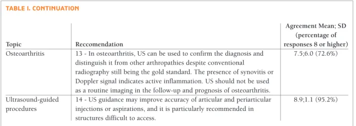
tAble I. contInuAtIon
Agreement Mean; SD (percentage of
Topic Reccomendation responses 8 or higher)
Osteoarthritis 13 - In osteoarthritis, US can be used to confirm the diagnosis and 7.5;6.0 (72.6%) distinguish it from other arthropathies despite conventional
radiography still being the gold standard. The presence of synovitis or Doppler signal indicates active inflammation. US should not be used as a routine imaging in the follow-up and prognosis of osteoarthritis.
Ultrasound-guided 14 - US guidance may improve accuracy of articular and periarticular 8.9;1.1 (95.2%) procedures injections or aspirations, and it is particularly recommended in
structures difficult to access.
RA – rheumatoid arthritis; US – ultrasound; GCA – giant cell arteritis; CPPD - calcium pyrophosphate dehydrate *Recommendation 8 did not achieve enough agreement to be supported.
doi: 10.1093/rheumatology/ket356. Epub 2013 Nov 5. 6. Zufferey P, Tamborrini G, Gabay C, Krebs A, Kyburz D,
Mi-chel B, et al. Recommendations for the use of ultrasound in rheumatoid arthritis: literature review and SONAR score ex-perience. Swiss Med Wkly. 2013 Dec 20;143:w13861. doi: 10.4414/smw.2013.13861. Review.
7. McAlindon T, Kissin E, Nazarian L, Ranganath V, Prakash S, Taylor M, et al. American College of Rheumatology report on reasonable use of musculoskeletal ultrasonography in rheu-matology clinical practice. Arthritis Care Res (Hoboken). 2012 Nov;64(11):1625-40. doi: 10.1002/acr.21836.
8. Klauser AS, Tagliafico A, Allen GM, Boutry N, Campbell R, Court-Payen M, et al. Clinical indications for musculoskele-tal ultrasound: a Delphi-based consensus paper of the Euro-pean Society of Musculoskeletal Radiology. Eur Radiol. 2012 May;22(5):1140-8. doi: 10.1007/s00330-011-2356-3. Epub 2012 Mar 28.
9. Finnoff JT, Hall MM, Adams E, Berkoff D, Concoff AL, Dex-ter W, et al. American Medical Society for Sports Medicine (AMSSM) position statement: interventional musculoskeletal ultrasound in sports medicine. PM R. 2015 Feb;7(2):151-68.e12. doi: 10.1016/j.pmrj.2015.01.003.
10. Pineda C, Reginato AM, Flores V, Aliste M, Alva M, Aragón-Laínez RA, et al; Pan-American League of Associations for Rheumatology (PANLAR) Ultrasound Study Group. Pan-Ame-rican League of Associations for Rheumatology (PANLAR) re-commendations and guidelines for musculoskeletal ultra-sound training in the Americas for rheumatologists. J Clin Rheumatol. 2010 Apr;16(3):113-8. doi: 10.1097/ RHU.0b013e3181d60053.
11. Terslev L, Hammer HB, Torp-Pedersen S, Szkudlarek M, Iag-nocco A, D'Agostino MA, et al. EFSUMB minimum training re-quirements for rheumatologists performing musculoskeletal ultrasound. Ultraschall Med. 2013 Oct;34(5):475-7. doi: 10.1055/s-0033-1335143. Epub 2013 May 21.
12. Cosgrove D, Piscaglia F, Bamber J, Bojunga J, Correas JM, Gil-ja OH, et al; EFSUMB. EFSUMB guidelines and recommen-dations on the clinical use of ultrasound elastography. Part 2: Clinical applications. Ultraschall Med. 2013
Jun;34(3):238-53. doi: 10.1055/s-0033-1335375. Epub 2013 Apr 19. 13. Möller I, Loza E, Uson J, Acebes C, Andreu JL, Battle E, et al.
Recomendaciones para el uso de la ecografía y la ressonância magnética en pacientes con artritis reumatoide. Reumatol Clin. 2016 14: 9-19
14. Filippucci E, Iagnocco A, Salaffi F, Cerioni A, Valesini G, Gras-si W. Power Doppler sonography monitoring of synovial per-fusion at the wrist joints in patients with rheumatoid arthritis treated with adalimumab. Ann Rheum Dis 2006; 65: 1433–1437.
15. Szkudlarek M, Klarlund M, Narvestad E, Court-Payen M, Strandberg C, Jensen KE, et al. Ultrasonography of the meta-carpophalangeal and proximal interphalangeal joints in rheu-matoid arthritis: A comparison with magnetic resonance ima-ging, conventional radiography and clinical examination. Arthritis Res Ther. 2006; 8:R52.
16. Krejza J, Kuryliszyn-Moskal A, Sierakowski S, et al. Ultraso-nography of the periarticular changes in patients with early ac-tive rheumatoid arthritis. Med Sc Monit 1998; 4:366–369 17. Ribbens C, André B, Marcelis S, Kaye O, Mathy L, Bonnet V
et al. Rheumatoid hand joint synovitis: gray-scale and power Doppler US quantifications following anti–tumor necrosis fac-tor– treatment: pilot study. Radiology 2003; 229:562–9. 18. Terslev L, von der Recke P, Savnik A, Koenig MJ, Bliddal H.
Diagnostic sensitivity and specificity of Doppler ultrasound in rheumatoid arthritis. J Rheumatol 2008; 35:49–53. 19. Wakefield RJ, Freeston JE, O’Connor P, Reay N, Budgen A,
Hensor EM, et al. The optimal assessment of the rheumatoid arthritis hindfoot: A comparative study of clinical examina-tion, ultrasound and high field MRI. Ann Rheum Dis. 2008; 67:1678
20. Amin MF, Ismail FM, El Shereef RR. The role of ultrasono-graphy in early detection and monitoring of shoulder erosions, and disease activity in rheumatoid arthritis patients; compa-rison with MRI examination. Acad Radiol. 2012;19:693–700 21. Hmamouchi I, Bahiri R, Srifi N, Aktaou S, Abouqal R, Hajjaj-Hassouni N. A comparison of ultrasound and clinical exami-nation in the detection of flexor tenosynovitis in early arthri-tis. BMC Musculoskelet Disord 2011;12:91
22. Haavardsholm EA, Ostergaard M, Hammer HB, Bøyesen P, Boonen A, van der Heijde D et al. Monitoring anti-TNF alp-ha treatment in rheumatoid arthritis: responsiveness of mag-netic resonance imaging and ultrasonography of the domi-nant wrist joint compared with conventional measures of di-sease activity and structural damage. Ann Rheum Dis 2009; 68:1572–1579.
23. Riente L, Delle Sedie A, Scirè CA, Filippucci E, Meenagh G, Iagnocco A et al. Ultrasound imaging for the rheumatologist. XXXI. Sonographic assessment of the foot in patients with rheumatoid arthritis. Clin Exp Rheumatol 2011;29:1–5. 24. Kunkel GA, Cannon GW, Clegg DO. Combined structural
and synovial assessment for improved ultrasound discrimi-nation of rheumatoid, osteoarthritic, and normal joints: a pi-lot study. Open Rheumatol J. 2012;6:199–206. 98. Milpi-lot F, Clavel G, Etchepare F, Gandjbakhch F, Grados F, Saraux A, et al. Musculoskeletal ultrasonography in healthy subjects and ultrasound criteria for early arthritis (the ESPOIR cohort). J Rheumatol. 2011;38:613–620
25. Sheane BJ, Beddy P, O’Connor M, Miller S, Cunnane G. Tar-geted ultrasound of the fifth metatarsophalangeal joint in an early inflammatory arthritis cohort. Arthritis Care Res (Ho-boken). 2009; 61:1004–1008.
26. Gutierrez M, Filippucci E, Ruta S, Salaffi F, Blasetti P, di Geso L, et al. Interobserver reliability of high-resolution ultraso-nography in the assessment of bone erosions in patients with rheumatoid arthritis: Experience of an intensive dedicated training programme. Rheumatology. 2011; 50:373–380. 27. Brown AK, Conaghan PG, Karim Z, Quinn MA, Ikeda K,
Pe-terfy CG, et al. An explanation for the apparent dissociation between clinical remission and continued structural deterio-ration in rheumatoid arthritis. Arthritis Rheum. 2008; 58:2958–2967.
28. Foltz V, Gandjbakhch F, Etchepare F, Rosenberg C, Tanguy ML, Rozenberg S, et al. Power Dopplerultrasound, butno-tlow-fieldmagnetic resonance imaging, predicts relapse and radiographic disease progression in rheumatoid arthritis pa-tients with low levels of disease activity. Arthritis Rheum. 2012; 64:67–76.
29. Yoshimi R, Hama M, Takase K, Ihata A, Kishimoto D, Terau-chi K, et al. Ultrasonography is a potent tool for the predic-tion of progressive joint destrucpredic-tion during clinical remission of rheumatoid arthritis. Mod Rheumatol. 2013; 23:456–465. 30. Naredo E, Valor L, de la Torre I, Martinez-Barrio J, Hinojosa M, Aramburu F, et al. Ultrasound joint inflammation in rheu-matoid arthritis in clinical remission: how many and which joints should be assessed? Arthritis Care Res (Hoboken). 2013; 65:512–517
31. Backhaus TM, Ohrndorf S, Kellner H, Strunk J, Hartung W, Sattler H, et al. The US7 score is sensitive to change in a lar-ge cohort of patients with rheumatoid arthritis over 12 months of therapy. Ann Rheum Dis. 2013; 72:1163–1169. 32. Boesen M, Boesen L, Jensen KE, Cimmino MA,
Torp-Peder-sen S, Terslev L, et al. Clinical outcome and imaging changes after intraarticular (IA) application of etanercept or methyl-prednisolone in rheumatoid arthritis: magnetic resonance imaging and ultrasound-Doppler show no effect of IA injec-tions in the wrist after 4 weeks. J Rheumatol. 2008; 35:584–591
33. Dougados M, Jousse-Joulin S, Mistretta F, d’Agostino MA, Backhaus M, Bentin J, et al. Evaluation of several
ultrasono-graphy scoring systems for synovitis and comparison to cli-nical examination: results from a prospective multicentre stu-dy of rheumatoid arthritis. Ann Rheum Dis. 2010; 69:828–833.
34. Hama M, Uehara T, Takase K, Ihata A, Ueda A, Takeno M, et al. Power Doppler ultrasonography is useful for assessing di-sease activity and predicting joint destruction in rheumatoid arthritis patients receiving tocilizumab—preliminary data. Rheumatol Int. 2012; 32:1327–1333.
35. Hammer HB, Sveinsson M, Kongtorp AK, Kvien TK. A 78-joints ultrasonographic assessment is associated with clinical assessments and is highly responsive to improvement in a longitudinal study of patients with rheumatoid arthritis star-ting adalimumab treatment. Ann Rheum Dis. 2010; 69:1349–1351.
36. Mandl P, Balint PV, Brault Y, Backhaus M, D’Agostino MA, Grassi W, et al. Metrologic properties of ultrasound versus clinical evaluation of synovitis in rheumatoid arthritis: results of a multicenter, randomized study. Arthritis Rheum. 2012; 64:1272–1282.
37. Conaghan PG, O’Connor P, McGonagle D, Astin P, Wakefield RJ, Gibbon WW et al. Elucidation of the relationship between synovitis and bone damage: a randomized magnetic reso-nance imaging study of individual joints in patients with ear-ly rheumatoid arthritis. Arthritis Rheum 2003; 48:64–71. 38. Lillegraven S, Bøyesen P, Berner Hammer H, Østergaard M,
Uhlig T, Sesseng S et al. Tenosynovitis of the extensor carpi ulnaris tendon predicts erosive progression in early rheuma-toid arthritis. Ann Rheum Dis 2011; 70:2049–2050 39. Kamishima T, Tanimura K, Shimizu M, Matsuhashi M, Fukae
J, Kon Y et al. Monitoring anti-interleukin 6 receptor antibo-dy treatment for rheumatoid arthritis by quantitative magne-tic resonance imaging of the hand and power Doppler ultra-sonography of the finger. Skeletal Radiol 2011; 40:745–755. 40. Hoving JL, Buchbinder R, Hall S, Lawler G, Coombs P, McNealy S et al. A comparison of magnetic resonance ima-ging, sonography, and radiography of the hand in patients with early rheumatoid arthritis. J Rheumatol 2004; 31:663–675.
41. Funck-Brentano T, Gandjbakhch F, Etchepare F, Jousse-Jou-lin S, Miquel A, Cyteval C, et al. Prediction of radiographic damage in early arthritis by sonographic erosions and power doppler signal: a longitudinal observational study. Arthritis Care Res (Hoboken). 2013;65:896–902.
42. Hooper L, Bowen CJ, Gates L, Culliford DJ, Ball C, Edwards CJ, et al. Prognostic indicators of foot-related disability in pa-tients with rheumatoid arthritis: results of a prospective three-year study. Arthritis Care Res (Hoboken). 2012; 64: 1116–1124.
43. Hoving JL, Buchbinder R, Hall S, Lawler G, Coombs P, McNealy S, et al. A comparison of magnetic resonance ima-ging, sonography, and radiography of the hand in patients with early rheumatoid arthritis. J Rheumatol. 2004; 31:663–675.
44. Reynolds PP, Heron C, Pilcher J, Kiely PD. Prediction of ero-sion progresero-sion using ultrasound in established rheumatoid arthritis: a 2-year follow-up study. Skeletal Radiol. 2009; 38:473–478.
45. Naredo E, Collado P, Cruz A, Palop MJ, Cabero F, Richi P, et al. Longitudinal power Doppler ultrasonographic assessment of joint inflammatory activity in early rheumatoid arthritis: