Research Article
EMMPRIN Expression in Oral Squamous Cell Carcinomas:
Correlation with Tumor Proliferation and Patient Survival
Luís Silva Monteiro,
1,2Maria Leonor Delgado,
1Sara Ricardo,
3,4Fernanda Garcez,
3Barbas do Amaral,
1,2,5José Júlio Pacheco,
1,2Carlos Lopes,
6and Hassan Bousbaa
1,7,81Molecular Oncology Group, Institute of Research and Advanced Training in Health Sciences and Technologies (IINFACTS),
Higher Institute of Health Sciences, CESPU, 4585-116 Paredes, Portugal
2Medicine and Oral Surgery Department, Higher Institute of Health Sciences, 4585-116 Paredes, Portugal 3Pathology Department, Higher School of Health of Vale do Sousa, 4585-116 Paredes, Portugal
4Differentiation and Cancer Group, Institute of Molecular Pathology and Immunology of the University of Porto, (IPATIMUP),
4200-465 Porto, Portugal
5Stomatology Department, Santo Ant´onio Hospital, Oporto Hospitalar Centre, 4099-001 Porto, Portugal 6Molecular Pathology and Immunology Department, Institute of Biomedical Sciences Abel Salazar (ICBAS),
Porto University, 4050-313 Porto, Portugal
7Centro de Qu´ımica Medicinal da Universidade do Porto (CEQUIMED-UP), Rua de Jorge Viterbo Ferreira 228,
4050-313 Porto, Portugal
8Centro Interdisciplinar de Investigac¸˜ao Marinha e Ambiental (CIIMAR/CIMAR), Universidade do Porto, Rua dos Bragas 289,
4050-123 Porto, Portugal
Correspondence should be addressed to Lu´ıs Silva Monteiro; [email protected] Received 14 February 2014; Revised 28 April 2014; Accepted 5 May 2014; Published 21 May 2014 Academic Editor: Mouldy Sioud
Copyright © 2014 Lu´ıs Silva Monteiro et al. This is an open access article distributed under the Creative Commons Attribution License, which permits unrestricted use, distribution, and reproduction in any medium, provided the original work is properly cited.
The aim of our study was to explore the clinicopathological and prognostic significance of extracellular matrix metalloproteinase inducer (EMMPRIN) expression in oral squamous cell carcinomas (OSCC), and its relation with the proliferative tumor status of OSCC. We examined EMMPRIN and Ki-67 proteins expression by immunohistochemistry in 74 cases with OSCC. Statistical analysis was conducted to examine their clinicopathological and prognostic significance in OSCC. EMMPRIN membrane expression was observed in all cases, with both membrane and cytoplasmic tumor expression in 61 cases (82.4%). EMMPRIN overexpression was observed in 56 cases (75.7%). Moderately or poorly differentiated tumors showed EMMPRIN overexpression more frequently than well-differentiated tumors(𝑃 = 0.002). Overexpression of EMMPRIN was correlated with high Ki-67 expression(𝑃 = 0.004). In the multivariate analysis, EMMPRIN overexpression reveals an adverse independent prognostic value for cancer-specific survival (CSS)(𝑃 = 0.034). Our results reveal that EMMPRIN protein is overexpressed in more than two-thirds of OSCC cases, especially in high proliferative and less differentiated tumors. The independent value of EMMPRIN overexpression in CSS suggests that this protein could be used as an important biological prognostic marker for patients with OSCC. Moreover, the high expression of EMMPRIN makes it a possible therapeutic target in OSCC patients.
1. Introduction
Oral cancer remains a major public health problem with almost 300,000 new cases worldwide [1,2]. New insights in cancer diagnosis and therapy have not changed significantly the survival rate for oral cancer (around 50%) during the last decades [1].
Oral tumorigenesis is a multistep process caused by accu-mulation of multiple genetic and epigenetic alterations [3]. The comprehension of the molecular pathways involved in this process may originate special biological markers able to differentiate tumors with a more or less aggressive behavior. These markers may contribute to identify and stratify patients with greater precision to the most appropriate treatment plan.
Volume 2014, Article ID 905680, 9 pages http://dx.doi.org/10.1155/2014/905680
apeutic targets.
Extracellular matrix metalloproteinase inducer (EMM-PRIN), also known as CD147, Basignin, M6, Neurothelin, or gp42, is a highly glycosylated transmembrane protein, mem-ber of the immunoglobulin superfamily of receptors, discov-ered by its capacity of inducing the expression of matrix met-alloproteinases [4]. It is present in epithelial cells, neuronal or nerve cells, myocardial cells, lymphoid cells, or germ cells and has an important role in several biological processes such as fetal development, retinal function, development of the nervous system and thymic T cell development [5]. EMMPRIN is expressed in several cancers including head and neck squamous-cell carcinomas, pancreatic adenocar-cinomas, kidney chromophobic caradenocar-cinomas, hepatocellular carcinomas, medullary breast adenocarcinomas, cervix carci-nomas, and glioblastomas [5]. EMMPRIN contributes to cell adhesion modulation, tumor growth, invasion, and angio-genesis [4–7] probably due to its association with several proteins implicated in different signaling pathways such as matrix metalloproteinases, ErbB, MAPK cascade proteins, monocarboxylate transporters (MCT), integrins, caveolin-1 (Cav-1), Tenascin (TN)-C, vascular endothelial growth factor (VEGF), urokinase-type plasminogen activator (uPA), and cyclophilins (Cyp) [4,6,8,9].
Previous reports have shown that EMMPRIN expression is associated with a high tumor aggressive behavior and with poor prognosis in several tumors [10–19]. However, in OSCC the prognostic significance of EMMPRIN is poorly studied. Moreover, the relation of this glycoprotein with the proliferative tumor capacity in patients with OSCC has not been reported.
We aimed in this study to evaluate the expression of EMMPRIN in patients with OSCC and investigate the asso-ciation of this glycoprotein with clinicopathological, tumor proliferation, and prognosis variables.
2. Material and Methods
2.1. Patient Recruitment. This retrospective study included
patients with newly diagnosed and consecutively treated primary OSCC at the Hospital de Santo Ant´onio (HSA), Porto, Portugal, between 2000 and 2006. The study was approved by the institutional review board of the hospital. From patient’s records, we obtained patient’s age, gender, tumor location, tumor stage (I–IV), primary treatment, histo-logical type, tumor grade, surgical margin status, and follow-up information.
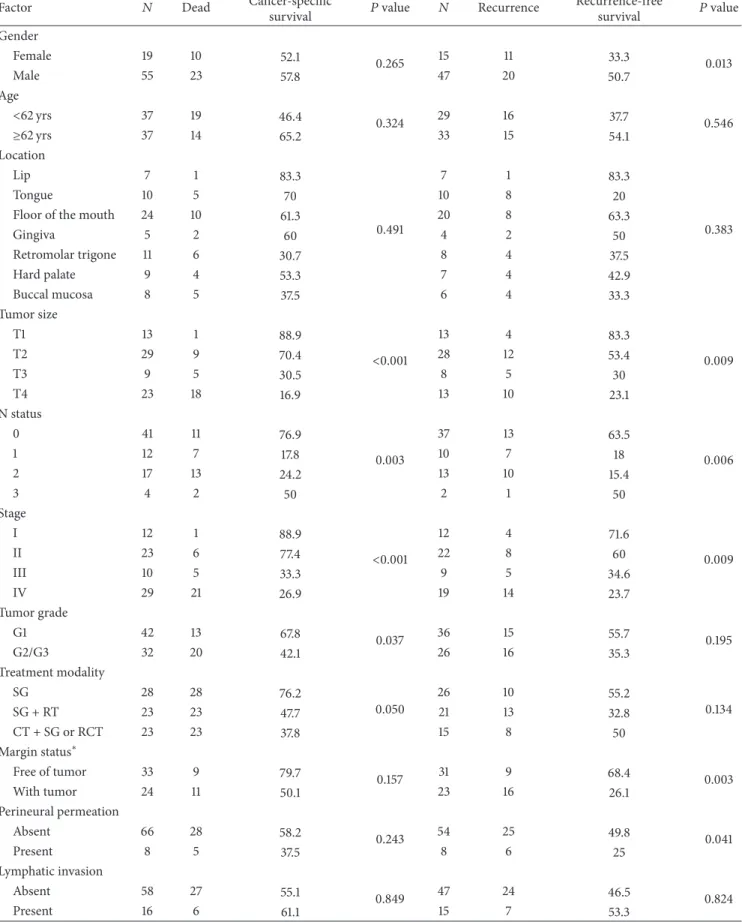
Patients were excluded if they lacked clinical and follow-up information or if their paraffin blocks lacked sufficient tumor tissue leaving 74 patients for this study, 55 men and 19 women, with a mean age of62.3 ± 15.3 years (range from 25 to 96 years).Table 1lists the clinicopathological features of these patients.
Tumor stage was classified according to the 7th edition of the classification of malignant tumors of American Joint Committee on Cancer [20]. For all tumors, 3𝜇m sections were cut and stained with haematoxylin-eosin (HE) to confirm the initial diagnosis. Tumor grade was reclassified
entiated (G1), moderately differentiated (G2), and poorly differentiated (G3) OSCC [21]. Inspection for possible pres-ence of tumor lymphatic invasion and perineural permeation reported as present or absent was performed on each sample.
2.2. Tissue Microarray (TMA) Construction.
Immunohisto-chemistry was performed on tumor tissues using TMA technology designed and constructed according to rules previously described [22]. Briefly, representative tumor areas were selected on haematoxylin and eosin-stained sections and marked on paraffin blocks, avoiding necrosis and keratin areas. Three cylindrical tissue cores (2 mm in diameter) were obtained from each selected specimen and transferred to a recipient paraffin block, using a microarray instrument (TMA Builder, Histopathology Ltd., Hungary). From each TMA block, 3𝜇m sections were cut and processed for immunohistochemistry.
2.3. Immunohistochemistry. TMA slides were deparaffinised
in xylene, dehydrated in an ethanol series, and rinsed in distilled water. Epitope retrieval treatment was performed using 0.01 M citrate buffer (pH 6.0) for CD147 and 0.01 M tri-sethylenediaminetetraacetic acid (EDTA) buffer (pH 9.0) for Ki-67 at high temperature (98∘C water bath during 30 minutes). After blocking endogenous peroxidase with meth-anol containing 0.3% hydrogen peroxide (H2O2) for 5 min, sections were incubated with a blocking solution made of 0.4% casein in trisbuffered saline (TBS) to reduce nonspecific binding. TMA slides were incubated with the primary mon-oclonal antibody (anti-CD147, clone AB1843, Novocastra, Newcastle upon Tyne, UK, diluted at 1 : 30; and anti-Ki-67, clone MIB1, Dako, Glostrup, Denmark, diluted at 1 : 10) during 60 minutes at room temperature. Then the slides were washed in TBS, followed by incubation with standard per-oxidase-labelled dextran polymer for visualization with diaminobenzidine as chromogen (NovoLink Polymer Detec-tion System, Novocastra, Leica Biosystems Newcastle Ltd.), according to the manufacturer’s instructions. TMA tissue sec-tions were lightly counterstained with Mayer haematoxylin for 2 min and cover-slipped. Positive (skin and oral mucosa) and negative (omission of primary antibody) controls were used in each staining run.
2.4. Evaluation of Immunohistochemical Expression. All
sam-ples were evaluated by two authors blinded to clinicopatho-logical characteristics. The discordant cases were reviewed under a multihead microscope to achieve a consensus. We used the higher score out of at least 2 of the 3 cores examined per case. EMMPRIN staining was evaluated on the basis of extent and intensity immunolabeling of tumor cells. The intensity of staining was scored as 0 (absent), 1 (weak), 2 (moderate), and 3 (strong). The extent of membrane tumor cells staining was semiquantitatively evaluated as 0 (no labelling or labelling in<10% of tumor cells); 1 (labelling in 10% to 24% of tumor cells); 2 (labelling in 25% to 49% of tumor cells); 3 (labelling in 50% to 74% of tumor cells); and 4 (labelling in 75% or more of tumor cells). The sum of the intensity and extent scores was used as the final score (0–7).
Table 1: Clinicopathological characteristics of the 74 patients with OSCC and their association with EMMPRIN and Ki-67 expressions.
Factor 𝑁 (%) EMMPRIN overexpression Ki-67 high expression
𝑁 (%) 𝑃 value 𝑁 (%) 𝑃 value All cases 74 (100%) 56 (75.7%) 38 (51.4%) Gender Female 19 (25.7%) 11 (63.2%) 0.140 8 (42.1%) 0.350 Male 55 (74.3%) 44 (80%) 30 (54.5%) Age <62 years 37 (50%) 30 (81.1%) 0.278 22 (59.5%) 0.163 ≥62 years 37 (50%) 26 (70.3%) 16 (43.2%) Location Labial mucosa 7 (9.5%) 6 (85.7%) 0.430 1 (14.3%) 0.396
Floor of the mouth 10 (13.5%) 7 (70%) 8 (80%)
Tongue 24 (32.4%) 15 (62.5%) 13 (54.2%) Buccal mucosa 5 (6.8%) 5 (100%) 2 (40%) Retromolar trigone 11 (14.9%) 8 (72.7%) 5 (45.5%) Hard palate 9 (12.2%) 8 (88.9%) 3 (55.6%) Gingiva 8 (10.8%) 7 (87.5%) 4 (50%) Tumor size T1 13 (17.6%) 8 (61.5%) 0.132 6 (46.2%) 0.396 T2 29 (39.2%) 20 (69%) 12 (41.4%) T3 9 (12.2%) 9 (100%) 6 (66.7%) T4 23 (31%) 19 (82.6%) 14 (60.9%) N status N0 41 (55.4%) 28 (68.3%) 0.349 16 (39%) 0.052 N1 12 (16.2%) 11 (91.7%) 7 (58.3%) N2 17 (23%) 14 (82.4%) 11 (64.7%) N3 4 (5.4%) 3 (75%) 4 (100%) Stage I 12 (16.2%) 8 (66.7%) 0.064 6 (50%) 0.080 II 23 (31.1%) 14 (60.9%) 7 (30.4%) III 10 (13.5%) 10 (100%) 7 (70%) IV 29 (39.2%) 24 (82.8%) 18 (62.1%) Treatment modality SG 28 (37.8%) 20 (71.4%) 0.633 12 (42.9%) 0.522 SG + RT 23 (31.3%) 17 (73.9%) 13 (56.5%) CT + SG or RCT 23 (31.3%) 19 (82.6%) 13 (56.5%) Tumor grade G1 42 (56.8%) 26 (61.9%) 0.002 16 (38.1%) 0.009 G2 + G3 32 (43.2%) 30 (93.8%) 22 (68.8%) Margin status∗ Free of tumor 33 (57.9%) 23 (69.7%) 0.423 14 (42.4%) 0.236 With tumor 24 (42.1%) 19 (79.2%) 14 (58.3%) Perineural permeation Absent 66 (89.2%) 50 (75.8%) 0.962 33 (50%) 0.504 Present 8 (10.8%) 6 (75%) 5 (62.5%) Lymphatic invasion Absent 58 (78.4%) 46 (79.3%) 0.165 30 (51.7%) 0.903 Present 16 (21.6%) 10 (62.5%) 8 (50%)
SG: surgery; RT: radiotherapy; CT: chemotherapy; RCT: radiochemotherapy.
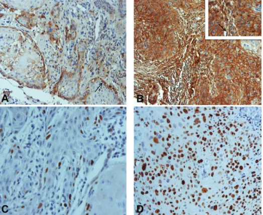
Figure 1: Immunohistochemical expression of EMMPRIN and Ki-67 in oral squamous cell carcinomas: (A) EMMPRIN score 1+ expression with predominantly peripheral distribution pattern (arrow) (×200); (B) EMMPRIN score 3+ expression with staining homogeneously distributed by the tumor islands (×100). Inset: higher magnification (×400). Note peritumoral fibroblast staining (arrow); (C) Ki-67 expression in less than 50% of tumor cells (×400); (D) Ki-67 expression in more than 50% of tumor cells (×400).
Tissues having a final score of 0-1 were considered negatives. Final scores of 2-3, 4-5, and 6-7 were considered 1+, 2+, and 3+, respectively. For data analysis, score 3+ was defined as EMMPRIN overexpression [12].
For Ki-67 evaluation, we considered the percentage of nuclear staining for scoring proliferative status. We classified tumors into two groups: low proliferative tumor (labelling from 0 to 49% of tumor cells) and high proliferative tumor (labelling in 50% or more of tumor cells) [23].
2.5. Statistical Analysis. Statistical analysis was carried out
using IBM SPSS Statistics version 21.0 software (IBM Cor-poration, NY, US). The associations between categorical vari-ables were evaluated by chi-square tests. Correlation between EMMPRIN and Ki-67 was measured by Spearman’s correla-tion coefficient. Cancer-specific survival (CSS) was defined as the time interval (months) between primary treatment and death from oral cancer or last follow-up. Recurrence-free sur-vival (RFS) was defined as the time interval (months) between primary treatment and the first recurrence (whether local, regional, or distant). The Kaplan-Meier method was used to plot survival curves and their prognostic effect was tested using the log-rank test. Variables with significant effects in the univariate analyses were entered into Cox proportional hazards model to investigate the independent effects of these variables. Differences were considered statistically significant at𝑃 < 0.05.
3. Results
3.1. EMMPRIN Expression. Immunohistochemistry was
per-formed in 74 human OSCC tissues to evaluate the extent and patterns of EMMPRIN protein expression. All cases presented membrane staining for EMMPRIN on tumor cells. Additionally, in 61 cases (82.4%), cytoplasmic expression was also observed. On the basis of EMMPRIN immuno-expression, cases were classified as 1+ in 2 (2.7%), 2+ in 16 (21.6%), and 3+ in 56 (75.7%) cases (Figures 1(A) and 1(B)). Staining of this protein was detected predominantly at the periphery of the tumor islands (45; 60.8%) or present homogenously within the tumor islands (29; 39.2%) (Figures 1(A) and1(B)). We observed also that EMMPRIN expression was seen in peritumoral fibroblasts in 65 (87.8%) cases (Figures 1(A) and 1(B)). Fibroblasts were identified by an experienced pathologist based on its histomorphological features. In cases in which difficulties existed in fibroblast identification, we recurred to coloration for vimentin and smooth muscle actin. Apparently, normal mucosa adjacent to primary tumor presented a strong EMMPRIN staining in basal and suprabasal epithelial layers.
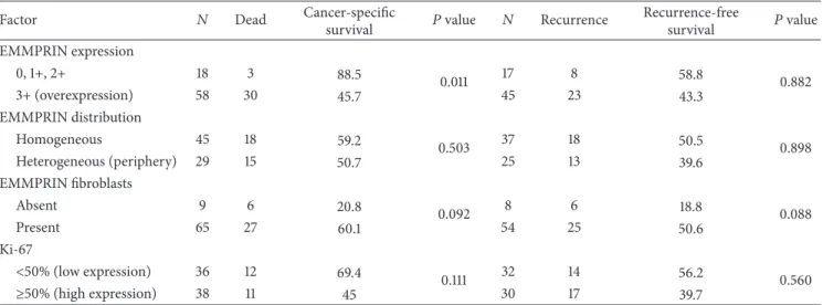
We compared EMMPRIN expression in OSCC tissue samples with patient clinicopathological variables. A posi-tive association of EMMPRIN expression with histological grade was noted where G2/G3 tumors presented EMMPRIN overexpression more often than G1 tumors (𝑃 = 0.002)
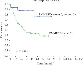
Cancer-specific survival C um. sur vi val Time (months) 1.0 0.9 0.8 0.7 0.6 0.5 0.4 0.3 0.2 0.1 0.0 0 12 24 36 48 60 72 84 96 108 120 132 144 156 0, 1+
EMMPRIN scores , and 2+
EMMPRIN score 3+
P = 0.011
Figure 2: Kaplan-Meier curves according to EMMPRIN expression demonstrating a worse cancer-specific survival in the OSCC patients with overexpression of this protein.
(Table 1). Although no significant association was found between EMMPRIN overexpression and clinical stage, when we divided clinical stage into initial stage (I/II) tumors and advanced stage tumors (III/IV), we observed a signif-icant association between EMMPRIN overexpression and advanced tumor stages (𝑃 = 0.015). No other significant relationship between EMMPRIN expression and the listed clinicopathological parameters was found. Moreover, we did not find significant relationship between EMMPRIN distri-bution pattern (homogeneously or peripherically), EMM-PRIN fibroblast staining, and the listed clinicopathological parameters.
3.2. EMMPRIN and Ki-67 Correlation. Ki-67 expression was
detected in 72 cases (97.3%). Thirty-six cases (48.6%) were classified as low proliferative tumors and 38 (51.4%) as high proliferative tumors (Figures1(C) and1(D)). The intensity of the marker was similar and homogeneous in almost all cases. The only association between Ki-67 and the clinicopatho-logical variables was observed with histoclinicopatho-logical grade (𝑃 = 0.009) (Table 1).
EMMPRIN was positively correlated with Ki-67 expres-sion (𝜌 = 0.33; 𝑃 = 0.004). Tumors with EMMPRIN over-expression (𝑛 = 56) presented high Ki-67 over-expression in 64.7% (𝑛 = 34) of the cases. By contrast, only 22.2% (4/18) of tumors without EMMPRIN overexpression expressed high levels of Ki-67 protein.
3.3. Survival Analysis. The mean follow-up for all patients
was 36.45 ± 31.7 months and mean follow-up for living patients was52.05±33.02 months. At the end of our study, 39 patients (52.7%) were alive without oral cancer, one patient (1.4%) was alive with oral cancer, 33 (44.6%) had died as a result of the oral cancer, and one patient (1.4%) had died as a result of cardiovascular disease. The cumulative 3-year cancer-specific survival (CSS) rate was 55.8% and recurrence-free survival (RFS) was 46.6%.
On a univariate analysis using the Kaplan-Meier method and log-rank test, we measured the influence of the clinical-pathological and immunoexpression variables on the sur-vival of patients with OSCC. EMMPRIN overexpression was statistically associated with a worse CSS (𝑃 = 0.011) (Figure 2; Table 2). Among the clinicopathological charac-teristics, tumor size (𝑃 < 0.001), 𝑁 status (𝑃 = 0.003), tumor stage (𝑃 < 0.001), treatment modality (𝑃 = 0.05), and histological grade (𝑃 = 0.037) were also statistically associated with a worse CSS (Table 2). As also described in Table 2, we observed a significant association between RFS and gender (𝑃 = 0.013), tumor size (𝑃 = 0.009), 𝑁 status (𝑃 = 0.006), tumor stage (𝑃 = 0.009), margin status (𝑃 = 0.003), and perineural permeation (𝑃 = 0.041).
In the multivariate analyses using Cox regression method, we found an association of EMMPRIN overexpression with poor survival (𝑃 = 0.034;Table 3), thus revealing EMMPRIN overexpression as an adverse independent prognostic factor for CSS in OSCC. In RFS, gender (𝑃 = 0.030) and margin status (𝑃 = 0.019) reveal an independent prognostic value for these tumors (Table 4).
4. Discussion
Recent studies have reported the biological and clinical role of EMMPRIN receptor in several cancers in the last decades [4, 5, 24]. However, the influence of this receptor in OSCC is poorly understood. In the present study, we aimed to evaluate the expression of EMMPRIN protein in OSCC and to analyse the correlation of this receptor with clinicopathological characteristics, tumor proliferation, and patient’s outcome.
Our study showed that EMMPRIN protein was present in all OSCC cases and overexpressed in more than two-thirds of the cases. This result is in accordance with the notably high expression of this glycoprotein in squamous cell carcinomas of head and neck region reported by Riethdorf et al. [5]. By analyzing EMMPRIN expression in multitumor TMAs, they observed expression in more than 95% of squamous cell carcinoma of oral cavity and 100% in squamous cell carci-nomas of salivary glands. Lower expression values have been reported by Gou et al. [25] in laryngeal carcinomas (87.5%), Zhu et al. [17] in esophageal squamous cell carcinomas (85%), and Huang et al. [11] in tongue squamous cell carcinomas (67%). They also observed a significantly higher expres-sion on tumor cells than in the noncancerous epithelium. Vigneswaran et al. [26] found a strong EMMPRIN expression in more than 90% of tumor cells in carcinoma in situ and early-invasive OSCC and also a significant higher expression compared with normal oral mucosa. The authors also found an increasing expression of this marker in oral leukoplakias gradually correlated with the degree of dysplasia, suggesting that EMMPRIN overexpression occurs at an early step of oral carcinogenesis and contributes to oral tumorigenesis. These data highlight the potential important role of EMMPRIN in OSCC.
We observed EMMPRIN expression in tumor cell mem-brane and also in the cytoplasm of some cases, in concor-dance with other studies [12,14,19]. Although our aim was
Table 2: Univariable analysis of cancer-specific and recurrence-free survivals (at 3-years of follow-up). Factor 𝑁 Dead Cancer-specific
survival 𝑃 value 𝑁 Recurrence
Recurrence-free survival 𝑃 value Gender Female 19 10 52.1 0.265 15 11 33.3 0.013 Male 55 23 57.8 47 20 50.7 Age <62 yrs 37 19 46.4 0.324 29 16 37.7 0.546 ≥62 yrs 37 14 65.2 33 15 54.1 Location Lip 7 1 83.3 0.491 7 1 83.3 0.383 Tongue 10 5 70 10 8 20
Floor of the mouth 24 10 61.3 20 8 63.3
Gingiva 5 2 60 4 2 50 Retromolar trigone 11 6 30.7 8 4 37.5 Hard palate 9 4 53.3 7 4 42.9 Buccal mucosa 8 5 37.5 6 4 33.3 Tumor size T1 13 1 88.9 <0.001 13 4 83.3 0.009 T2 29 9 70.4 28 12 53.4 T3 9 5 30.5 8 5 30 T4 23 18 16.9 13 10 23.1 N status 0 41 11 76.9 0.003 37 13 63.5 0.006 1 12 7 17.8 10 7 18 2 17 13 24.2 13 10 15.4 3 4 2 50 2 1 50 Stage I 12 1 88.9 <0.001 12 4 71.6 0.009 II 23 6 77.4 22 8 60 III 10 5 33.3 9 5 34.6 IV 29 21 26.9 19 14 23.7 Tumor grade G1 42 13 67.8 0.037 36 15 55.7 0.195 G2/G3 32 20 42.1 26 16 35.3 Treatment modality SG 28 28 76.2 0.050 26 10 55.2 0.134 SG + RT 23 23 47.7 21 13 32.8 CT + SG or RCT 23 23 37.8 15 8 50 Margin status∗ Free of tumor 33 9 79.7 0.157 31 9 68.4 0.003 With tumor 24 11 50.1 23 16 26.1 Perineural permeation Absent 66 28 58.2 0.243 54 25 49.8 0.041 Present 8 5 37.5 8 6 25 Lymphatic invasion Absent 58 27 55.1 0.849 47 24 46.5 0.824 Present 16 6 61.1 15 7 53.3
Table 2: Continued. Factor 𝑁 Dead Cancer-specific
survival 𝑃 value 𝑁 Recurrence
Recurrence-free survival 𝑃 value EMMPRIN expression 0, 1+, 2+ 18 3 88.5 0.011 17 8 58.8 0.882 3+ (overexpression) 58 30 45.7 45 23 43.3 EMMPRIN distribution Homogeneous 45 18 59.2 0.503 37 18 50.5 0.898 Heterogeneous (periphery) 29 15 50.7 25 13 39.6 EMMPRIN fibroblasts Absent 9 6 20.8 0.092 8 6 18.8 0.088 Present 65 27 60.1 54 25 50.6 Ki-67 <50% (low expression) 36 12 69.4 0.111 32 14 56.2 0.560 ≥50% (high expression) 38 11 45 30 17 39.7
SG: surgery; RT: radiotherapy; CT: chemotherapy; RCT: radiochemotherapy.
∗Not determined in the 17 cases.
Table 3: Multivariable analysis of cancer-specific survival on vari-ables with significant effect in univariable analysis.
Variablea 𝑃 value HR 95% CI Stage 0.205 1.640 0.763–3.524 T status 0.075 1.824 0.941–3.532 N status 0.353 0.799 0.497–1.283 Treatment modality 0.673 1.110 0.685–1.797 Tumor grade 0.411 1.405 0.624–3.160 EMMPRIN expression 0.034 3.894 1.106–13.709
HR: hazard ratio; CI: confidence interval for HR.
aVariables included in multivariable Cox regression analysis using enter
method; stage (ordinal variable); T status (ordinal variable); N status (ordinal variable); treatment modality (ordinal variable); tumor grade, G2 + G3 versus G1 (reference category); EMMPRIN expression, positive versus negative (reference category).
to analyse the expression of EMMPRIN in tumour cells, we detected the presence of this receptor in peritumoral fibro-blasts as described by Vigneswaran et al. [26]. Furthermore, EMMPRIN expression showed a predominantly periph-eric/basal distribution pattern in the tumor islands in most of our cases. This was reported in other works suggesting a more frequent distribution of this receptor in tumor cells with a more proliferative phenotype [11,26].
In order to assess the relationship of EMMPRIN with proliferative activity, we evaluated the expression of Ki-67 in these tumors and found a significant correlation between these two proteins. To our knowledge, this correlation has not been reported in OSCC, although Yang et al. [14] described a positive association between EMMPRIN expression and Ki-67 index labelling and also with tumor size in adenoid cystic carcinomas. Similar results were reported by Zheng et al. [27] showing a positive correlation between the two markers in gastric carcinomas. These results are in line with ours, suggesting that EMMPRIN might be relevant for the tumor proliferation and tumor growth of OSCC. Furthermore,
Table 4: Multivariable analysis of recurrence-free survival on variables with significant effect in univariable analysis.
Variablea 𝑃 value HR 95% CI Gender 0.030 2.849 1.110–7.315 Stage 0.384 1.589 0.560–4.512 T status 0.365 1.372 0.693–2.718 N status 0.700 0.836 0.337–2.077 Margin status 0.019 3.081 1.205–7.879 Perineural permeation 0.498 1.454 0.492–4.294
HR: hazard ratio; CI: confidence interval for HR.
aVariables included in multivariable Cox regression analysis using enter
method; gender, female versus male (reference category); stage (ordinal variable); T status (ordinal variable); N status (ordinal variable); margin status, with tumor versus without tumor (reference category); perineural permeation, present versus absent (reference category).
knockdown of EMMPRIN in head and neck carcinomas decreased cellular proliferation and tumor growth in vitro and in vivo analyses [28–30]. Mechanisms involved in tumor proliferation via EMMPRIN are poorly understood, but some authors have described the role of this receptor in association with cyclophilin A in the activation of ERK1/2 and p38 pathways [31].
We found that EMMPRIN expression was significantly associated with histological grade. Moderately or poorly dif-ferentiated tumors showed more EMMPRIN overexpression than well-differentiated tumors. Zhu et al. [17] observed the same positive association in 86 esophageal squamous cell carcinomas. Clinical stage and tumor size have been posi-tively related with EMMPRIN expression in several cancers including head and neck cancers [11, 12, 25]. We observed that EMMPRIN overexpression was more frequently found in patients with advanced clinical stage (III/IV), emphasizing the biological significance of this marker to tumor growth and progression of OSCC. We did not find any other significant relation with other clinicopathological parameters although
[32].
The influence of EMMPRIN expression on patient’s sur-vival has been reported in glioblastomas, seminomas, and other cancers including tongue, salivary gland, esophageal, ovary, colorectal, breast, bladder, and lung cancers [11–15, 17, 19, 33–36]. In our univariate analysis, we found that cases with EMMPRIN overexpression were associated with a lower CSS (𝑃 = 0.011) additionally with other clinical variables such as TNM and clinical stage. Nevertheless, in the multivariate analysis for CSS, EMMPRIN protein was the only independent prognostic factor (𝑃 = 0.034), revealing the adverse independent impact of EMMPRIN overexpression on the survival of patients with OSCC. To our knowledge, this is the first report of the independent prognostic value of EMMPRIN in a cohort of patients with squamous cell carcinoma of the oral cavity. Previously, Huang et al. [11] described the independent significant influence of this recep-tor in the overall survival of patients with squamous cell carcinomas of the tongue. This could be an important result suggesting the use of this receptor as a prognostic biomarker in OSCC. Interestingly, some studies report that EMMPRIN might be even a predictive marker of chemoresistance in head and neck carcinomas [8]. The influence of this receptor on patient’s prognostic could be related to the multiple biological functions of this protein on tumor cells such as proliferation, migration, invasion, angiogenesis, and dissemination on OSCC [4]. Studies have described the role of EMMPRIN in the stimulation of several metalloproteinases and proangio-nenic factors from tumor and adjacent stroma cells that could contribute to tumor multistep pathogenesis [27]. It would be interesting to analyse the relationship of EMMPRIN expres-sion and molecules involved in different pathways, such as EGFR, MMP’s, and VEGFR’s, in a larger sample of OSCC.
The understanding of the different pathways involved in oral tumorigenesis could reveal new candidate target molecules for anticancer drugs. Anti-EGFR targeted ther-apies are currently available for head and neck cancer but with modest results [37]. New anticancer therapies directed to molecular targets on oral cancer cells are needed and some molecules have been proposed, including EMMPRIN recep-tor [5,9,11,38]. The high expression of this receptor in OSCC, the cell membrane location, the biological role on tumor growth, invasion, dissemination, and the influence in the patient’s prognosis make EMMPRIN a strong candidate for a potential molecular target for monoclonal therapies against this receptor in OSCC. Anti-EMMPRIN molecular therapies showed growth inhibitory effect on head and neck squamous cell carcinoma, alone and in combination with radiotherapy in vitro and in vivo [39]. Sweeny et al. [40] reported a promising extracellular drug conjugate (EDC22), capable of inhibiting HNSCC cell proliferation in vitro and in vivo, with better results than with radiation or cisplatin monotherapy.
In conclusion, our results reveal that EMMPRIN protein is frequently overexpressed in OSCC, especially in high proliferative tumors, suggesting that it might be involved in the growth of these tumors. The independent value of EMMPRIN overexpression in CSS indicates that this protein could be used as an important biological prognostic marker
right decision as to the appropriate treatment. Furthermore, the high expression of this receptor could be regarded as potential therapeutic target against OSCC.
Conflict of Interests
There is no potential conflict of interests.
Acknowledgments
This work was supported by Grants (01-GCD-CICS-09; 02-GCD-CICS-09; and 05-GCD-CICS-2011) from Cooperativa de Ensino Superior Polit´ecnico e Universit´ario (CESPU). The authors would like to thank the Departments of Pathology of Hospital de Santo Ant´onio and CESPU and Professor Oliveira Torres.
References
[1] S. Warnakulasuriya, “Global epidemiology of oral and oropha-ryngeal cancer,” Oral Oncology, vol. 45, no. 4-5, pp. 309–316, 2009.
[2] A. Jemal, F. Bray, M. M. Center, J. Ferlay, E. Ward, and D. Forman, “Global cancer statistics,” CA Cancer Journal for
Clini-cians, vol. 61, no. 2, pp. 69–90, 2011.
[3] W. C. Hahn and R. A. Weinberg, “Rules for making human tumor cells,” The New England Journal of Medicine, vol. 347, no. 20, pp. 1593–1603, 2002.
[4] K. T. Iacono, A. L. Brown, M. I. Greene, and S. J. Saouaf, “CD147 immunoglobulin superfamily receptor function and role in pathology,” Experimental and Molecular Pathology, vol. 83, no. 3, pp. 283–295, 2007.
[5] S. Riethdorf, N. Reimers, V. Assmann et al., “High incidence of EMMPRIN expression in human tumors,” International Journal
of Cancer, vol. 119, no. 8, pp. 1800–1810, 2006.
[6] S. Zucker, M. Hymowitz, E. E. Rollo et al., “Tumorigenic potential of extracellular matrix metalloproteinase inducer,” The
American Journal of Pathology, vol. 158, no. 6, pp. 1921–1928,
2001.
[7] Y. Tang, M. T. Nakada, P. Kesavan et al., “Extracellular matrix metalloproteinase inducer stimulates tumor angiogenesis by elevating vascular endothelial cell growth factor and matrix metalloproteinases,” Cancer Research, vol. 65, no. 8, pp. 3193– 3199, 2005.
[8] Z. Huang, L. Wang, Y. Wang et al., “Overexpression of CD147 contributes to the chemoresistance of head and neck squamous cell carcinoma cells,” Journal of Oral Pathology & Medicine, vol. 42, no. 7, pp. 541–546, 2013.
[9] S. Suzuki and K. Ishikawa, “Combined inhibition of EMMPRIN and epidermal growth factor receptor prevents the growth and migration of head and neck squamous cell carcinoma cells,”
International Journal of Oncology, vol. 44, no. 3, pp. 912–917,
2014.
[10] J. Afonso, A. Longatto-Filho, F. Baltazar et al., “CD147 overex-pression allows an accurate discrimination of bladder cancer patients’ prognosis,” European Journal of Surgical Oncology, vol. 37, no. 9, pp. 811–817, 2011.
[11] Z. Huang, H. Huang, H. Li, W. Chen, and C. Pan, “EMMPRIN expression in tongue squamous cell carcinoma,” Journal of Oral
[12] S. Piao, S. Zhao, F. Guo et al., “Increased expression of CD147 and MMP-9 is correlated with poor prognosis of salivary duct carcinoma,” Journal of Cancer Research and Clinical Oncology, vol. 138, no. 4, pp. 627–635, 2012.
[13] A. Stenzinger, D. Wittschieber, M. von Winterfeld et al., “High extracellular matrix metalloproteinase inducer/CD147 expres-sion is strongly and independently associated with poor prog-nosis in colorectal cancer,” Human Pathology, vol. 43, no. 9, pp. 1471–1481, 2012.
[14] X. Yang, J. Dai, T. Li et al., “Expression of EMMPRIN in adenoid cystic carcinoma of salivary glands: correlation with tumor progression and patients’ prognosis,” Oral Oncology, vol. 46, no. 10, pp. 755–760, 2010.
[15] S. Zhao, W. Ma, M. Zhang et al., “High expression of CD147 and MMP-9 is correlated with poor prognosis of triple-negative breast cancer (TNBC) patients,” Medical Oncology , vol. 30, no. 1, article 335, 2013.
[16] S. Zhu, D. Chu, Y. Zhang et al., “EMMPRIN/CD147 expression is associated with disease-free survival of patients with colorec-tal cancer,” Medical Oncology, vol. 30, no. 1, article 369, 2013. [17] S. Zhu, Y. Li, L. Mi et al., “Clinical impact of HAb18G/CD147
expression in esophageal squamous cell carcinoma,” Digestive
Diseases and Sciences, vol. 56, no. 12, pp. 3569–3576, 2011.
[18] M. Yang, Y. Yuan, H. Zhang et al., “Prognostic significance of CD147 in patients with glioblastoma,” Journal of
Neuro-Onco-logy, vol. 115, no. 1, pp. 19–26, 2013.
[19] X. Y. Xu, N. Lin, Y. M. Li et al., “Expression of HAb18G/CD147 and its localization correlate with the progression and poor prognosis of non-small cell lung cancer,” Pathology—Research
and Practice, vol. 209, no. 6, pp. 345–352, 2013.
[20] M. Brandwein-Gensler and R. V. Smith, “Prognostic indicators in head and neck oncology including the new 7th edition of the AJCC staging system,” Head and Neck Pathology, vol. 4, no. 1, pp. 53–61, 2010.
[21] L. Barnes, J. W. Eveson, P. Reichart, and D. Sidransky, Pathology
and Genetics of Head and Neck Tumours, World Health
Orga-nization Classification of Tumours, IARC Press, Lyon, France, 2005.
[22] L. S. Monteiro, M. Diniz-Freitas, T. Garcia-Caballero, J. Forteza, and M. Fraga, “EGFR and Ki-67 expression in oral squamous cell carcinoma using tissue microarray technology,” Journal of
Oral Pathology & Medicine, vol. 39, no. 7, pp. 571–578, 2010.
[23] J. Carlos de Vicente, A. Herrero-Zapatero, M. F. Fresno, and J. S. L´opez-Arranz, “Expression of cyclin D1 and Ki-67 in squamous cell carcinoma of the oral cavity: clinicopathological and prognostic significance,” Oral Oncology, vol. 38, no. 3, pp. 301–308, 2002.
[24] Y. Li, J. Xu, and L. Chen, “HAb18G (CD147), a cancer-associated biomarker and its role in cancer detection,” Histopathology, vol. 54, no. 6, pp. 677–687, 2009.
[25] X. Gou, H. Chen, F. Jin et al., “Expressions of CD147, MMP-2 and MMP-9 in laryngeal carcinoma and its correlation with poor prognosis,” Pathology & Oncology Research, vol. 20, no. 2, pp. 475–481, 2014.
[26] N. Vigneswaran, S. Beckers, S. Waigel et al., “Increased EMM-PRIN (CD 147) expression during oral carcinogenesis,”
Experi-mental and Molecular Pathology, vol. 80, no. 2, pp. 147–159, 2006.
[27] H. C. Zheng, H. Takahashi, Y. Murai et al., “Upregulated EMM-PRIN/CD147 might contribute to growth and angiogenesis of gastric carcinoma: a good marker for local invasion and prog-nosis,” The British Journal of Cancer, vol. 95, no. 10, pp. 1371– 1378, 2006.
[28] L. Sweeny, Z. Liu, B. D. Bush et al., “CD147 and AGR2 expression promote cellular proliferation and metastasis of head and neck squamous cell carcinoma,” Experimental Cell Research, vol. 318, no. 14, pp. 1788–1798, 2012.
[29] X. Yang, P. Zhang, Q. Ma et al., “EMMPRIN silencing inhibits proliferation and perineural invasion of human salivary ade-noid cystic carcinoma cells in vitro and in vivo,” Cancer Biology
& Therapy, vol. 13, no. 2, pp. 85–91, 2012.
[30] C. Zhu, Y. Pan, B. He et al., “Inhibition of CD147 gene expres-sion via RNA interference reduces tumor cell invaexpres-sion, tumori-genicity and increases chemosensitivity to cisplatin in laryngeal carcinoma Hep2 cells,” Oncology Reports, vol. 25, no. 2, pp. 425– 432, 2011.
[31] M. Li, Q. Zhai, U. Bharadwaj et al., “Cyclophilin A is overex-pressed in human pancreatic cancer cells and stimulates cell proliferation through CD147,” Cancer, vol. 106, no. 10, pp. 2284– 2294, 2006.
[32] C. Huang, Z. Sun, Y. Sun et al., “Association of increased ligand cyclophilin A and receptor CD147 with hypoxia, angiogenesis, metastasis and prognosis of tongue squamous cell carcinoma,”
Histopathology, vol. 60, no. 5, pp. 793–803, 2012.
[33] C. Pinheiro, A. Longatto-Filho, K. Sim˜oes et al., “The prognostic value of CD147/EMMPRIN is associated with monocarboxylate transporter 1 co-expression in gastric cancer,” European Journal
of Cancer, vol. 45, no. 13, pp. 2418–2424, 2009.
[34] B. Davidson, I. Goldberg, A. Berner, G. B. Kristensen, and R. Reich, “EMMPRIN (extracellular matrix metalloproteinase inducer) is a novel marker of poor outcome in serous ovarian carcinoma,” Clinical and Experimental Metastasis, vol. 20, no. 2, pp. 161–169, 2003.
[35] Y. J. Xue, Q. Lu, and Z. X. Sun, “CD147 overexpression is a prog-nostic factor and a potential therapeutic target in bladder can-cer,” Medical Oncology, vol. 28, no. 4, pp. 1363–1372, 2011. [36] X. C. Bi, J. M. Liu, H. C. He et al., “Extracellular matrix
metallo-proteinase inducer: a novel poor prognostic marker for human seminomas,” Clinical and Translational Oncology, vol. 14, no. 3, pp. 190–196, 2012.
[37] G. Rabinowits and R. I. Haddad, “Overcoming resistance to EGFR inhibitor in head and neck cancer: a review of the literature,” Oral Oncology, vol. 48, no. 11, pp. 1085–1089, 2012. [38] L. S. Monteiro, M. L. Delgado, S. Ricardo et al., “Phosphorylated
mammalian target of rapamycin is associated with an adverse outcome in oral squamous cell carcinoma,” Oral Surgery, Oral
Medicine, Oral Pathology, Oral Radiology, vol. 115, no. 5, pp. 638–
645, 2013.
[39] N. R. Dean, J. R. Newman, E. E. Helman et al., “Anti-EMMPRIN monoclonal antibody as a novel agent for therapy of head and neck cancer,” Clinical Cancer Research, vol. 15, no. 12, pp. 4058– 4065, 2009.
[40] L. Sweeny, Y. E. Hartman, K. R. Zinn et al., “A novel extracellular drug conjugate significantly inhibits head and neck squamous cell carcinoma,” Oral Oncology, vol. 49, no. 10, pp. 991–997, 2013.
Submit your manuscripts at
http://www.hindawi.com
Hindawi Publishing Corporation
http://www.hindawi.com Volume 2014
Anatomy
Research International
Hindawi Publishing Corporation
http://www.hindawi.com Volume 2014
Hindawi Publishing Corporation http://www.hindawi.com
International Journal of
Volume 2014
Zoology
Hindawi Publishing Corporation
http://www.hindawi.com Volume 2014 Molecular Biology International
Hindawi Publishing Corporation
http://www.hindawi.com Volume 2014
The Scientific
World Journal
Hindawi Publishing Corporationhttp://www.hindawi.com Volume 2014
Hindawi Publishing Corporation
http://www.hindawi.com Volume 2014
Bioinformatics
Advances inMarine Biology
Journal ofHindawi Publishing Corporation
http://www.hindawi.com Volume 2014 Hindawi Publishing Corporation
http://www.hindawi.com Volume 2014
Signal Transduction
Journal ofHindawi Publishing Corporation
http://www.hindawi.com Volume 2014
BioMed
Research International
Evolutionary Biology International Journal of
Hindawi Publishing Corporation
http://www.hindawi.com Volume 2014
Hindawi Publishing Corporation
http://www.hindawi.com Volume 2014 Biochemistry Research International
Archaea
Hindawi Publishing Corporation
http://www.hindawi.com Volume 2014
Hindawi Publishing Corporation
http://www.hindawi.com Volume 2014
Genetics
Research International
Hindawi Publishing Corporation
http://www.hindawi.com Volume 2014
Advances in
Virology
Hindawi Publishing Corporation http://www.hindawi.com
Nucleic Acids
Journal ofVolume 2014
Stem Cells
International
Hindawi Publishing Corporation
http://www.hindawi.com Volume 2014
Hindawi Publishing Corporation
http://www.hindawi.com Volume 2014
Enzyme
Research
Hindawi Publishing Corporation
http://www.hindawi.com Volume 2014
International Journal of