BIOMEDICAL SCIENCES
AND
CLINICAL INVESTIGATION
www.bjournal.com.br
www.bjournal.com.br
Institutional Sponsors
The Brazilian Journal of Medical and Biological Research is partially financed by
Braz J Med Biol Res, October 2009, Volume 42(10) 882-891
High expression of the circadian gene
mPer2
diminishes the
radiosensitivity of NIH 3T3 cells
Brazilian Journal of Medical and Biological Research (2009) 42: 882-891 ISSN 0100-879X
High expression of the circadian gene
mPer2
diminishes the radiosensitivity
of NIH 3T3 cells
L. Chang, Y.Y. Liu, B. Zhu, Y. Li, H. Hua, Y.H. Wang,
J. Zhang, Z. Jiang and Z.R. Wang
Health Ministry Key Laboratory of Chronobiology, West China Medical Center, Sichuan University, Chengdu, China
Abstract
Period2 is a core circadian gene, which not only maintains the circadian rhythm of cells but also regulates some organic func-tions. We investigated the effects of mPeriod2 (mPer2) expression on radiosensitivity in normal mouse cells exposed to 60
Co-γ-rays. NIH 3T3 cells were treated with 12-O-tetradecanoylphorbol-13-acetate (TPA) to induce endogenous mPer2 expression
or transfected with pcDNA3.1(+)-mPer2 and irradiated with 60Co-γ-rays, and then analyzed by several methods such as flow
cytometry, colony formation assay, RT-PCR, and immunohistochemistry. Flow cytometry and colony formation assay revealed
that irradiated NIH 3T3 cells expressing high levels of mPer2 showed a lower death rate (TPA: 24 h 4.3% vs 12 h 6.8% and control 9.4%; transfection: pcDNA3.1-mPer2 3.7% vspcDNA3.1 11.3% and control 8.2%), more proliferation and clonogenic survival (TPA: 121.7 ± 6.51 vs 66.0 ± 3.51 and 67.7 ± 7.37; transfection: 121.7 ± 6.50 vs 65.3 ± 3.51 and 69.0 ± 4.58) both
when treated with TPA and transfected with mPer2. RT-PCR analysis showed an increased expression of bax, bcl-2, p53, c-myc, mre11, and nbs1, and an increased proportionality of bcl-2/bax in the irradiated cells at peak mPer2 expression compared with cells at trough mPer2 expression and control cells. However, no significant difference in rad50 expression was observed
among the three groups of cells. Immunohistochemistry also showed increased protein levels of P53, BAX and proliferating
cell nuclear antigen in irradiated cells with peak mPer2 levels. Thus, high expression of the circadian gene mPer2 may reduce
the radiosensitivity of NIH 3T3 cells. For this effect, mPer2 may directly or indirectly regulate the expressions of cell
prolifera-tion- and apoptosis-related genes and DNA repair-related genes.
Key words: Circadian; mPer2; Radiation; Cell death; Proliferation; DNA repair
Introduction
Correspondence: Z.R. Wang, Health Ministry Key Laboratory of Chronobiology, West China Medical Center, Sichuan University,
No.17 Section 3 South Renming Road, Chengdu, 610041, China. Fax: +86-028-8550-3204. E-mail: [email protected]
Research supported by the China National Natural Science Foundation (Nos. 30871357 and 30700393) and by CMB (No. 88-486).
Received January 23, 2009. Accepted July 22, 2009. Available online September 4, 2009.
Radiobiology studies have shown that ionizing radiation
is a DNA-damaging agent inducing cell death, gene muta -tions and chromosome aberra-tions even at low doses (1). Most of these reactions are induced by hydroxyl radicals (indirect effects) and by one-electron oxidation (direct ef-fects) resulting from exposure to ionizing radiation (2). Cell
DNA molecules are important targets of radiation injury (3,4). Activation of the damage checkpoint occurs in response
to many types of genomic lesions, including double-strand
DNA breaks, single-strand DNA breaks and chemical modi
-fication of DNA by UV and γ irradiation (5,6). The cell cycle
potentially stalls at several phases to provide ample time
for the cell to repair DNA lesions before the S-phase (G1 arrest) and/or mitosis (G2 arrest). When cells recognize
DNA injuries, especially double-strand DNA breaks, they activate the DNA damage checkpoint and repair the dam
-age. When cells fail to fully repair the disordered DNA they activate the apoptotic cell death pathways. This fine tuning of the balance between DNA repair and apoptosis may be mediated by the DNA-binding properties of the related
proteins and by their transactivation of gene transcription
such as the P53 protein (7-9).
role in controlling circadian rhythms, but also participate in other physiological and pathological activities, such as drug dependence, tumor development and radiation response (13-15).
The Period2 (Per2) gene, an indispensable component of the circadian clock, not only modulates circadian oscilla-tions, but also regulates other organic functions. mPeriod2
(mPer2) gene-deficient mice are cancer prone. After γ radiation, these mice showed a marked increase in tumor development and reduced thymocyte apoptosis. Temporal expression of the genes involved in cell cycle regulation and tumor suppression, such as Cyclin D1, Cyclin A, Mdm-2, and Gadd45, were reportedly altered in mPer2 mutant mice (16). The mPer2 gene may play an important role in
tumor suppression by regulating DNA damage-response
pathways.
It previous studies from our laboratory, Zhang et al. (17) observed that high expression of the circadian gene mPer2
mightreduce the radiosensitivity of irradiated mouse tumor
cells such as LLC and EMT6 cells. In the present study,
we focus on the effects of the circadian gene mPer2 on irradiated normal cells such as NIH 3T3 cells. We treated
NIH 3T3 cells with 12-O-tetradecanoylphorbol-13-acetate (TPA) to induce endogenous circadian mPer2 expression, or established mPer2-overexpressing cells by transfecting
pcDNA3.1(+)-mPer2 into NIH 3T3 cells. We then exposed the cells to 60Co-γ-rays, assessed the effects of mPer2
on cell death, proliferation and clonogenic survival after radiation, and explored the possible mechanism involved. We trust that the present study will offer a new theory for radiochronotherapy and provide a new way to protect normal cells against radiation injury.
Material and Methods
Cell culture
NIH 3T3 cells were maintained in Dulbecco’s modified Eagle’s medium (DMEM; Hyclone, USA) supplemented with antibiotics (BioWhitaker, USA) and 10% fetal calf serum (Hyclone) in a humidified atmosphere of 95% air and 5% CO2 at 37°C.
Induction of rhythm by TPA treatment
TPA, the classic protein kinase C activator, was pur
-chased from Promega (USA). We treated NIH 3T3 cells
with 100 nM TPA to induce endogenous circadian mPer2 expression. At time zero, TPA was added to the medium, which was replaced with serum-free DMEM after 2 h. TPA
treatment without serum can trigger the induction of the circadian oscillation of expression of some genes, includ-ing mPer2, with an approximate period of 24 h in NIH 3T3
cells. And the trough level of mPer2 mRNA occurs 12 h after TPA treatment and peaks 24 h after TPA treatment (18).
Cells were then divided into three radiotreatment groups: a)
control group (cells irradiated without TPA treatment), b) TPA
12-h group (cells irradiated at 12 h after TPA treatment with
trough mPer2 level), and c) TPA 24-h group (cells irradiated at 24 h after TPA treatment with peak mPer2 level).
Cell transfection
The eukaryotic expression vector pcDNA3.1(+)-mPer2 containing a cDNA copy of mPer2 (GenBank No.
NM_011066) was used in this study. The mPer2 genewas
confirmed as being in frame with no mutations by DNA
sequencing. The cells were transfected with the indicated plasmids using the lipofectamine 2000 transfection reagent
(Invitrogen, USA). Cell lysates were prepared 48 h later for
the examination of protein expression and radiotreatment. The cells were divided into three groups: a) control group
(cells without transfection), b) pcDNA3.1 group (cells trans
-fected with the empty vector pcDNA3.1), and c) pcDNA3.1-mPer2 group (cells transfected with pcDNA3.1-mPer2).
Antibodies
Mouse antibodies against mPER2, P53, BAX and proliferating cell nuclear antigen (PCNA) were purchased from Sigma (USA). Rabbit anti-goat IgG and horseradish
peroxidase-conjugated secondary antibodies were
pur-chased from Santa Cruz Biotechnology (USA).
Western blot analysis
At 48 h after transfection, cells were lysed with cold RIPA lysis buffer (Sigma-Aldrich, USA) containing protease inhibi
-tors, and proteins were collected by centrifugation. Protein
concentrations were determined by the bicinchoninic acid
assay (Pierce, USA) and transferred electro-phoretically to a polyvinylidene difluoride membrane (Pierce). Detection
was carried out using an enhanced chemiluminescence
reagent (Pierce).
Radiotreatment
All cell groups were irradiated with γ-rays, with a total absorbed dose of 4 Gy, using a 60Co teletherapy machine
(Phoenix, Japan). Cells were exposed at a dose rate of
115.38 cGy/min in an exposure field of 25 x 25 cm, with spacing of 80 cm. Cells were then processed for flow
cytometry and colony formation assay to test cell death, proliferation and clonogenic survival.
Flow cytometry
To determine the expression of mPER2 protein, the cells were harvested at 48 h after transfection, fixed in 70%
ethanol for 30 min at 4°C, and incubated with 0.1% saponin for 20 min. They were then incubated with primary antibodies
(Sigma) at 1:200 dilution for 30 min and with
fluorescein-isothiocyanate-conjugated secondary antibodies
(Chemi-con, USA) at 1:150 dilution for 30 min, and analyzed by flow cytometry (Beckman Coulter Elite ESP, USA).
884 L. Chang et al.
saline (PBS), fixed in 70% ethanol for 30 min at 4°C, treated with 50 μg/mL RNase A (Sigma), stained with 50 μg/mL
propidium iodide for 20 min at 4°C without light, and
ana-lyzed by flow cytometry using an instrument equipped with a 488-nm argon laser for the determination of DNA synthesis
and cell cycle status. Data were collected in linear mode and analyzed with the Multicycle Software (Beckman Coulter,
USA). Apoptotic cells with degraded DNA appear as cells with hypodiploid DNA content and are represented in so-called "sub-G1" peaks on DNA histograms. Four distinct phases were recognized by flow cytometry in a proliferating cell population, including the G0/G1, S- (DNA synthesis phase), G2 and M-phases (mitosis).
Colony formation assay
Six hours after radiotreatment, all cells were digested
with trypsin, statically cultured in DMEM in the cell culture
incubator (SANYO, Japan) for 14 days, washed with PBS, fixed with methanol, stained for 15 min and washed again. The preparation was then photographed with the Omegapic
formatter and analyzed.
RT-PCR
Total RNA of cells treated by TPA was isolated with Trisol reagent (Invitrogen) 30 min after irradiation. RT-PCR
for mouse bax, bcl-2, p53, c-myc, rad50, mre11,nbs1, and
GAPDH mRNA was carried out. The primer sequences were 5’-GATGCGTCCACCAAGAA-3’ and 5’-AGTAGAAG AGGGCAACCAC-3’ for bax, 5’-CCCAAGGGAAGACG ATG-3’ and 5’-GAGCGGGTAGGGAAAGA-3’ for bcl-2,
5’-CCCAAGGGAAGACGATG-3’ and 5’-GAGCGGGTAG GGAAAGA-3’for c-myc, 5’-GCAACGAGCCCTCAACA-3’ and 5’-GGACCCACGGATGAACCT-3’ for p53, 5’-TTTGG
CGGAGTACCTATC-3’ and 5’-CACCACTCGGTAGTTGT AAT-3’ for rad50, 5’-GGCGAAGCAGTTCAAGAG-3’ and 5’-GGCTGTTGTCGGGTAGAT-3’ for mre11, 5’-GGAAGCC GACACCTCATC-3’ and 5’-CACAATCATTTACGCACAG-3’
for nbs1, and 5’-TCACTGCCACCCAGAAGA-3’ and
5’-AAGTCGCAGGAGACAACC-3’ for GAPDH. RT-PCR
products were detected by 1% agarose electrophoresis and analyzed according to the integral optical density method
with a Gel-Pro analyzer. Lane-to-lane variation in the amount of loaded mRNA was controlled internally by normalizing
the level of each geneto that of GAPDH.
Immunohistochemistry
Six hours after irradiation, the cells treated with TPA
were used for immunohistochemistry. The test was carried out using the avidin-biotin complex method. The numbers
of P53-, BAX- and PCNA-positive and -negative NIH 3T3 cells were determined in four random fields at 100X and 400X magnification, and the percentages of positive cells
were calculated.
Statistical analysis
Data are reported as means ± SD. One-way ANOVA was used to compare difference among groups and P
values of less than 0.05 were considered to be statistically
significant.
Results
The effects of radiation on NIH 3T3 cells treated with TPA
Parental NIH 3T3 cells normally produce very low and
barely detectable levels of mPer2. In the present study,
NIH 3T3 cells, TPA treatment was used to induce endog -enous mPer2 expression and to trigger the induction of the circadian oscillation of expression of various clock and clock-related genes, including mPer2. The expression level of mPer2 mRNA oscillated within an approximate period of 24 h, with the trough occurring after 12 h and the peak
after 24 h of TPA treatment.
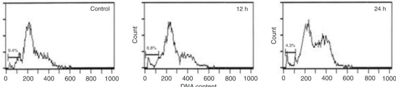
The death and proliferation of cells irradiated after TPA treatment were determined by flow cytometry. The cells
irradiated at peak mPer2 expression had much lower apop-totic peaks than cells irradiated at trough mPer2 expression
and than control cells irradiated without TPA treatment (P < 0.01; Figure 1).
Figure 1. Apoptosis in NIH 3T3 cells irradiated after 12-O-tetradecanoylphorbol-13-acetate (TPA) treatment. Control: cells irradiated
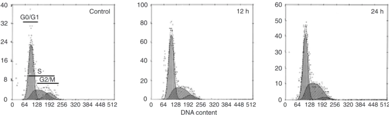
The G0/G1, S-, G2, and M-phases of the cell cycle
were also analyzed by flow cytometry, with the following cell percentages being observed in each phase: control
group, G0/G1 (59.3 ± 1.07%), S (29.2 ± 0.30%), and G2/M (11.7 ± 0.21%); TPA 12-h group, G0/G1 (58.8 ± 0.91%), S (32.2 ± 0.66%) and G2/M (9.4 ± 0.47%); TPA 24-h group, G0/G1 (53.2 ± 0.73%), S (42.7 ± 0.43%), and G2/M (4.1 ± 0.34%). The S-phase fraction was greater in TPA 24-h group cells than in TPA 12-h group cells and control cells (P < 0.01; Figure 2). Clonogenic
survival was determined by the colony formation assay. The colony-forming efficiency was significantly higher
in TPA 24-h group cells than in TPA 12-h group cells and control cells (P < 0.01; Figure 3). Irradiated NIH
3T3 cells expressing high levels of mPer2 showed less cell death and more cell proliferation and clonogenic survival. Three independent experiments demonstrated similar results.
Role of radiation in NIH 3T3 cells with up-regulated mPer2
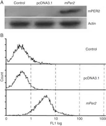
We established mPer2-overexpressing cells by
transfecting pcDNA3.1(+)-mPer2 into NIH 3T3 cells. Successful transfection of the mPer2 gene using the lipofectamine 2000 reagent was confirmed by Western
blotting (Figure 4A). It was also evident by flow cytom
-etry that the fluorescence intensity of mPER2 protein expression in pcDNA3.1(+)-mPer2-transfected cells was
Figure 2. The cell cycle in NIH 3T3 cells irradiated after 12-O-tetradecanoylphorbol-13-acetate (TPA) treatment. Control: cells irradi
-ated without TPA treatment; 12 h: cells irradi-ated 12 h after TPA treatment; 24 h: cells irradi-ated 24 h after TPA treatment. G0/G1, S and G2/M phases are indicated. The S-phase fraction of the TPA 24-h group was significantly higher than that of the TPA 12-h group and of control cells (P < 0.01, one-way ANOVA).
Figure 3. Colony formation of
12-O-tetradecanoylphorbol-13-acetate (TPA)-treated NIH 3T3 cells after irradiation. A,Control:
cells irradiated without TPA treatment; 12 h: cells irradiated 12 h after TPA treatment; 24 h: cells irradiated 24 h after TPA treat -ment. B, High expression of mPER2 dramatically increased the
clonogenic survival of irradiated NIH 3T3 cells. The
886 L. Chang et al.
higher than in pcDNA3.1-transfected cells and in control cells without transfection (Figure 4B).
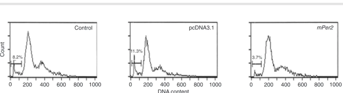
The effect of mPer2 on the death, proliferation and clonogenic survival of cells irradiated after transfection was
determined by flow cytometry and the colony formation as -say. The results revealed that the apoptotic peak was lower in mPer2 overexpressing cells than in empty vector cells
and control cells (P < 0.01; Figure 5). Analysis of cell cycle distribution showed: control group, G0/G1 (46.3 ± 1.21%), S (40.9 ± 1.67%), and G2/M (12.8 ± 0.78%); pcDNA3.1 group, G0/G1 (55.6 ± 0.84%), S (39.0 ± 0.56%), and G2/M (5.4 ± 0.41%); pcDNA3.1-mPer2 group, G0/G1 (45.9 ± 1.04%), S (46.5 ± 0.72%), and G2/M (7.6 ± 0.54%). The
S-phase fraction of mPer2 overexpressing cells was higher
than that of empty vector cells and control cells (P < 0.01; Figure 6). mPER2 dramatically increased the clonogenic
survival of irradiated NIH 3T3 cells. The colony-forming
ef-ficiency of mPer2 overexpressing cells was also higher than
that of other cells (P < 0.01; Figure 7). Three independent
experiments demonstrated that irradiated NIH 3T3 cells overexpressing mPer2 also showed less cell death and more cell proliferation and clonogenic survival.
mPer2 up-regulated the expressions of cell
proliferation- and apoptosis-related genes and DNA repair-related genes and proteins
We chose RT-PCR analysis and immunohistochemis -try to study the mechanism of the effects of mPer2 gene expression on the response of NIH 3T3 cells to radiation.
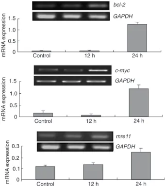
RT-PCR analysis showed that the mRNA levels of bax,
bcl-2, p53, c-myc, mre11, and nbs1 were up-regulated in the irradiated cells at peak mPer2 expression compared with cells at trough mPer2 expression and control cells, with the expression of c-myc and bcl-2 being particularly
up-regulated. However, there was no significant difference
in rad50 level among the three cell groups (Figure 8). Im -munohistochemistry also showed increased protein levels
of P53, BAX and PCNA in irradiated NIH 3T3 cells with high mPER2 expression (Figure 9). Percentages of positive cells were: P53, control (34.1 ± 2.7%), 12 h (40.2 ± 3.9%), and 24 h (61.3 ± 3.6%); BAX, control (20.7 ± 4.1%), 12 h (22.8 ± 4.3%), and 24 h (37.1 ± 6.4%); PCNA, control (29.4 ±
3.2%), 12 h (33.7 ± 5.6%), and 24 h (87.3 ± 4.7%). Three independent experiments demonstrated similar results.
Discussion
Circadian clocks are molecular time-keeping mecha-nisms that reside in a diverse range of cell types in a variety of organisms. The primary role of these cell-autonomous clocks is to maintain their own approximately 24-h molecu-lar rhythms and to drive the rhythmic expression of genes involved in physiology, metabolism and behavior (19,20). The circadian clock is composed of multiple, single-cell circadian oscillators, which, when synchronized, generate coordinated circadian outputs that regulate overt rhythms.
Eight clock genes involved in the interacting transcriptional-/
translational-feedback loops that compose the molecular clockwork have been cloned (21).
Parental NIH 3T3 fibroblasts normally produce very low and barely detectable levels of circadian genes. TPA
treatment is as effective as serum shock in triggering the induction of circadian gene expression in cultured cells, including mPer2. As a core circadian gene, mPer2 not only maintains the circadian rhythm of cells, but also sustains
the normal cell cycle. After TPA treatment, parental NIH 3T3
cells present circadian oscillation of mPer2 expression within an approximate period of 24 h, with the trough at zeitgeber time 12 (ZT12)and the peak at ZT24 (18,22,23).
In the present study, we treated NIH 3T3 cells with TPA
to induce endogenous mPer2 expression, and cells were irradiated at ZT12 with trough mPer2 level and at ZT24 with peak mPer2 level. The death, proliferation and clonogenic
survival of irradiated cells were determined by flow cytom -Figure 4. Detection of mPER2 expression by Western blot
analysis and flow cytometry. Control: cells without transfection; pcDNA3.1: cells transfected with pcDNA3.1; mPer2: cells
trans-fected with pcDNA3.1-mPer2. A, Western blot analysis of mPER2
expression. The mPER2 expression of mPer2 group cells was
significantly higher than that of pcDNA3.1 group cells and of con -trol cells. B, Flow cytometric analysis of mPER2 expression. The
fluorescence intensity of mPER2 expression in mPer2 group cells
Figure 7. Colony formation of transfected NIH 3T3 cells after irradiation. A, Control: cells without transfection; pcDNA3.1:
cells transfected with pcDNA3.1; mPer2: cells transfected with
pcDNA3.1-mPer2. B, mPER2 dramatically increased the clono -genic survival of irradiated NIH 3T3 cells. The colony-forming
ef-ficiency of mPer2 overexpressing cells was significantly higher
than that of empty vector cells and of control cells (P < 0.01, one-way ANOVA).
Figure 6. The cell cycle in NIH 3T3 cells irradiated after transfection. Control: cells without transfection; pcDNA3.1: cells transfected
with pcDNA3.1; mPer2: cells transfected with pcDNA3.1-mPer2. G0/G1, S and G2/M phases are indicated. The S-phase fraction of mPer2 overexpressing cells was higher than that of empty vector cells and of control cells (P < 0.01, one-way ANOVA).
Figure 5. Apoptosis in NIH 3T3 cells irradiated after transfection. Control: cells without transfection; pcDNA3.1: cells transfected with
pcDNA3.1; mPer2: cells transfected with pcDNA3.1-mPer2. The percentage of sub-G1 cells undergoing apoptosis is indicated by the
bar. The apoptotic peak of mPer2 overexpressing cells was lower than that of empty vector cells and of control cells (P < 0.01, one-way
888 L. Chang et al.
Figure 9. Expression of P53 (A), BAX (B) and proliferating cell number antigen (PCNA; C) proteins determined by immunocytochem
-istry (100X, 400X magnification). Control: cells irradiated without 12-O-tetradecanoylphorbol-13-acetate (TPA) treatment; 12 h: cells irradiated 12 h after TPA treatment; 24 h: cells irradiated 24 h after TPA treatment. P53, BAX and PCNA levels were up-regulated in
cells irradiated with peak mPer2 level (24-h group) compared to cells irradiated with trough mPer2 level (12-h group) and control cells.
Magnification bar = 200 μm.
Figure 8. RT-PCR analysis of mouse bax, bcl-2, p53, c-myc, rad50, mre11,
and nbs1 genes (RT-PCR electropherogram and integral optical density ratio
results). Control: cells irradiated without 12-O-tetradecanoylphorbol-13-acetate (TPA) treatment; 12 h: cells irradiated 12 h after TPA treatment with trough
mPer2 level; 24 h: cells irradiated 24 h after TPA treatment with peak mPer2
level. The mRNA levels of mouse bax, bcl-2, p53, c-myc, mre11, and nbs1
etry and colony formation assay. The results of the present study showed that high expression of mPer2 might reduce cell death and enhance cell proliferation of irradiated NIH
3T3 cells. However, after TPA treatment, parental NIH 3T3
cells presented circadian expression not only of mPer2, but also of other clock and clock-related genes such as mPer1
and DBP (18). Therefore, the radiobiological effects may result from mPer2 or other clock and clock-related genes. In order to assess the effects of mPer2 expression on the
irradiated cells, we transfected pcDNA3.1(+)-mPer2 into NIH 3T3 cells. The results showed that overexpression of
mPER2 in NIH 3T3 cells resulted in reduced cell death and
enhanced cell proliferation after radiation, suggesting that overexpressed mPer2 may diminish the radiosensitivity of NIH 3T3 cells.
Fu et al. (16) reported that the loss of mPer2 function
resulted in increased tumor development and deficiencies in response to DNA damage in mice, suggesting that the mPer2 gene functions in tumor suppression by regulating
DNA damage-responsive pathways. Compared with
wild-type mice, mPer2 mutant mice showed a neoplastic growth
phenotype and an increased sensitivity to γ radiation in thy -mocytes. Zhang et al. (17) also found that high expression of the circadian gene mPer2 might diminish radiosensitivity
of irradiated mouse tumor cells. LLC and EMT6 cells with
high mPer2 expression exposed to 60Co-γ-rays presented
reduced DNA damage, increased survival and clone-forming
rate, which suggested that mPer2 might protect cells against
the radiation injury of γ-rays and increase the survival rate
of tumor cells. The present study demonstrated that high expression of mPer2 could diminish the radiosensitivity of NIH 3T3 cells, with reduced cell death, enhanced cell proliferation and increased clonogenic survival. Moreover, tumor cells and NIH 3T3 cells were irradiated at different
times after TPA induction, i.e., at 12 h (trough time of mPer2
expression) and at 24 h (peak time) after induction, whereas LLC cells were irradiated at 18 h (trough time) and 30 h (peak time) after induction. The present research combined
with the previous research of Fu et al. (16) and Zhang et al. (17) would contribute to protecting normal cells against radiation injury in cancer radiotherapy. We propose that radiotherapy could be used against cancer at a certain time of Per2 expression for the best ratio of tumor suppression and normal tissue protection, which can kill tumor cells and protect normal cells maximally. However, our hypothesis needs further study.
Without radiation, overexpression of mPER2 results in
reduced proliferation and rapid apoptosis of tumor cells, but not of NIH 3T3 cells, suggesting that mPer2 may play an important role in tumor suppression by inducing apoptotic cell death (24). However, irradiated NIH 3T3 cells with
mPER2 overexpression showed reduced cell death, sug -gesting that mPer2 may affect the radiation-induced cell death by other ways.
In response to ionizing radiation, cells immediately
activate a series of biochemical pathways that promote cell survival while maintaining genetic integrity. The main cellular defense system against ionizing radiation exposure is composed of two distinct types of biochemical pathways,
i.e., the DNA damage cell cycle checkpoint pathways and the DNA repair pathways (25). Arrest of replicative DNA synthesis after DNA damage is thought to occur to provide ample time for the cell to repair DNA lesions before the S-phase (G1 arrest) and/or mitosis (G2 arrest).
The RAD50-MRE11-NBS1 (MRN) complex plays an important role in the repair of DNA damage caused by
radiation. The complex practically participates in all repair mechanisms, especially homologous recombination and non-homologous end-joining, which are the most important
mechanisms in the repair of DNA double-strand breaks (26).
In the present study, the levels of rad50 expression of the various cell groups were not different, in agreement with reports showing that rad50 expression is persistent and
stable (27). As the core part of the MRN complex, MRE11
has not only nuclease activity, but also a connective effect
on RAD50 and NBS1, and its expression changes rapidly
after radiation (28). NBS1 is also an important part of the MRN complex (29). The irradiated NIH 3T3 cells expressing high levels of mPer2 showed an increased expression of
mre1l and nbs1, whichsuggested that mPer2 could enhance
the function of DNA damage repair MRN complex. c-Myc, a proto-oncogene, plays an important role in both cell proliferation and apoptosis (30,31). Circadian regulators may target genes that are controlled by c-myc. c-Myc itself is also controlled by the circadian clock, and the level of
c-myc mRNA oscillated in 24-h light/dark cycles in wild-type mouse livers, peaking at ZT14 (16). Gamma radiation may
increase the expression of c-myc, whose overexpression could drive cells to progress through the cell cycle in the
presence of genomic DNA damage in order to improve the efficiency of DNA repair (32). It has also been reported
that c-myc directly regulates the transcription of the nbs1 gene involved in DNA double-strand break repair (33). The present study showed that c-myc expression increasedin irradiated NIH 3T3 cells with high expression of mPer2, thus suppressing cell death and enhancing cell repair.
BCL-2 and BAX are members of the BCL-2 family
that is a key regulator of the mitochondrial response to apoptotic signals in the intrinsic pathway (34,35). Bcl-2
is an antiapoptotic gene acting as a potent suppressor of apoptosis by blocking the release of cytochrome c, whereas
bax is a proapoptotic gene with oppose functions acting as a promoter of cell death. The ratio of antiapoptotic-to-proapoptotic molecules determines the response to a death signal (36). In the present study, the high expression of
mPer2 increased the BCL-2/BAX ratio, thus suppressing
cell death and enhancing cell growth.
Wild-type P53 protein levels rise dramatically after
exposure to ionizing radiation. This rise results from as yet
890 L. Chang et al.
undergone by the P53 protein such as phosphorylation, bind
-ing to other proteins, or oligomerization. At subsequent end points of DNA-damage, a prolonged half-life was observed as well as increased DNA-binding activity of the P53 protein
and enhanced transcriptional transactivation activity driven
by this protein (37,38). P53 can keep genomic stability by mediating apoptosis and DNA repair (7-9,39) and can induce a transient arrest of the cell cycle at G1, so that the cells will have time to repair damaged DNA. Activated p53 can also
eliminate cells through mechanisms involving prolonged
arrest at G1 or apoptosis. Our study showed an increased
expression of p53 in irradiated NIH 3T3 cells at peak mPer2
expression, with decreased cell death. This indicated that
a high expression of mPER2 up-regulated P53 expression, which could enhance DNA repair.
PCNA is a protein that acts as a processivity factor for DNA polymerase delta in eukaryotic cells. PCNA is an es -sential factor in cell proliferation and can be used as an index
to evaluate cell proliferation (40). Since DNA polymerase delta is involved in resynthesis of excised damaged DNA strands during DNA repair, PCNA is important for both DNA
synthesis and DNA repair. PCNA is also involved in the DNA damage tolerance pathway known as post-replication
repair. In the present study, the irradiated cells with high expression of mPer2 showed enhanced cell proliferation,
and the increase in PCNA staining after DNA damage could be a result of DNA repair.
After radiation, the high expression of mPer2 in NIH 3T3 cells results in reduced cell death and enhanced cell proliferation and clonogenic survival, which means that high expression of the circadian gene mPer2 diminishes
radiosensitivity of NIH 3T3 cells. A high expression of mPer2
may up-regulate the expression of apoptosis-related genes, with an increased proportionality of bcl-2/bax, and may reduce cell death and enhance cell proliferation to dimin-ish radiosensitivity. Moreover, mPer2 may also up-regulate
DNA repair-related genes to increase DNA-repair efficiency after radiation to diminish radiosensitivity. Future research
should be focused on studying the detailed mechanisms by which the circadian clock controls genes related to radiosensitivity.
References
1. Lorimore SA, Wright EG. Radiation-induced genomic in
-stability and bystander effects: related inflammatory-type responses to radiation-induced stress and injury? A review.
Int J Radiat Biol 2003; 79: 15-25.
2. Cadet J, Bellon S, Douki T, Frelon S, Gasparutto D, Muller E, et al. Radiation-induced DNA damage: formation, measure -ment, and biochemical features. J Environ Pathol Toxicol Oncol 2004; 23: 33-43.
3. Bourguignon MH, Gisone PA, Perez MR, Michelin S, Dubner D, Giorgio MD, et al. Genetic and epigenetic features in ra
-diation sensitivity. Part I: cell signalling in ra-diation response.
Eur J Nucl Med Mol Imaging 2005; 32: 229-246.
4. Kent CR, Eady JJ, Ross GM, Steel GG. The comet moment as a measure of DNA damage in the comet assay. Int J Radiat Biol 1995; 67: 655-660.
5. Willers H, Dahm-Daphi J, Powell SN. Repair of radiation damage to DNA. Br J Cancer 2004; 90: 1297-1301.
6. Wilson GD. Radiation and the cell cycle, revisited. Cancer Metastasis Rev 2004; 23: 209-225.
7. Biard DS, Martin M, Rhun YL, Duthu A, Lefaix JL, May E, et
al. Concomitant p53 gene mutation and increased radiosen-sitivity in rat lung embryo epithelial cells during neoplastic development. Cancer Res 1994; 54: 3361-3364.
8. Kastan MB, Onyekwere O, Sidransky D, Vogelstein B, Craig
RW. Participation of p53 protein in the cellular response to
DNA damage. Cancer Res 1991; 51: 6304-6311.
9. Vogelstein B, Lane D, Levine AJ. Surfing the p53 network.
Nature 2000; 408: 307-310.
10. Albrecht U, Eichele G. The mammalian circadian clock. Curr Opin Genet Dev 2003; 13: 271-277.
11. Pittendrigh CS. Temporal organization: reflections of a Dar -winian clock-watcher. Annu Rev Physiol 1993; 55: 16-54.
12. Richter HG, Torres-Farfan C, Rojas-Garcia PP, Campino C, Torrealba F, Seron-Ferre M. The circadian timing system:
making sense of day/night gene expression. Biol Res 2004; 37: 11-28.
13. Andretic R, Chaney S, Hirsh J. Requirement of circadian
genes for cocaine sensitization in Drosophila. Science 1999; 285: 1066-1068.
14. Filipski E, King VM, Li X, Granda TG, Mormont MC, Liu X, et
al. Host circadian clock as a control point in tumor progres-sion. J Natl Cancer Inst 2002; 94: 690-697.
15. Haus E. Chronobiology of the mammalian response to ioniz
-ing radiation. Potential applications in oncology. Chronobiol Int 2002; 19: 77-100.
16. Fu L, Pelicano H, Liu J, Huang P, Lee C. The circadian gene Period2 plays an important role in tumor suppression and DNA damage response in vivo. Cell 2002; 111: 41-50. 17. Zhang J, Zhu B, Liu Y, Jiang Z, Wang Y, Li Y, et al. High
expression of circadian gene mPer2 diminishes radiosensi -tivity of tumor cells. Cancer Biother Radiopharm 2008; 23: 561-570.
18. Akashi M, Nishida E. Involvement of the MAP kinase cas -cade in resetting of the mammalian circadian clock. Genes Dev 2000; 14: 645-649.
19. Andretic R, Hirsh J. Circadian modulation of dopamine
receptor responsiveness in Drosophila melanogaster. Proc Natl Acad Sci U S A 2000; 97: 1873-1878.
20. Dunlap JC. Molecular bases for circadian clocks. Cell 1999; 96: 271-290.
21. Reppert SM, Weaver DR. Molecular analysis of mammalian circadian rhythms. Annu Rev Physiol 2001; 63: 647-676.
22. Balsalobre A, Damiola F, Schibler U. A serum shock induces
circadian gene expression in mammalian tissue culture cells.
Cell 1998; 93: 929-937.
24. Hua H, Wang Y, Wan C, Liu Y, Zhu B, Yang C, et al. Circadian
gene mPer2 overexpression induces cancer cell apoptosis.
Cancer Sci 2006; 97: 589-596.
25. Li L, Story M, Legerski RJ. Cellular responses to ionizing radiation damage. Int J Radiat Oncol Biol Phys 2001; 49: 1157-1162.
26. Khanna KK, Jackson SP. DNA double-strand breaks: signal -ing, repair and the cancer connection. Nat Genet 2001; 27: 247-254.
27. Dolganov GM, Maser RS, Novikov A, Tosto L, Chong S, Bressan DA, et al. Human Rad50 is physically associated
with human Mre11: identification of a conserved multiprotein complex implicated in recombinational DNA repair. Mol Cell Biol 1996; 16: 4832-4841.
28. D’Amours D, Jackson SP. The Mre11 complex: at the cross
-roads of DNA repair and checkpoint signalling. Nat Rev Mol Cell Biol 2002; 3: 317-327.
29. Kobayashi J, Antoccia A, Tauchi H, Matsuura S, Komatsu K. NBS1 and its functional role in the DNA damage response.
DNA Repair 2004; 3: 855-861.
30. Blackwell TK, Huang J, Ma A, Kretzner L, Alt FW, Eisenman
RN, et al. Binding of myc proteins to canonical and non-Binding of myc proteins to canonical and
non-canonical DNA sequences. Mol Cell Biol 1993; 13: 5216-5224.
31. Evan GI, Vousden KH. Proliferation, cell cycle and apoptosis
in cancer. Nature 2001; 411: 342-348.
32. Bil’din VN, Seregina TB, Pospelova TV. [The regulation of
DNA repair processes in mammalian cells. II. The repair of DNA radiation damage in NIH 3T3 murine cells transformed
by the v-myc oncogene]. Tsitologiia 1991; 33: 39-47.
33. Chiang YC, Teng SC, Su YN, Hsieh FJ, Wu KJ. c-Myc di -rectly regulates the transcription of the NBS1 gene involved
in DNA double-strand break repair. J Biol Chem 2003; 278: 19286-19291.
34. Adams JM, Cory S. The Bcl-2 protein family: arbiters of cell
survival. Science 1998; 281: 1322-1326.
35. Cory S, Adams JM. The Bcl2 family: regulators of the cellular
life-or-death switch. Nat Rev Cancer 2002; 2: 647-656. 36. Tsujimoto Y. Cell death regulation by the Bcl-2 protein family
in the mitochondria. J Cell Physiol 2003; 195: 158-167.
37. Fei P, El-Deiry WS. P53 and radiation responses. Oncogene
2003; 22: 5774-5783.
38. Li CY, Nagasawa H, Dahlberg WK, Little JB. Diminished
capacity for p53 in mediating a radiation-induced G1 arrest
in established human tumor cell lines. Oncogene 1995; 11: 1885-1892.
39. Benchimol S. p53-dependent pathways of apoptosis. Cell Death Differ 2001; 8: 1049-1051.
40. Takasaki Y, Kaneda K, Takeuchi K, Matsudaira R, Matsishita